Shang Xian-Jin, He Cai-Feng, Tang Biao, Chang Xiao-Li, Ci Chao, Sang Hong
Department of Neurology, Yijishan Hospital of Wannan Medical College, Wuhu, 241001, People's Republic of China.
Jinling Hospital Department of Dermatology, Nanjing Medical University, Nanjing, 210002, People's Republic of China.
Dermatol Ther (Heidelb). 2020 Apr;10(2):273-283. doi: 10.1007/s13555-020-00361-3. Epub 2020 Mar 2.
Many studies have explored the imaging characteristics of patients with neurosyphilis, but no systematic study has been made on the neuroimaging changes after anti-syphilitic treatment. The purpose of this study was to examine neuroimaging differences before and after treatment, comparing patients with asymptomatic and symptomatic neurosyphilis.
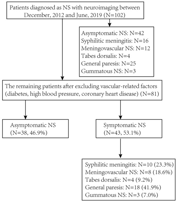
A total of 102 patients with neurosyphilis, including 60 cases of symptomatic neurosyphilis and 42 cases of asymptomatic neurosyphilis, were identified between December 2012 and June 2019. Their demographics, medical histories, serological tests of peripheral blood and cerebrospinal fluid, and especially neuroimaging features before and after anti-syphilitic treatment were collected and analyzed.
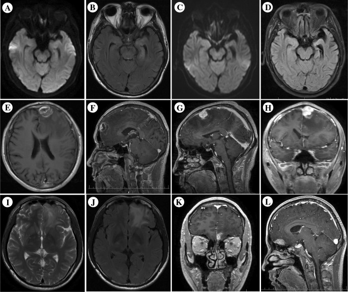
The patients presented with variable clinical and neuroimaging features, including cerebral infarction or hemorrhage, atrophy, demyelination, arteritis, encephalitis, and hippocampal sclerosis. A total of 29 neuroradiological re-examinations were performed in 19 patients treated with anti-syphilitic medicine. The results indicated that some patients still presented neuroradiological progression after treatment, including 42.1% showing infarction lesions, 47.4% mild to severe brain atrophy, and 15.8% white matter demyelination.
The clinical and neuroimaging features of neurosyphilis patients are diverse, and their follow-up neuroimaging continued to show progression even with standardized treatment.
许多研究探讨了神经梅毒患者的影像学特征,但尚未对抗梅毒治疗后的神经影像学变化进行系统研究。本研究的目的是检查治疗前后的神经影像学差异,比较无症状和有症状神经梅毒患者。
2012年12月至2019年6月期间共确诊102例神经梅毒患者,其中有症状神经梅毒60例,无症状神经梅毒42例。收集并分析了他们的人口统计学资料、病史、外周血和脑脊液的血清学检查结果,尤其是抗梅毒治疗前后的神经影像学特征。
患者表现出多样的临床和神经影像学特征,包括脑梗死或出血、萎缩、脱髓鞘、动脉炎、脑炎和海马硬化。19例接受抗梅毒药物治疗的患者共进行了29次神经放射学复查。结果表明,部分患者治疗后仍有神经放射学进展,包括42.1%出现梗死灶,47.4%出现轻度至重度脑萎缩,15.8%出现白质脱髓鞘。
神经梅毒患者的临床和神经影像学特征多样,即使经过标准化治疗,其随访神经影像学仍显示有进展。