• 文献检索
  • 文档翻译
  • 深度研究
  • 学术资讯
  • Suppr Zotero 插件Zotero 插件
  • 邀请有礼
  • 套餐&价格
  • 历史记录
应用&插件
Suppr Zotero 插件Zotero 插件浏览器插件Mac 客户端Windows 客户端微信小程序
定价
高级版会员购买积分包购买API积分包
服务
文献检索文档翻译深度研究API 文档MCP 服务
关于我们
关于 Suppr公司介绍联系我们用户协议隐私条款
关注我们

Suppr 超能文献

核心技术专利:CN118964589B侵权必究
粤ICP备2023148730 号-1Suppr @ 2026

文献检索

告别复杂PubMed语法,用中文像聊天一样搜索,搜遍4000万医学文献。AI智能推荐,让科研检索更轻松。

立即免费搜索

文件翻译

保留排版,准确专业,支持PDF/Word/PPT等文件格式,支持 12+语言互译。

免费翻译文档

深度研究

AI帮你快速写综述,25分钟生成高质量综述,智能提取关键信息,辅助科研写作。

立即免费体验

一种用于精确识别肾移植排斥反应的多模态计算机辅助诊断系统:初步结果。

A multimodal computer-aided diagnostic system for precise identification of renal allograft rejection: Preliminary results.

作者信息

Shehata Mohamed, Shalaby Ahmed, Switala Andrew E, El-Baz Maryam, Ghazal Mohammed, Fraiwan Luay, Khalil Ashraf, El-Ghar Mohamed Abou, Badawy Mohamed, Bakr Ashraf M, Dwyer Amy, Elmaghraby Adel, Giridharan Guruprasad, Keynton Robert, El-Baz Ayman

机构信息

BioImaging Lab, Department of Bioengineering, University of Louisville, Louisville, KY, 40208, USA.

Electrical and Computer Engineering Department, Abu Dhabi University, Abu Dhabi, 59911, UAE.

出版信息

Med Phys. 2020 Jun;47(6):2427-2440. doi: 10.1002/mp.14109. Epub 2020 Apr 3.

DOI:10.1002/mp.14109
PMID:32130734
原文链接:https://pmc.ncbi.nlm.nih.gov/articles/PMC8524762/
Abstract

PURPOSE

Early assessment of renal allograft function post-transplantation is crucial to minimize and control allograft rejection. Biopsy - the gold standard - is used only as a last resort due to its invasiveness, high cost, adverse events (e.g., bleeding, infection, etc.), and the time for reporting. To overcome these limitations, a renal computer-assisted diagnostic (Renal-CAD) system was developed to assess kidney transplant function.

METHODS

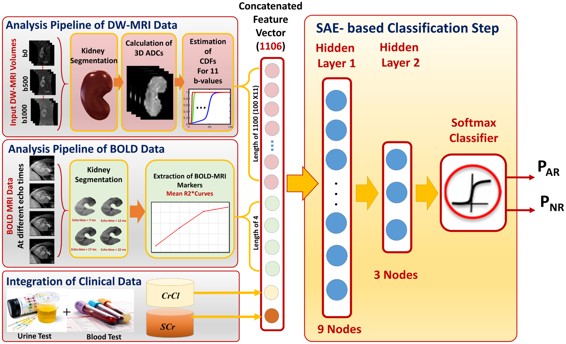
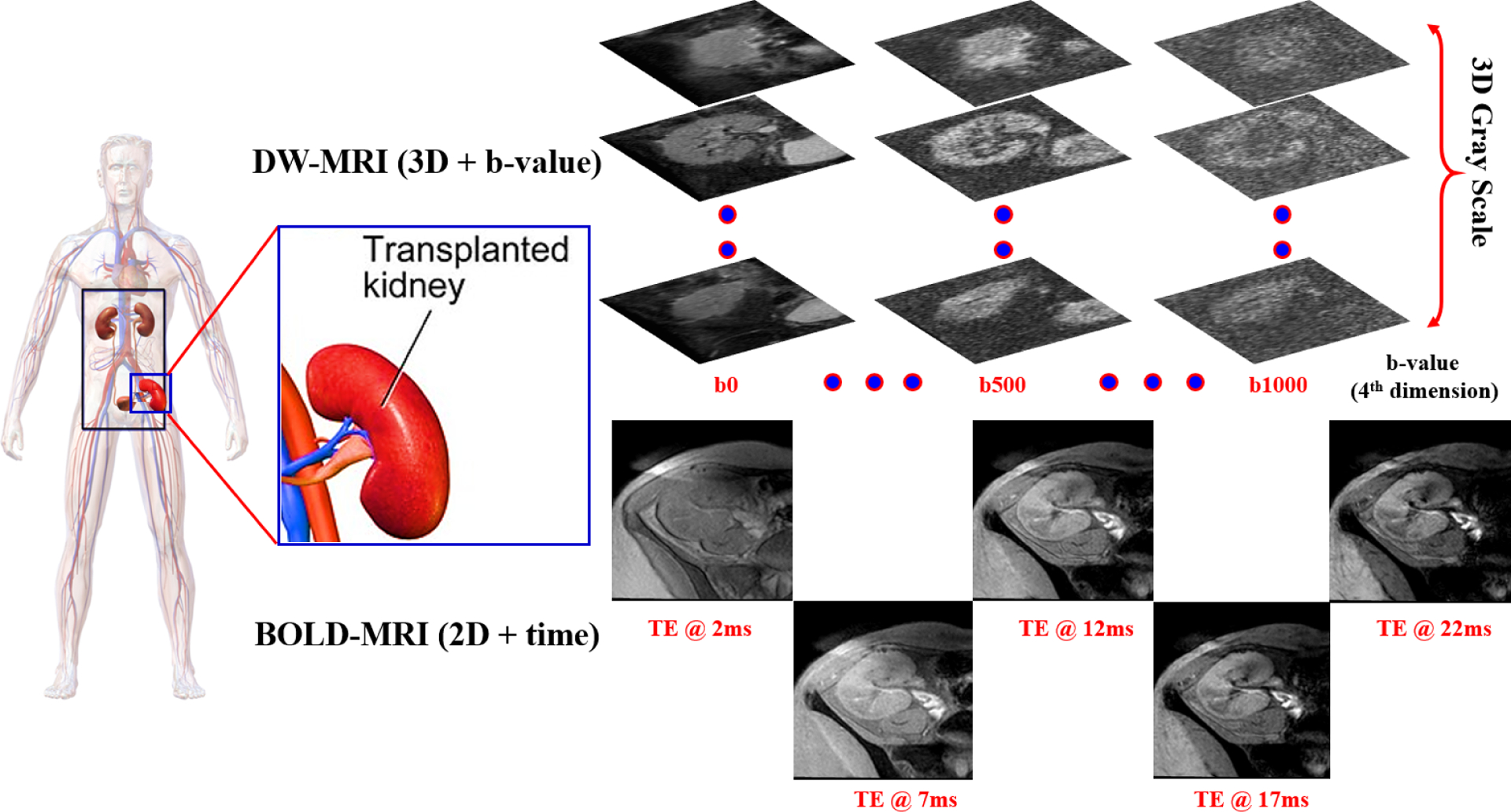
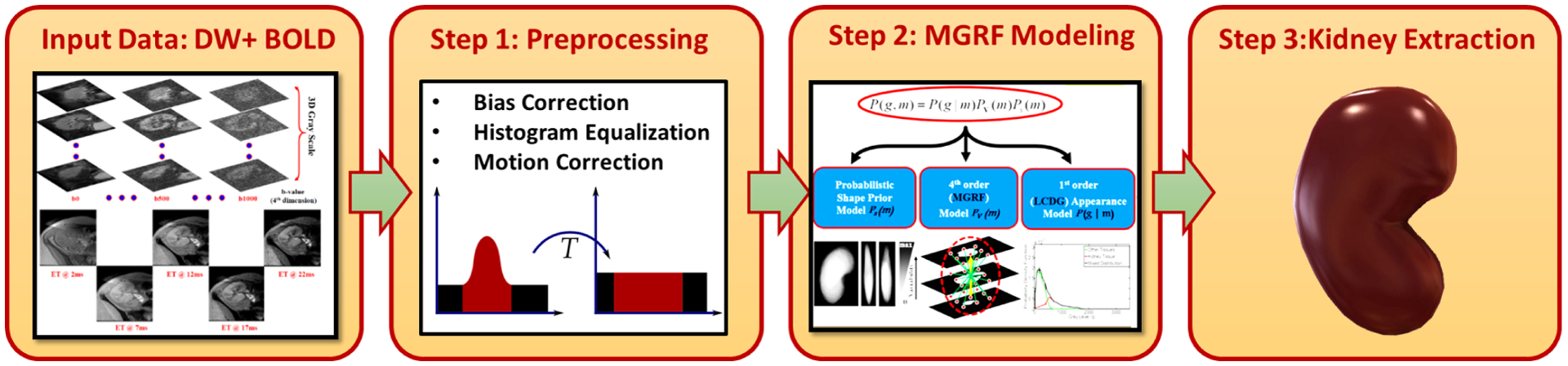
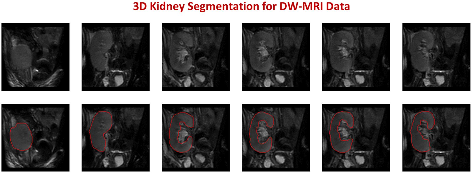
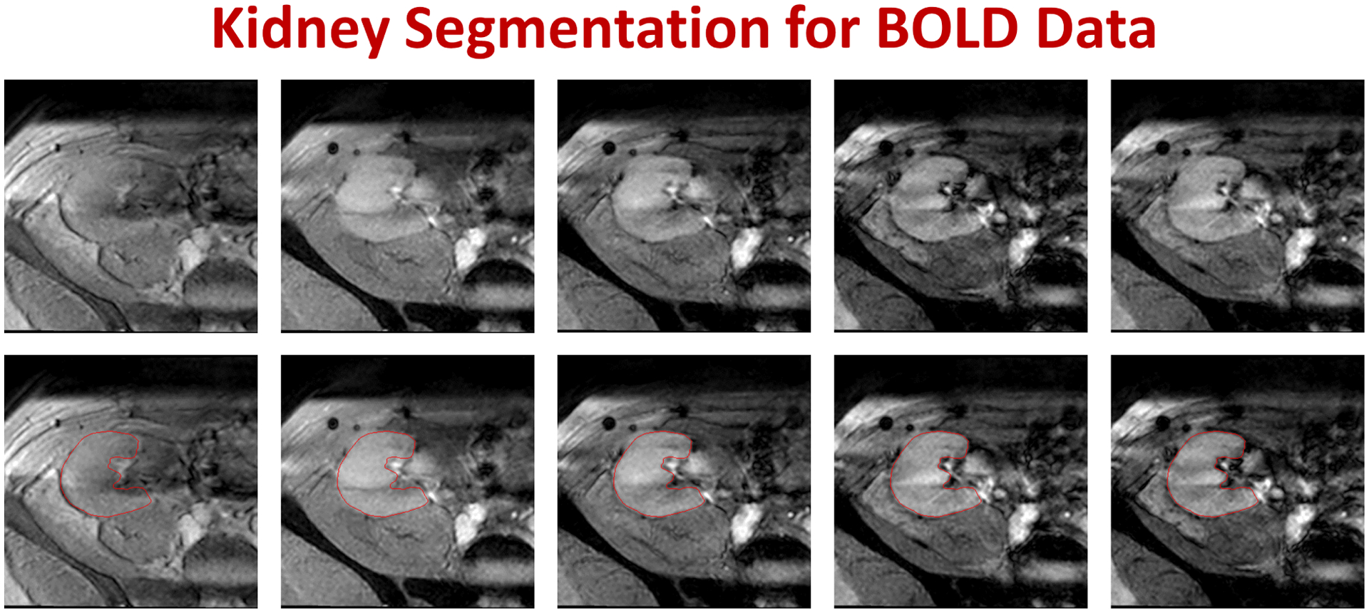
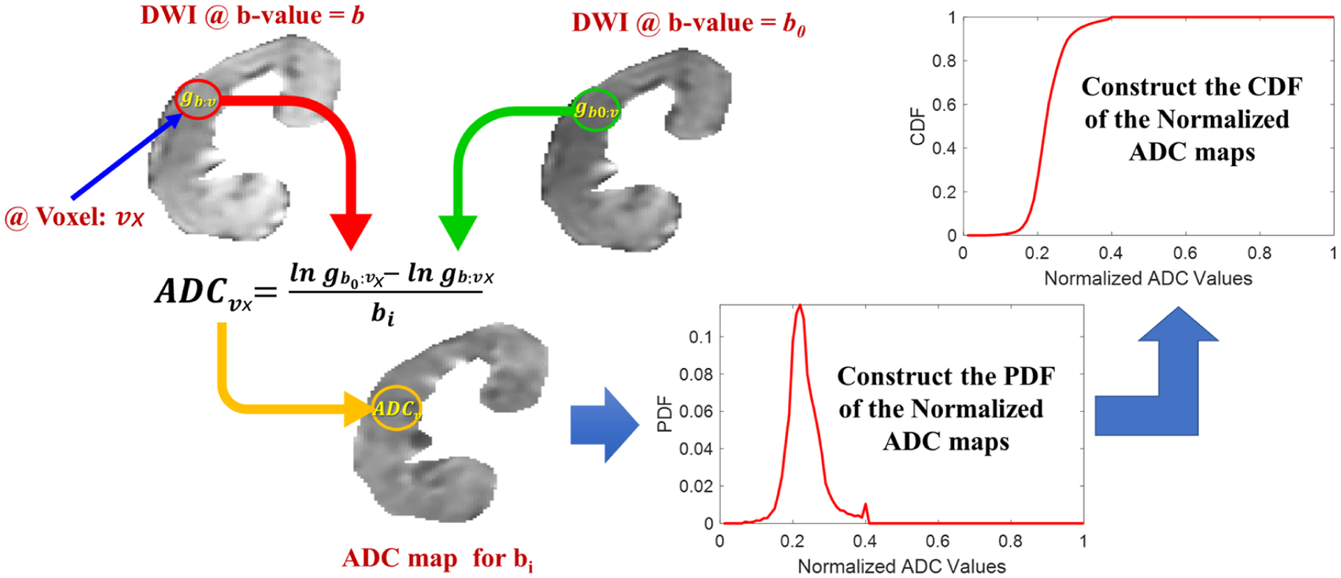
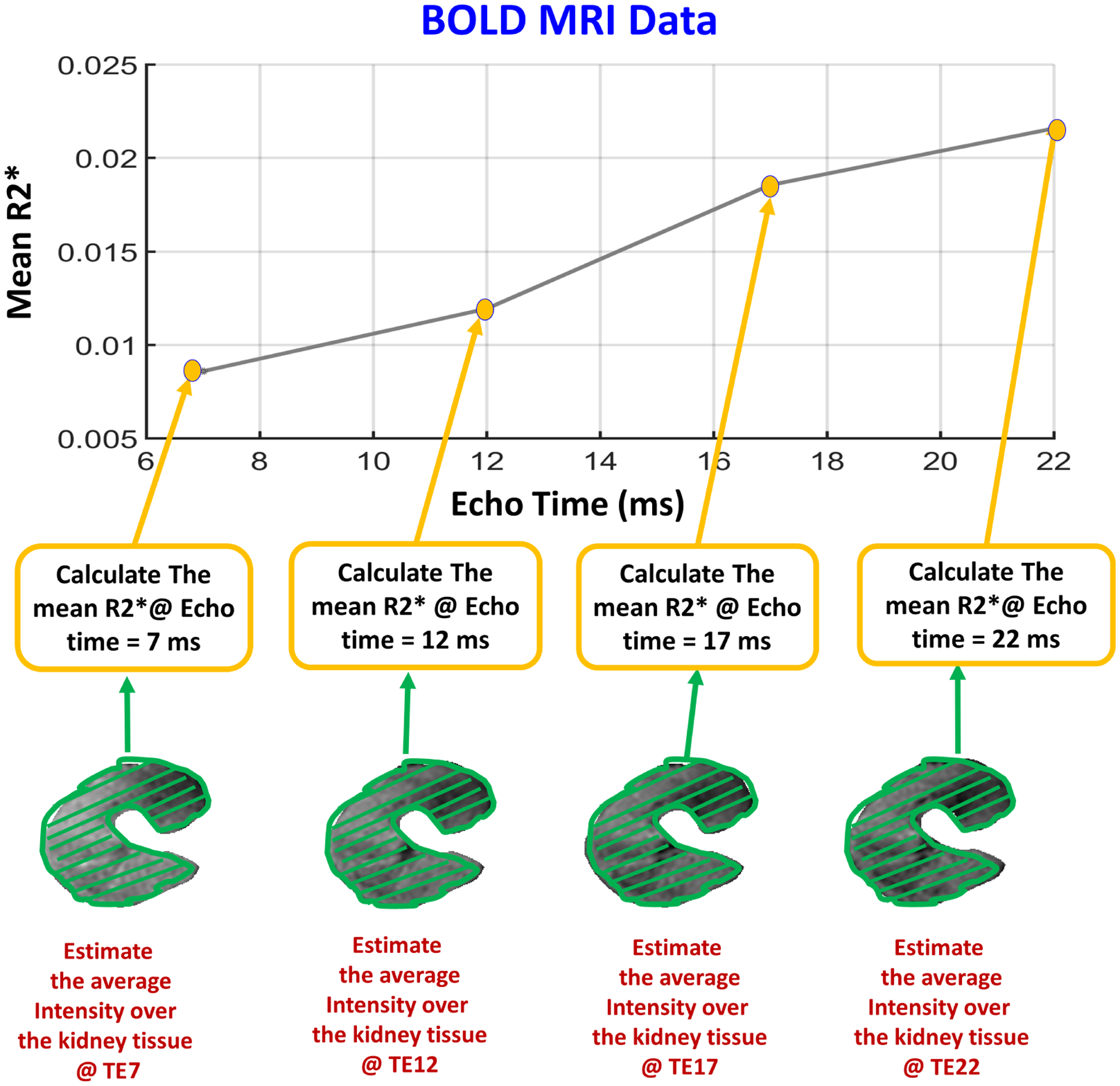
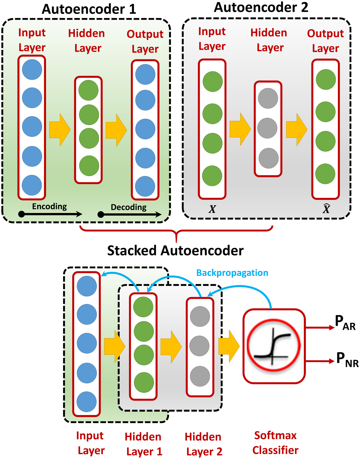
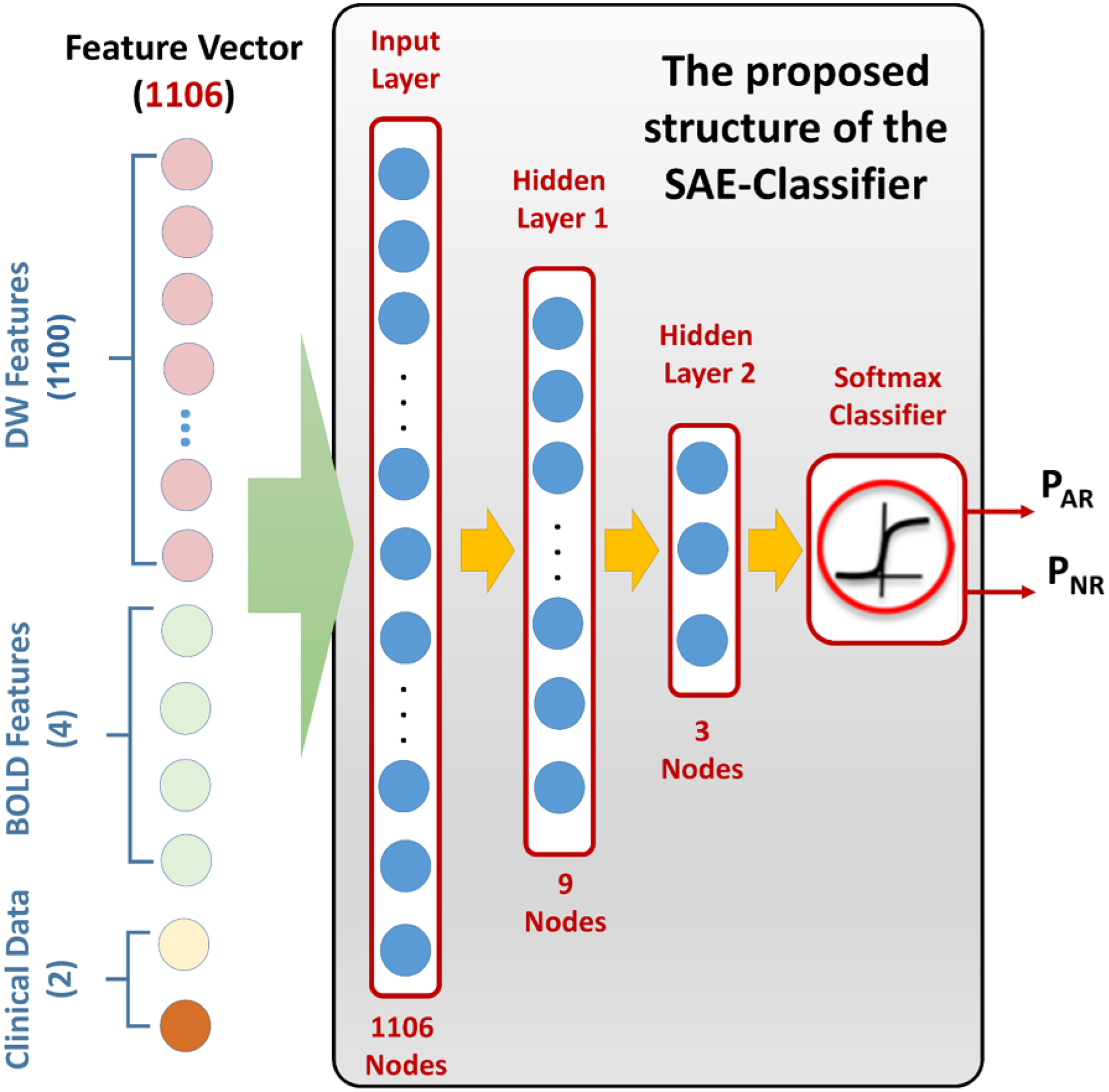
The developed Renal-CAD system integrates data collected from two image-based sources and two clinical-based sources to assess renal transplant function. The imaging sources were the apparent diffusion coefficients (ADCs) extracted from 47 diffusion-weighted magnetic resonance imaging (DW-MRI) scans at 11 different b-values (b0, b50, b100, ..., b1000 s/mm ), and the transverse relaxation rate (R2*) extracted from 30 blood oxygen level-dependent MRI (BOLD-MRI) scans at 5 different echo times (TEs = 2, 7, 12, 17, and 22 ms). Serum creatinine (SCr) and creatinine clearance (CrCl) were the clinical sources for kidney function evaluation. The Renal-CAD system initially performed kidney segmentation using the level-set method, followed by estimation of the ADCs from DW-MRIs and the R2* from BOLD-MRIs. ADCs and R2* estimates from 30 subjects that have both types of scans were integrated with their associated SCr and CrCl. The integrated biomarkers were then used as our discriminatory features to train and test a deep learning-based classifier, namely stacked autoencoders (SAEs) to differentiate non-rejection (NR) from acute rejection (AR) renal transplants.

RESULTS

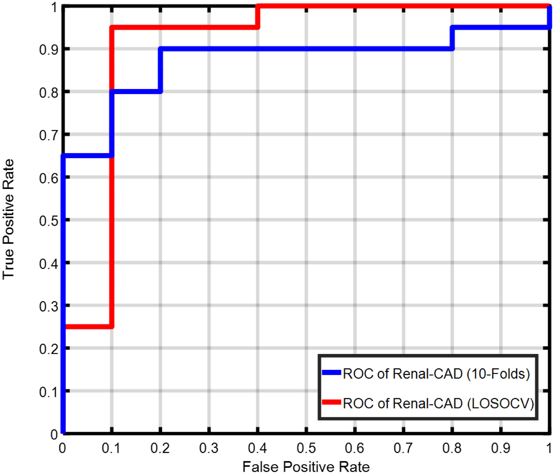
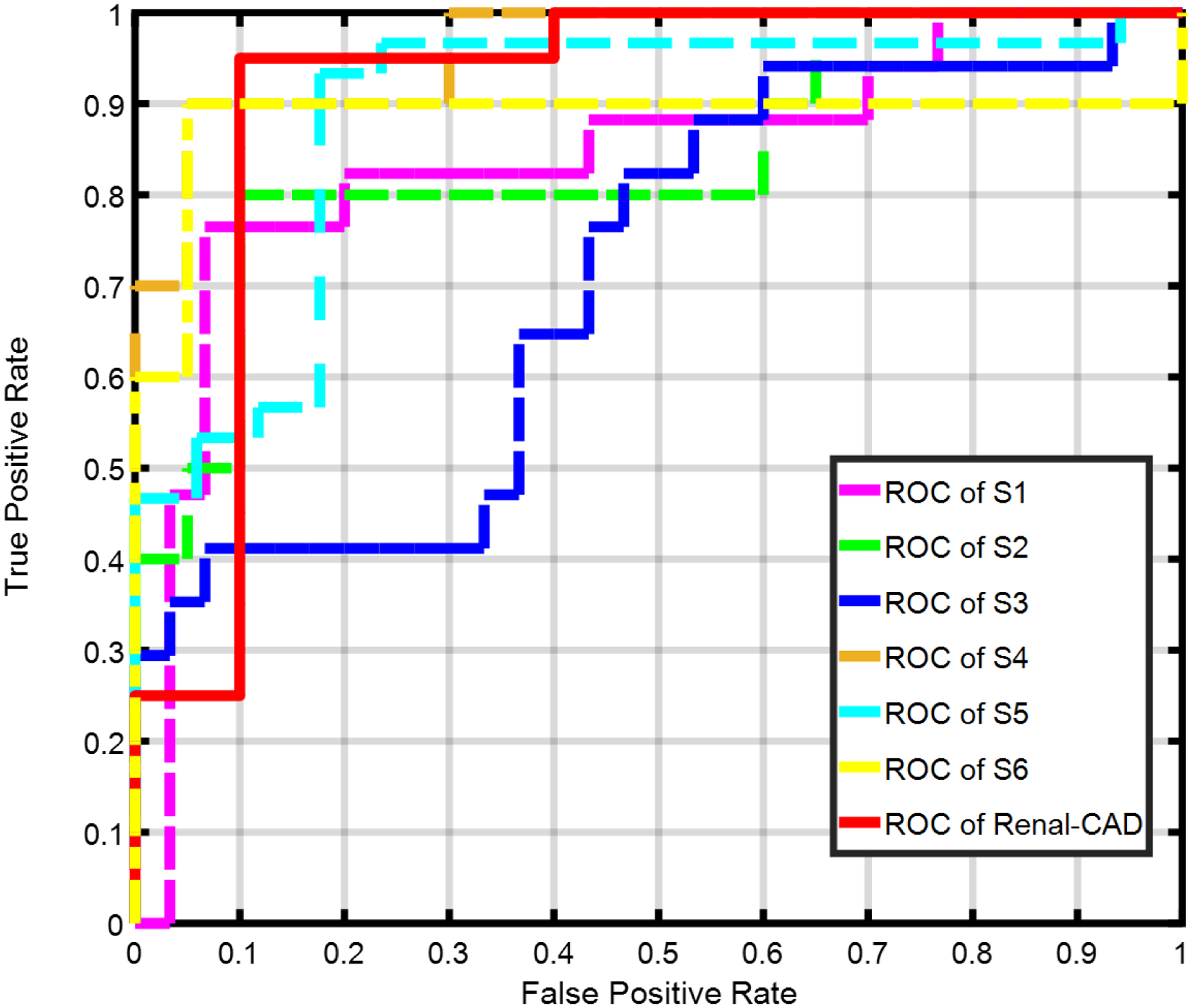
Using a leave-one-subject-out cross-validation approach along with SAEs, the Renal-CAD system demonstrated 93.3% accuracy, 90.0% sensitivity, and 95.0% specificity in differentiating AR from NR. Robustness of the Renal-CAD system was also confirmed by the area under the curve value of 0.92. Using a stratified tenfold cross-validation approach, the Renal-CAD system demonstrated its reproducibility and robustness by a diagnostic accuracy of 86.7%, sensitivity of 80.0%, specificity of 90.0%, and AUC of 0.88.

CONCLUSION

The obtained results demonstrate the feasibility and efficacy of accurate, noninvasive identification of AR at an early stage using the Renal-CAD system.

摘要

目的

肾移植术后早期评估移植肾功能对于最小化和控制移植肾排斥反应至关重要。活检——金标准——由于其侵入性、高成本、不良事件(如出血、感染等)以及报告所需时间,仅作为最后手段使用。为克服这些局限性,开发了一种肾计算机辅助诊断(Renal-CAD)系统来评估肾移植功能。

方法

所开发的Renal-CAD系统整合了从两个基于图像的来源和两个基于临床的来源收集的数据,以评估肾移植功能。成像来源是从11个不同b值(b0、b50、b100、...、b1000 s/mm²)的47次扩散加权磁共振成像(DW-MRI)扫描中提取的表观扩散系数(ADC),以及从5个不同回波时间(TE = 2、7、12、17和22 ms)的30次血氧水平依赖性功能磁共振成像(BOLD-MRI)扫描中提取的横向弛豫率(R2*)。血清肌酐(SCr)和肌酐清除率(CrCl)是评估肾功能的临床来源。Renal-CAD系统最初使用水平集方法进行肾脏分割,随后从DW-MRI估计ADC,从BOLD-MRI估计R2*。将30名同时进行了两种扫描的受试者的ADC和R2*估计值与其相关的SCr和CrCl进行整合。然后将整合后的生物标志物用作我们的鉴别特征,以训练和测试基于深度学习的分类器,即堆叠自编码器(SAE),以区分非排斥(NR)和急性排斥(AR)肾移植。

结果

使用留一法交叉验证方法结合SAE,Renal-CAD系统在区分AR和NR方面表现出93.3%的准确率、90.0%的灵敏度和95.0%的特异性。曲线下面积值为0.92也证实了Renal-CAD系统的稳健性。使用分层十折交叉验证方法,Renal-CAD系统通过86.7%的诊断准确率、80.0%的灵敏度、90.0%的特异性和0.88的AUC证明了其可重复性和稳健性。

结论

所得结果证明了使用Renal-CAD系统在早期准确、无创识别AR的可行性和有效性。

https://cdn.ncbi.nlm.nih.gov/pmc/blobs/d515/8524762/cdee21130664/nihms-1746470-f0012.jpg
https://cdn.ncbi.nlm.nih.gov/pmc/blobs/d515/8524762/2bd521a63847/nihms-1746470-f0002.jpg
https://cdn.ncbi.nlm.nih.gov/pmc/blobs/d515/8524762/ac7bc6932b39/nihms-1746470-f0003.jpg
https://cdn.ncbi.nlm.nih.gov/pmc/blobs/d515/8524762/a9bfb66594c1/nihms-1746470-f0004.jpg
https://cdn.ncbi.nlm.nih.gov/pmc/blobs/d515/8524762/3f31363201b5/nihms-1746470-f0005.jpg
https://cdn.ncbi.nlm.nih.gov/pmc/blobs/d515/8524762/d10e3f0a1f1b/nihms-1746470-f0006.jpg
https://cdn.ncbi.nlm.nih.gov/pmc/blobs/d515/8524762/576d61b660fd/nihms-1746470-f0007.jpg
https://cdn.ncbi.nlm.nih.gov/pmc/blobs/d515/8524762/84467acb78d1/nihms-1746470-f0008.jpg
https://cdn.ncbi.nlm.nih.gov/pmc/blobs/d515/8524762/10e3ebf14303/nihms-1746470-f0009.jpg
https://cdn.ncbi.nlm.nih.gov/pmc/blobs/d515/8524762/4053eed81499/nihms-1746470-f0010.jpg
https://cdn.ncbi.nlm.nih.gov/pmc/blobs/d515/8524762/28abf965c115/nihms-1746470-f0011.jpg
https://cdn.ncbi.nlm.nih.gov/pmc/blobs/d515/8524762/cdee21130664/nihms-1746470-f0012.jpg

相似文献

1
A multimodal computer-aided diagnostic system for precise identification of renal allograft rejection: Preliminary results.一种用于精确识别肾移植排斥反应的多模态计算机辅助诊断系统:初步结果。
Med Phys. 2020 Jun;47(6):2427-2440. doi: 10.1002/mp.14109. Epub 2020 Apr 3.
2
A DEEP LEARNING-BASED CAD SYSTEM FOR RENAL ALLOGRAFT ASSESSMENT: DIFFUSION, BOLD, AND CLINICAL BIOMARKERS.一种基于深度学习的肾移植评估计算机辅助诊断系统:扩散、血氧水平依赖性功能磁共振成像及临床生物标志物
Proc Int Conf Image Proc. 2020 Oct;2020:355-359. doi: 10.1109/ICIP40778.2020.9190818. Epub 2020 Sep 30.
3
EARLY ASSESSMENT OF RENAL TRANSPLANTS USING BOLD-MRI: PROMISING RESULTS.使用血氧水平依赖性功能磁共振成像(BOLD-MRI)对肾移植进行早期评估:取得了有前景的结果。
Proc Int Conf Image Proc. 2019 Sep;2019:1395-1399. doi: 10.1109/ICIP.2019.8803042. Epub 2019 Aug 26.
4
Computer-Aided Diagnostic System for Early Detection of Acute Renal Transplant Rejection Using Diffusion-Weighted MRI.基于磁共振弥散加权成像的急性肾移植排斥反应早期诊断的计算机辅助诊断系统。
IEEE Trans Biomed Eng. 2019 Feb;66(2):539-552. doi: 10.1109/TBME.2018.2849987. Epub 2018 Jun 25.
5
A Novel CNN-Based CAD System for Early Assessment of Transplanted Kidney Dysfunction.一种基于卷积神经网络的新型 CAD 系统,用于早期评估移植肾功能障碍。
Sci Rep. 2019 Apr 11;9(1):5948. doi: 10.1038/s41598-019-42431-3.
6
Statistical analysis of ADCs and clinical biomarkers in detecting acute renal transplant rejection.检测急性肾移植排斥反应中表观扩散系数(ADCs)和临床生物标志物的统计分析
Br J Radiol. 2017 Dec;90(1080):20170125. doi: 10.1259/bjr.20170125. Epub 2017 Sep 13.
7
Detection of renal allograft rejection using blood oxygen level-dependent and diffusion weighted magnetic resonance imaging: a retrospective study.利用血氧水平依赖和扩散加权磁共振成像检测肾移植排斥反应:一项回顾性研究。
BMC Nephrol. 2014 Oct 1;15:158. doi: 10.1186/1471-2369-15-158.
8
The Use of Diffusion Tensor Imaging in the Identification of Acute Rejection and Chronic Allograft Nephropathy After Renal Transplantation.弥散张量成像在肾移植后急性排斥反应和慢性移植肾肾病中的应用。
J Magn Reson Imaging. 2024 Jun;59(6):2082-2088. doi: 10.1002/jmri.29042. Epub 2023 Oct 9.
9
Functional evaluation of transplanted kidneys with diffusion-weighted and BOLD MR imaging: initial experience.磁共振扩散加权成像及血氧水平依赖性功能磁共振成像对移植肾的功能评估:初步经验
Radiology. 2006 Dec;241(3):812-21. doi: 10.1148/radiol.2413060103.
10
Functional MRI evaluation of blood oxygen dependent (BOLD) in renal allograft dysfunction: a prospective study.功能磁共振成像评估肾移植功能障碍中的血氧依赖(BOLD):一项前瞻性研究。
Acta Radiol. 2024 May;65(5):397-405. doi: 10.1177/02841851231217052. Epub 2023 Dec 25.

引用本文的文献

1
Present and Future Applications of Artificial Intelligence in Kidney Transplantation.人工智能在肾脏移植中的当前及未来应用
J Clin Med. 2024 Oct 5;13(19):5939. doi: 10.3390/jcm13195939.
2
Radiologic evaluation of the kidney transplant donor and recipient.肾移植供体和受体的放射学评估。
Abdom Radiol (NY). 2025 Jan;50(1):272-289. doi: 10.1007/s00261-024-04477-4. Epub 2024 Jul 10.
3
Towards a safe and efficient clinical implementation of machine learning in radiation oncology by exploring model interpretability, explainability and data-model dependency.

本文引用的文献

1
Renal blood oxygenation level-dependent magnetic resonance imaging to measure renal tissue oxygenation: a statement paper and systematic review.肾血氧水平依赖磁共振成像测量肾组织氧合:一份声明文件和系统评价。
Nephrol Dial Transplant. 2018 Sep 1;33(suppl_2):ii22-ii28. doi: 10.1093/ndt/gfy243.
2
3D kidney segmentation from abdominal diffusion MRI using an appearance-guided deformable boundary.基于外观引导的可变形边界的腹部弥散 MRI 三维肾脏分割
PLoS One. 2018 Jul 13;13(7):e0200082. doi: 10.1371/journal.pone.0200082. eCollection 2018.
3
BOLD magnetic resonance imaging in nephrology.
通过探索模型的可解释性、可解释性和数据-模型依赖性,实现机器学习在放射肿瘤学中的安全高效临床应用。
Phys Med Biol. 2022 May 27;67(11). doi: 10.1088/1361-6560/ac678a.
4
A DEEP LEARNING-BASED CAD SYSTEM FOR RENAL ALLOGRAFT ASSESSMENT: DIFFUSION, BOLD, AND CLINICAL BIOMARKERS.一种基于深度学习的肾移植评估计算机辅助诊断系统:扩散、血氧水平依赖性功能磁共振成像及临床生物标志物
Proc Int Conf Image Proc. 2020 Oct;2020:355-359. doi: 10.1109/ICIP40778.2020.9190818. Epub 2020 Sep 30.
5
Artificial intelligence and kidney transplantation.人工智能与肾脏移植
World J Transplant. 2021 Jul 18;11(7):277-289. doi: 10.5500/wjt.v11.i7.277.
6
Artificial intelligence and machine learning for medical imaging: A technology review.人工智能和机器学习在医学成像中的应用:技术综述。
Phys Med. 2021 Mar;83:242-256. doi: 10.1016/j.ejmp.2021.04.016. Epub 2021 May 9.
肾脏病学中的血氧水平依赖性功能磁共振成像
Int J Nephrol Renovasc Dis. 2018 Mar 13;11:103-112. doi: 10.2147/IJNRD.S112299. eCollection 2018.
4
Functional Evaluation of Transplanted Kidneys with Reduced Field-of-View Diffusion-Weighted Imaging at 3T.3T 下小视野扩散加权成像评估移植肾功能。
Korean J Radiol. 2018 Mar-Apr;19(2):201-208. doi: 10.3348/kjr.2018.19.2.201. Epub 2018 Feb 22.
5
US Renal Data System 2017 Annual Data Report: Epidemiology of Kidney Disease in the United States.美国肾脏数据系统2017年年报:美国肾脏疾病流行病学
Am J Kidney Dis. 2018 Mar;71(3 Suppl 1):A7. doi: 10.1053/j.ajkd.2018.01.002.
6
Selection for biopsy of kidney transplant patients by diffusion-weighted MRI.通过弥散加权 MRI 对肾移植患者进行活检选择。
Eur Radiol. 2017 Oct;27(10):4336-4344. doi: 10.1007/s00330-017-4814-z. Epub 2017 Apr 3.
7
Diffusion-Weighted imaging and diffusion tensor imaging detect delayed graft function and correlate with allograft fibrosis in patients early after kidney transplantation.扩散加权成像和扩散张量成像可检测肾移植术后早期患者的移植肾功能延迟,并与同种异体移植纤维化相关。
J Magn Reson Imaging. 2016 Jul;44(1):112-21. doi: 10.1002/jmri.25158. Epub 2016 Jan 18.
8
Renal Blood Oxygenation Level-dependent Imaging in Longitudinal Follow-up of Donated and Remaining Kidneys.捐赠肾和剩余肾的纵向随访中的肾脏血氧水平依赖成像。
Radiology. 2016 Jun;279(3):795-804. doi: 10.1148/radiol.2015150370. Epub 2016 Jan 8.
9
Deep Learning of Part-Based Representation of Data Using Sparse Autoencoders With Nonnegativity Constraints.基于非负稀疏自动编码器的数据的基于部分表示的深度学习。
IEEE Trans Neural Netw Learn Syst. 2016 Dec;27(12):2486-2498. doi: 10.1109/TNNLS.2015.2479223. Epub 2015 Oct 28.
10
Diffusion weighted magnetic resonance imaging and its recent trend-a survey.磁共振扩散加权成像及其近期趋势——一项综述
Quant Imaging Med Surg. 2015 Jun;5(3):407-22. doi: 10.3978/j.issn.2223-4292.2015.03.01.