Sharma Sudhir Kumar, Vijay Sauparnika, Gore Sangram, Dore Timothy M, Jagannathan Ramesh
Engineering Division, New York University Abu Dhabi, PO Box 129188, Abu Dhabi, UAE.
Science Division, New York University Abu Dhabi, PO Box 129188, Abu Dhabi, UAE.
ACS Omega. 2020 Feb 18;5(8):4024-4031. doi: 10.1021/acsomega.9b03589. eCollection 2020 Mar 3.
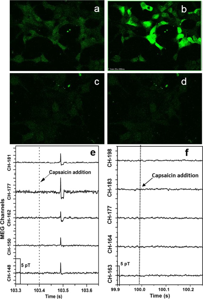
The cellular-level process of ion transport is known to generate a magnetic field. A noninvasive magnetoencephalography (MEG) technique was used to measure the magnetic field emanating from HeLa, HEK293, and H9c2(2-1) rat cardiac cells. The addition of a nonlethal dose of ionomycin to HeLa and capsaicin to TRPV1-expressing HEK293 cells resulted in a sudden change in the magnetic field signal consistent with Ca influx, which was also observed by confocal fluorescence microscopy under the same conditions. In contrast, addition of capsaicin to TRPV1-expressing HEK293 cells containing an optimum amount of a TRPV1 antagonist (ruthenium red), resulted in no detectable magnetic or fluorescent signals. These signals confirmed that the measured MEG signals are due to cellular ion transport through the cell membrane. In general, there is evidence that ion channel/transporter activation and ionic flux are linked to cancer. Therefore, our work suggests that MEG could represent a noninvasive method for detecting cancer.
已知离子运输的细胞水平过程会产生磁场。采用一种非侵入性的脑磁图(MEG)技术来测量源自HeLa细胞、HEK293细胞和H9c2(2-1)大鼠心肌细胞的磁场。向HeLa细胞添加非致死剂量的离子霉素,向表达TRPV1的HEK293细胞添加辣椒素,导致磁场信号突然变化,这与钙离子内流一致,在相同条件下通过共聚焦荧光显微镜也观察到了这种情况。相比之下,向含有最佳量TRPV1拮抗剂(钌红)的表达TRPV1的HEK293细胞添加辣椒素,未检测到可测量的磁场或荧光信号。这些信号证实,所测量的MEG信号是由于细胞离子通过细胞膜的运输所致。总体而言,有证据表明离子通道/转运体激活和离子通量与癌症有关。因此,我们的研究表明,MEG可能代表一种检测癌症的非侵入性方法。