• 文献检索
  • 文档翻译
  • 深度研究
  • 学术资讯
  • Suppr Zotero 插件Zotero 插件
  • 邀请有礼
  • 套餐&价格
  • 历史记录
应用&插件
Suppr Zotero 插件Zotero 插件浏览器插件Mac 客户端Windows 客户端微信小程序
定价
高级版会员购买积分包购买API积分包
服务
文献检索文档翻译深度研究API 文档MCP 服务
关于我们
关于 Suppr公司介绍联系我们用户协议隐私条款
关注我们

Suppr 超能文献

核心技术专利:CN118964589B侵权必究
粤ICP备2023148730 号-1Suppr @ 2026

文献检索

告别复杂PubMed语法,用中文像聊天一样搜索,搜遍4000万医学文献。AI智能推荐,让科研检索更轻松。

立即免费搜索

文件翻译

保留排版,准确专业,支持PDF/Word/PPT等文件格式,支持 12+语言互译。

免费翻译文档

深度研究

AI帮你快速写综述,25分钟生成高质量综述,智能提取关键信息,辅助科研写作。

立即免费体验

弥漫性食管壁内假性憩室病的临床和内镜特征。

Clinical and endoscopic characteristics of diffuse esophageal intramural pseudo-diverticulosis.

机构信息

Center for Internal Medicine II, Brandenburg Medical School Theodor Fontane, Brandenburg an der Havel, Germany.

Zentrum für Innere Medizin II, Hochschulklinikum Brandenburg der MHB, Hochstr. 29, 14770, Brandenburg an der Havel, Germany.

出版信息

Esophagus. 2020 Oct;17(4):492-501. doi: 10.1007/s10388-020-00729-6. Epub 2020 Mar 11.

DOI:10.1007/s10388-020-00729-6
PMID:32162106
原文链接:https://pmc.ncbi.nlm.nih.gov/articles/PMC7497296/
Abstract

INTRODUCTION

With 250 published cases worldwide, diffuse esophageal intramural pseudo-diverticulosis (DEIPD) is a poorly understood disease. The aim of this study was to determine the prevalence of DEIPD in our own population, identify risk factors and clinical symptoms, and characterize its typical endoscopic signs.

METHODS

Retrospective search in our center's endoscopic and clinical database. Reviewing of all cases by re-examining stored endoscopic photographs. Reviewing of all cases regarding age, sex, risk factors, comorbidities, histology, and clinical symptoms.

RESULTS

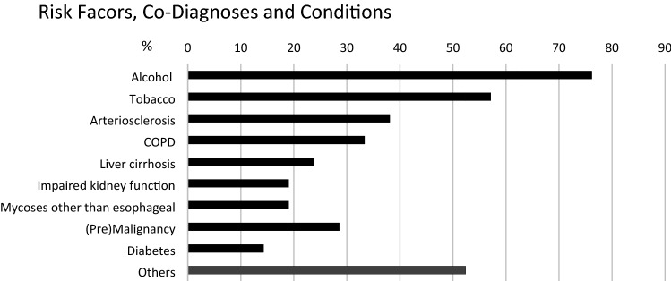
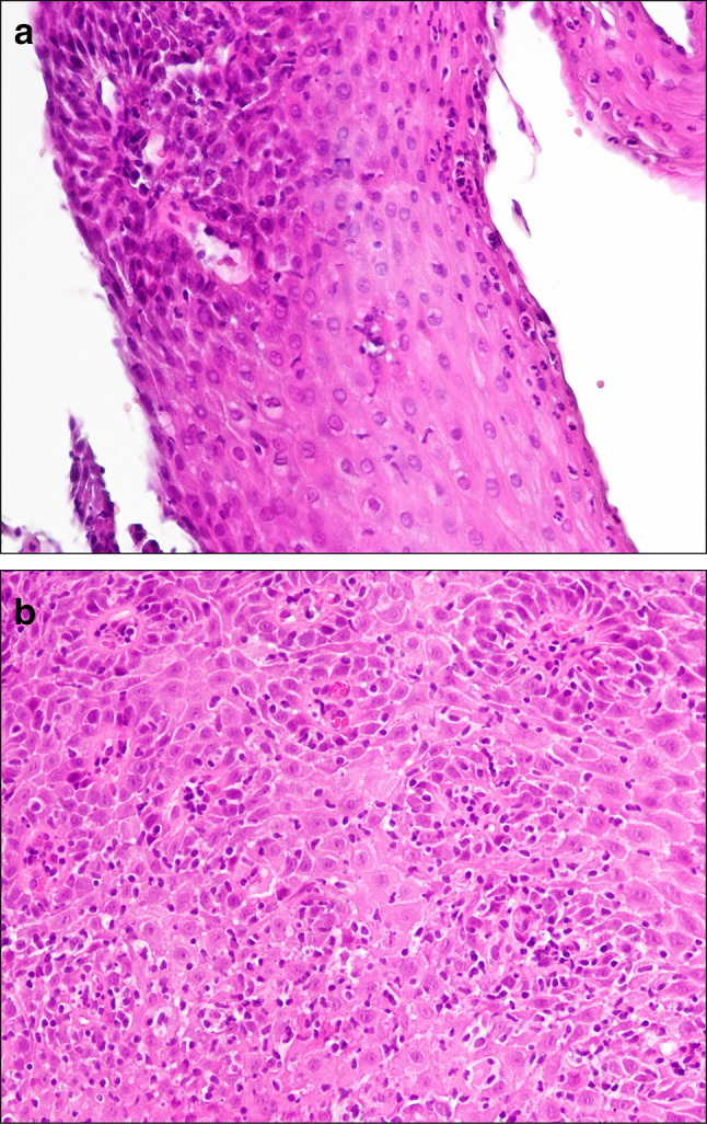
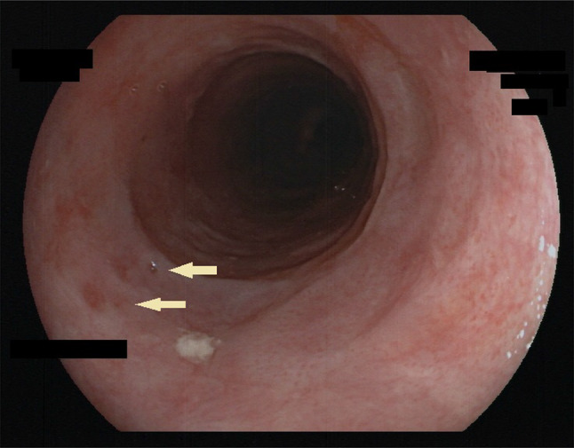
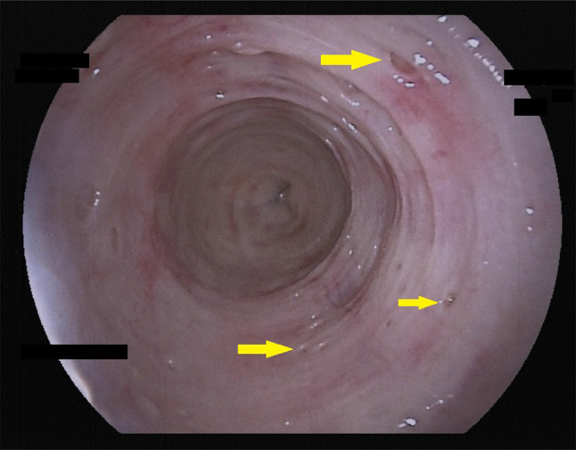
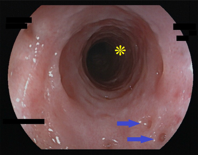
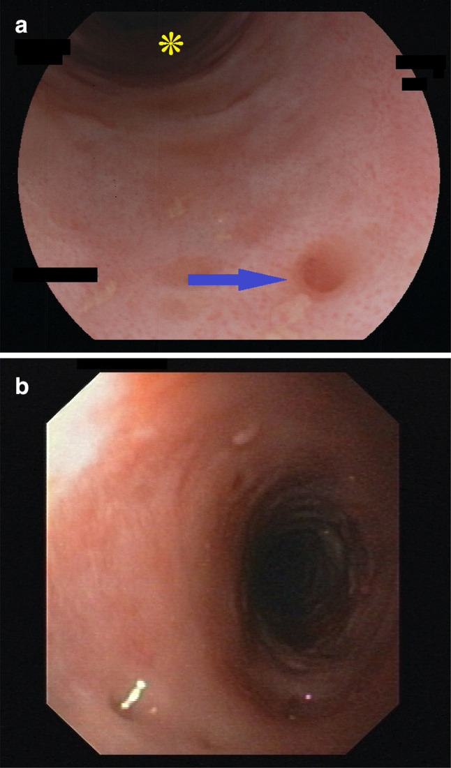
In a population of 150.000 we found 21 cases of DEIPD. Mean age was 56 ± 10 years. 86% were males, 76% had alcohol abuse, 57% had nicotine abuse, 38% had arteriosclerosis, 33% had COPD, 29% had malignancies, 24% had liver cirrhosis, 19% had impaired kidney function, and 15% had diabetes. Dysphagia was present in 62% and food bolus impaction (single or repeated) in 48%. Endoscopically, 95% of patients had multiple (> 4), small (0.25-2.5 mm) pseudodiverticle openings in the esophageal wall. In 62%, openings were aligned longitudinally. 86% showed edematous swelling of mucosa ("frosted glass look"), 76% showed a fine-grained pattern of small (10-100 µm) red dots ("faux uni pattern"), and 76% had a rigid, narrow lumen with multiple rings ("trachealization").

CONCLUSION

With a prevalence of approximately 5 to 50/100.000, DEIPD may be more frequent than previously estimated. It preferably affects middle-aged male alcoholics. Key symptoms are chronic dysphagia and food impaction. Typical endoscopic findings are multiple, small, longitudinally aligned pseudodiverticle openings, frosted glass look, faux uni pattern, and trachealization of the esophagus.

摘要

简介

全球已有 250 例病例报道,弥漫性食管壁内假性憩室病(DEIPD)是一种了解甚少的疾病。本研究的目的是确定我们人群中 DEIPD 的患病率,确定其危险因素和临床症状,并描述其典型的内镜表现。

方法

在我们中心的内镜和临床数据库中进行回顾性搜索。通过重新检查存储的内镜照片来重新检查所有病例。回顾所有病例的年龄、性别、危险因素、合并症、组织学和临床症状。

结果

在 150000 人的人群中,我们发现了 21 例 DEIPD。平均年龄为 56±10 岁。86%为男性,76%有酗酒,57%有尼古丁滥用,38%有动脉硬化,33%有 COPD,29%有恶性肿瘤,24%有肝硬化,19%有肾功能不全,15%有糖尿病。62%的患者有吞咽困难,48%的患者有食物团块嵌塞(单发或多发)。内镜下,95%的患者食管壁有多个(>4 个)、小(0.25-2.5 毫米)假性憩室开口。62%的开口呈纵向排列。86%的患者黏膜呈水肿肿胀(“磨砂玻璃样外观”),76%的患者有细小(10-100 微米)红点的细颗粒状模式(“假性 uni 样外观”),76%的患者食管腔狭窄,有多个环形(“气管样化”)。

结论

DEIPD 的患病率约为 5 至 50/100000,可能比以前估计的更为常见。它主要影响中年男性酗酒者。主要症状是慢性吞咽困难和食物嵌塞。典型的内镜表现是多个、小的、纵向排列的假性憩室开口、磨砂玻璃样外观、假性 uni 样外观和食管气管样化。

https://cdn.ncbi.nlm.nih.gov/pmc/blobs/364c/7497296/a0af5c66e9ee/10388_2020_729_Fig7_HTML.jpg
https://cdn.ncbi.nlm.nih.gov/pmc/blobs/364c/7497296/cad7d3c384bd/10388_2020_729_Fig1_HTML.jpg
https://cdn.ncbi.nlm.nih.gov/pmc/blobs/364c/7497296/43405422269b/10388_2020_729_Fig2_HTML.jpg
https://cdn.ncbi.nlm.nih.gov/pmc/blobs/364c/7497296/d082061ee665/10388_2020_729_Fig3_HTML.jpg
https://cdn.ncbi.nlm.nih.gov/pmc/blobs/364c/7497296/ef16cfef01b2/10388_2020_729_Fig4_HTML.jpg
https://cdn.ncbi.nlm.nih.gov/pmc/blobs/364c/7497296/8cc8f00e8a5d/10388_2020_729_Fig5_HTML.jpg
https://cdn.ncbi.nlm.nih.gov/pmc/blobs/364c/7497296/c3d67b4a105f/10388_2020_729_Fig6_HTML.jpg
https://cdn.ncbi.nlm.nih.gov/pmc/blobs/364c/7497296/a0af5c66e9ee/10388_2020_729_Fig7_HTML.jpg

相似文献

1
Clinical and endoscopic characteristics of diffuse esophageal intramural pseudo-diverticulosis.弥漫性食管壁内假性憩室病的临床和内镜特征。
Esophagus. 2020 Oct;17(4):492-501. doi: 10.1007/s10388-020-00729-6. Epub 2020 Mar 11.
2
Chronic fibrosing esophagitis with diffuse esophageal intramural pseudo-diverticulosis.慢性纤维性食管炎伴弥漫性食管壁内假性憩室病
JGH Open. 2022 May 11;6(5):287-291. doi: 10.1002/jgh3.12750. eCollection 2022 May.
3
Esophageal intramural pseudodiverticulosis treated by endoscopic balloon dilatation.内镜下球囊扩张术治疗食管壁内假性憩室病
Tokai J Exp Clin Med. 2014 Sep 20;39(3):137-40.
4
[Dysphagia and recurrent esophageal stenosis associated with intramural pseudodiverticulosis of the esophagus. A case report].[食管壁内假性憩室病相关的吞咽困难和复发性食管狭窄。病例报告]
Rev Med Brux. 2010 Nov-Dec;31(6):529-32.
5
[Esophageal intramural pseudodiverticulosis].[食管壁内假性憩室病]
Praxis (Bern 1994). 2007 Sep 12;96(37):1385-9. doi: 10.1024/1661-8157.96.37.1385.
6
Candida albicans: a cause or a consequence of esophageal intramural pseudo-diverticulosis.白色念珠菌:食管壁内假性憩室病的病因还是后果?
Pan Afr Med J. 2019 Aug 2;33:280. doi: 10.11604/pamj.2019.33.280.19601. eCollection 2019.
7
Esophageal intramural pseudodiverticulosis in a patient with food bolus impaction.一名患有食物团块嵌塞患者的食管壁内假性憩室病
Rev Gastroenterol Peru. 2019 Oct-Dec;39(4):362-363.
8
Pneumomediastinum as a complication of esophageal intramural pseudodiverticulosis.纵隔气肿作为食管壁内假性憩室病的一种并发症。
Acta Gastroenterol Belg. 2018 Jul-Sep;81(3):433-435.
9
Not always eosinophilic esophagitis - intramural pseudodiverticulosis of the esophagus - a case report and literature review.并非总是嗜酸性粒细胞性食管炎——食管壁内假性憩室病——一例报告及文献综述
Z Gastroenterol. 2020 Dec;58(12):1201-1207. doi: 10.1055/a-1288-1521. Epub 2020 Nov 5.
10
Esophageal intramural pseudodiverticulosis with esophageal strictures successfully treated with dilation therapy.伴有食管狭窄的食管壁内假性憩室病经扩张治疗成功治愈。
Ann Thorac Surg. 2006 Sep;82(3):1119-21. doi: 10.1016/j.athoracsur.2006.01.062.

引用本文的文献

1
A Rare Endoscopic Finding - Swiss Cheese Esophagus.一种罕见的内镜检查发现——瑞士奶酪样食管。
GE Port J Gastroenterol. 2023 Jul 7;31(2):139-141. doi: 10.1159/000531168. eCollection 2024 Apr.
2
A Complex Presentation of Esophageal Pseudodiverticulosis With Candida Albicans Esophagitis in a 68-Year-Old Female.一名68岁女性食管假性憩室合并白色念珠菌食管炎的复杂病例
Cureus. 2024 Feb 29;16(2):e55286. doi: 10.7759/cureus.55286. eCollection 2024 Feb.
3
Esophageal intramural pseudodiverticulosis.食管壁内假性憩室病

本文引用的文献

1
Candida albicans: a cause or a consequence of esophageal intramural pseudo-diverticulosis.白色念珠菌:食管壁内假性憩室病的病因还是后果?
Pan Afr Med J. 2019 Aug 2;33:280. doi: 10.11604/pamj.2019.33.280.19601. eCollection 2019.
2
"Trachealization" of the Esophagus.食管的“气管化”
N Engl J Med. 2019 Jan 10;380(2):177. doi: 10.1056/NEJMicm1807124.
3
Lymphocytic esophagitis: an update on histologic diagnosis, endoscopic findings, and natural history.淋巴细胞性食管炎:组织学诊断、内镜表现和自然史的最新进展。
World J Gastroenterol. 2024 Jan 14;30(2):137-145. doi: 10.3748/wjg.v30.i2.137.
4
Intrapapillary capillary loop abundance on nonmagnified blue laser imaging in esophageal intramural pseudodiverticulosis.食管壁内假性憩室病非放大蓝色激光成像时乳头内毛细血管襻丰度
Porto Biomed J. 2023 Jun 23;8(3):e211. doi: 10.1097/j.pbj.0000000000000211. eCollection 2023 May-Jun.
5
Symptomatic esophageal intramural pseudodiverticulosis without stricture: A case report.无症状性无狭窄食管壁内假性憩室病:一例报告。
SAGE Open Med Case Rep. 2023 Feb 17;11:2050313X221140244. doi: 10.1177/2050313X221140244. eCollection 2023.
6
Esophageal Intramural Pseudodiverticulosis: A Histopathological and Immunohistochemical Study of 2 Cases.食管壁内假性憩室病:2例组织病理学和免疫组织化学研究
Case Rep Gastroenterol. 2022 Apr 28;16(1):270-277. doi: 10.1159/000524496. eCollection 2022 Jan-Apr.
7
Chronic fibrosing esophagitis with diffuse esophageal intramural pseudo-diverticulosis.慢性纤维性食管炎伴弥漫性食管壁内假性憩室病
JGH Open. 2022 May 11;6(5):287-291. doi: 10.1002/jgh3.12750. eCollection 2022 May.
8
Epidermoid Metaplasia in Diffuse Esophageal Intramural Pseudodiverticulosis.弥漫性食管壁内假性憩室病中的表皮样化生
Case Rep Gastroenterol. 2022 Feb 28;16(1):94-96. doi: 10.1159/000522184. eCollection 2022 Jan-Apr.
9
Development of Epidermoid Metaplasia of the Mucosa in Association with Esophageal Intramural Pseudodiverticulosis and Candidiasis.黏膜表皮样化生伴食管壁内假性憩室病和念珠菌病的发展
Case Rep Gastroenterol. 2021 Aug 2;15(2):709-714. doi: 10.1159/000518023. eCollection 2021 May-Aug.
Ann N Y Acad Sci. 2018 Dec;1434(1):185-191. doi: 10.1111/nyas.13710. Epub 2018 May 24.
4
The microscopic anatomy of the esophagus including the individual layers, specialized tissues, and unique components and their responses to injury.食管的微观解剖结构,包括各个层次、特殊组织以及独特的组成部分及其对损伤的反应。
Ann N Y Acad Sci. 2018 Dec;1434(1):304-318. doi: 10.1111/nyas.13705. Epub 2018 May 15.
5
Understanding fibrosis in eosinophilic esophagitis: Are we there yet?理解嗜酸性粒细胞性食管炎中的纤维化:我们做到了吗?
J Leukoc Biol. 2018 Jul;104(1):31-40. doi: 10.1002/JLB.5MR1017-395R. Epub 2018 Jan 29.
6
Rings in the esophagus are not always eosinophilic esophagitis: Case series of ring forming lymphocytic esophagitis and review of the literature.食管环并不总是嗜酸性粒细胞性食管炎:环状淋巴细胞性食管炎病例系列及文献综述
Endosc Int Open. 2017 Jun;5(6):E484-E488. doi: 10.1055/s-0043-106579. Epub 2017 May 31.
7
Esophageal Intramural Pseudodiverticulosis and Concomitant Eosinophilic Esophagitis: A Case Series.食管壁内假性憩室病和伴发嗜酸性食管炎:病例系列。
Can J Gastroenterol Hepatol. 2016;2016:1761874. doi: 10.1155/2016/1761874. Epub 2016 Aug 28.
8
Does the healing of the esophageal mucosa improve the function of the esophageal submucosal and salivary glands?食管黏膜的愈合是否能改善食管黏膜下和唾液腺的功能?
Ann N Y Acad Sci. 2016 Sep;1380(1):155-161. doi: 10.1111/nyas.13187. Epub 2016 Aug 17.
9
Severity of endoscopically identified esophageal rings correlates with reduced esophageal distensibility in eosinophilic esophagitis.内镜检查发现的食管环的严重程度与嗜酸性食管炎中食管扩张性降低相关。
Endoscopy. 2016 Sep;48(9):794-801. doi: 10.1055/s-0042-107340. Epub 2016 May 20.
10
[S3-guidelines "sedation in gastrointestinal endoscopy" 2014 (AWMF register no. 021/014)].[2014年《S3指南:胃肠内镜检查中的镇静》(德国医学专业协会注册编号:021/014)]
Z Gastroenterol. 2015 Aug;53(8):E1. doi: 10.1055/s-0035-1553971. Epub 2015 Oct 8.