Batchelder Kendra A, Tanenbaum Aaron B, Albert Seth, Guimond Lyne, Kestener Pierre, Arneodo Alain, Khalil Andre
CompuMAINE Lab, University of Maine, Orono, Maine, United States of America; Department of Mathematics and Statistics, University of Maine, Orono, Maine, United States of America; Institute for Molecular Biophysics, University of Maine, Orono, Maine, United States of America.
Commissariat a l'Energie Atomique, Gif-sur-Yvette, France.
PLoS One. 2014 Sep 15;9(9):e107580. doi: 10.1371/journal.pone.0107580. eCollection 2014.
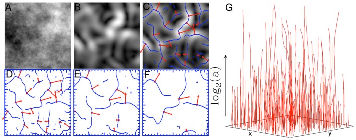
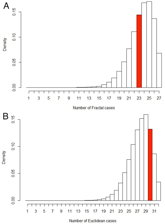
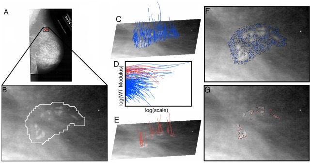
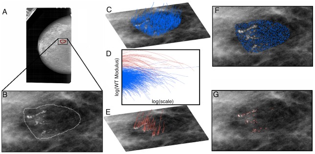
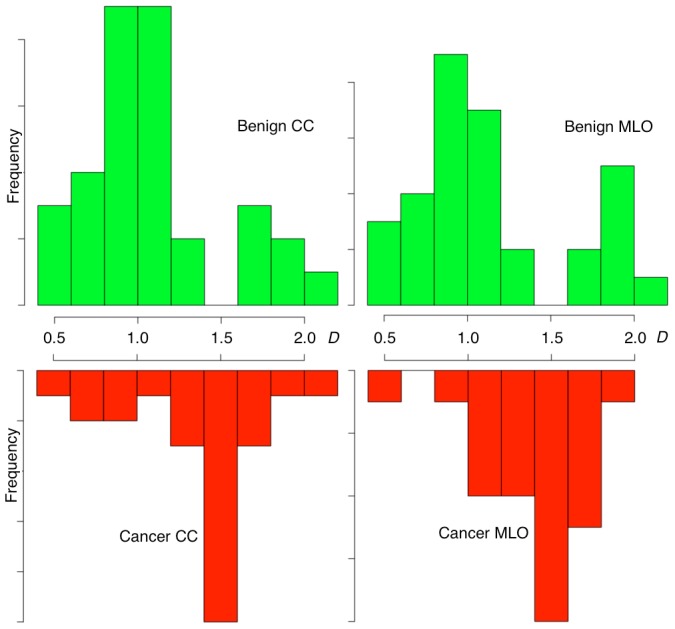
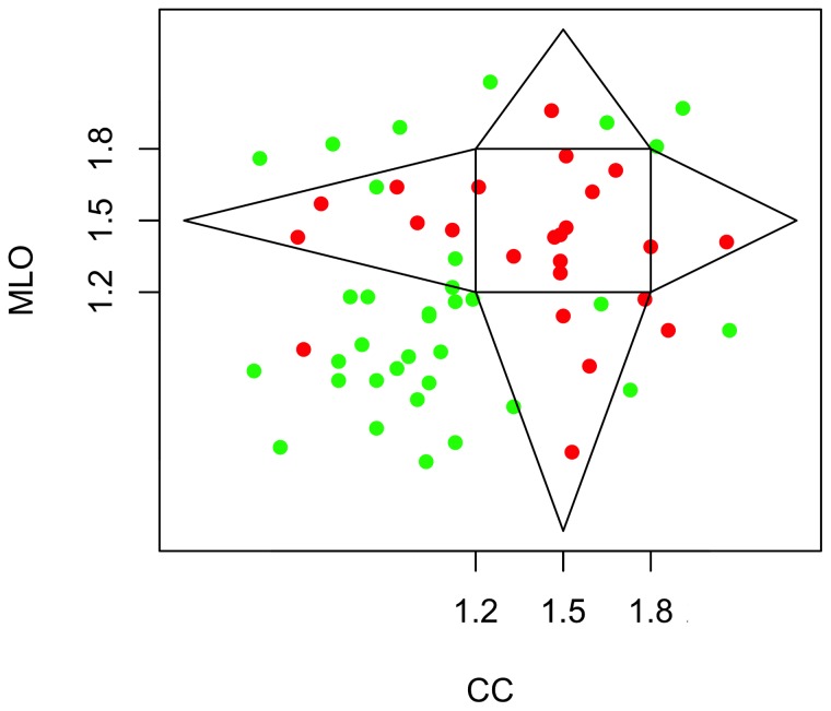
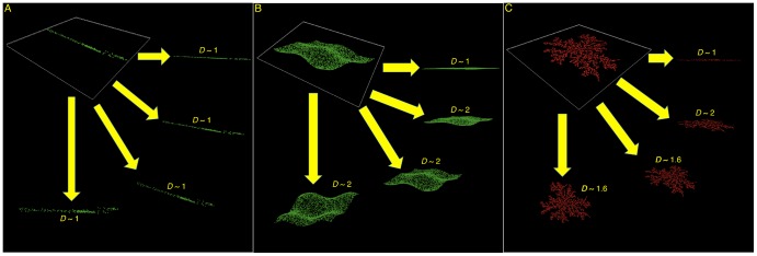
The 2D Wavelet-Transform Modulus Maxima (WTMM) method was used to detect microcalcifications (MC) in human breast tissue seen in mammograms and to characterize the fractal geometry of benign and malignant MC clusters. This was done in the context of a preliminary analysis of a small dataset, via a novel way to partition the wavelet-transform space-scale skeleton. For the first time, the estimated 3D fractal structure of a breast lesion was inferred by pairing the information from two separate 2D projected mammographic views of the same breast, i.e. the cranial-caudal (CC) and mediolateral-oblique (MLO) views. As a novelty, we define the "CC-MLO fractal dimension plot", where a "fractal zone" and "Euclidean zones" (non-fractal) are defined. 118 images (59 cases, 25 malignant and 34 benign) obtained from a digital databank of mammograms with known radiologist diagnostics were analyzed to determine which cases would be plotted in the fractal zone and which cases would fall in the Euclidean zones. 92% of malignant breast lesions studied (23 out of 25 cases) were in the fractal zone while 88% of the benign lesions were in the Euclidean zones (30 out of 34 cases). Furthermore, a Bayesian statistical analysis shows that, with 95% credibility, the probability that fractal breast lesions are malignant is between 74% and 98%. Alternatively, with 95% credibility, the probability that Euclidean breast lesions are benign is between 76% and 96%. These results support the notion that the fractal structure of malignant tumors is more likely to be associated with an invasive behavior into the surrounding tissue compared to the less invasive, Euclidean structure of benign tumors. Finally, based on indirect 3D reconstructions from the 2D views, we conjecture that all breast tumors considered in this study, benign and malignant, fractal or Euclidean, restrict their growth to 2-dimensional manifolds within the breast tissue.
二维小波变换模极大值(WTMM)方法用于检测乳腺钼靶片中人类乳腺组织中的微钙化(MC),并表征良性和恶性MC簇的分形几何特征。这是在对一个小数据集进行初步分析的背景下,通过一种划分小波变换空间尺度骨架的新方法完成的。首次通过将来自同一乳房的两个单独的二维投影乳腺钼靶视图(即头尾位(CC)和内外斜位(MLO)视图)的信息配对,推断出乳腺病变的估计三维分形结构。作为一项创新,我们定义了“CC-MLO分形维数图”,其中定义了一个“分形区”和“欧几里得区”(非分形)。对从具有已知放射科诊断结果的乳腺钼靶数字数据库中获得的118幅图像(59例,25例恶性和34例良性)进行分析,以确定哪些病例将被绘制在分形区,哪些病例将落在欧几里得区。所研究的恶性乳腺病变中有92%(25例中的23例)位于分形区,而88%的良性病变位于欧几里得区(34例中的30例)。此外,贝叶斯统计分析表明,在95%的可信度下,分形乳腺病变为恶性的概率在74%至98%之间。或者,在95%的可信度下,欧几里得乳腺病变为良性的概率在76%至96%之间。这些结果支持了这样一种观点,即与良性肿瘤侵入性较小的欧几里得结构相比,恶性肿瘤的分形结构更有可能与向周围组织的侵袭行为相关。最后,基于从二维视图进行的间接三维重建,我们推测本研究中考虑的所有乳腺肿瘤,无论是良性还是恶性,分形还是欧几里得,都将其生长限制在乳腺组织内的二维流形上。