Xi Xuehua, Gao Luying, Wu Qiong, Fang Shibao, Xu Jingzhu, Liu Ruyu, Yang Xiao, Zhu Shenling, Zhao Ruina, Lai Xingjian, Zhang Xiaoyan, Zhang Bo, Jiang Yuxin
Department of Ultrasound, China-Japan Friendship Hospital, Beijing, China.
Department of Ultrasound, Peking Union Medical College Hospital, Chinese Academy of Medical Sciences & Peking Union Medical College, Beijing, China.
Front Oncol. 2020 Feb 27;10:112. doi: 10.3389/fonc.2020.00112. eCollection 2020.
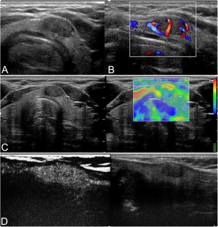
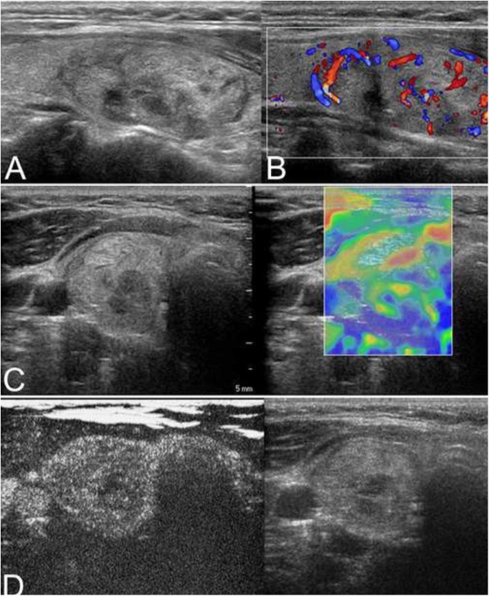
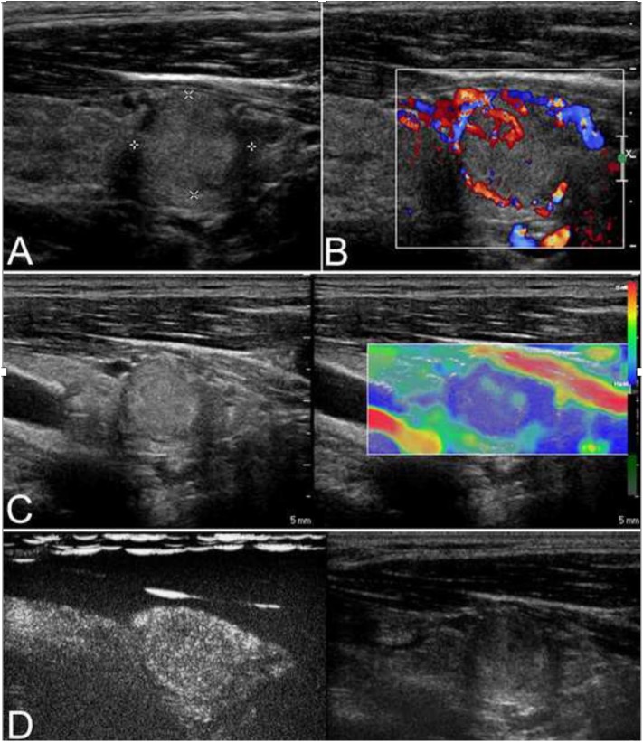
According to the 2015 American Thyroid Association (ATA), referred risk stratification and thyroid nodules with intermediate- and low-suspicion patterns are difficult to diagnose. The objective of this study is to evaluate the diagnostic performance of contrast-enhanced ultrasonography (CEUS) and elastosonography (ES) for the differentiation of these thyroid nodules. From November 2011 to June 2016, a total of 163 thyroid nodules with intermediate- and low-suspicion patterns in 150 consecutive patients at our hospital were studied before surgery. With surgical pathology as the standard, the diagnostic value of CEUS and ES was analyzed. There were 29 (17.8%) malignant lesions and 134 (82.2%) benign lesions. The enhancement patterns of CEUS, the echogenicity, and the elastography were significantly different between malignant and benign lesions ( < 0.05). Heterogenous enhancement was more common in malignant nodules, and the sensitivity, specificity, positive predictive value, negative predictive value, and odds ratio were 51.7, 88.1, 48.4, 89.4, and 10.1%, respectively. The diagnostic accuracy of CEUS was better than the conventional ultrasound [area under the curve (AUC), 0.729 vs. 0.616, = 0.021]. The enhancement patterns of CEUS were helpful in the differential diagnosis of thyroid nodules with intermediate and low suspicion.
根据2015年美国甲状腺协会(ATA)的报告,风险分层以及具有中等和低可疑特征的甲状腺结节难以诊断。本研究的目的是评估超声造影(CEUS)和弹性成像(ES)对这些甲状腺结节鉴别的诊断性能。2011年11月至2016年6月,对我院150例连续患者中的163个具有中等和低可疑特征的甲状腺结节进行术前研究。以手术病理为标准,分析CEUS和ES的诊断价值。其中有29个(17.8%)恶性病变和134个(82.2%)良性病变。恶性和良性病变之间,CEUS的增强模式、回声性和弹性成像有显著差异(<0.05)。不均匀增强在恶性结节中更常见,其灵敏度、特异度、阳性预测值、阴性预测值和比值比分别为51.7%、88.1%、48.4%、89.4%和10.1%。CEUS的诊断准确性优于传统超声[曲线下面积(AUC),0.729对0.616,=0.021]。CEUS的增强模式有助于对中等和低可疑的甲状腺结节进行鉴别诊断。