Liu Yingying, Dou Yafang, Lu Fang, Liu Lei
Institutes of Biomedical Sciences, Fudan University School of Basic Medical Sciences.
Department of Radiology, Shanghai Shuguang Hospital Affiliated to TCM University, Shanghai, China.
Medicine (Baltimore). 2020 Mar;99(11):e19251. doi: 10.1097/MD.0000000000019251.
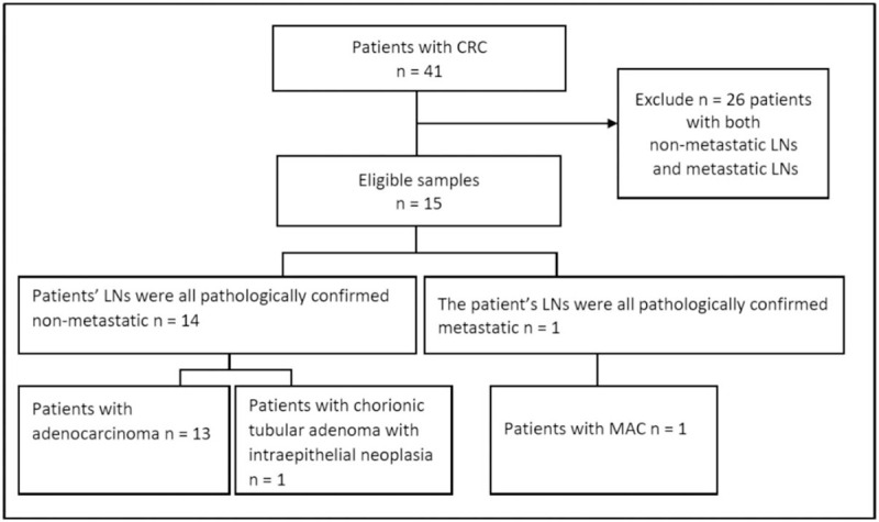
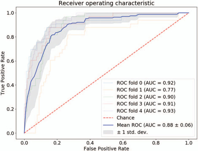
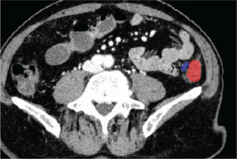
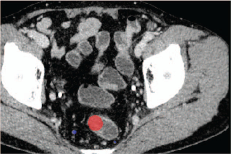
Lymph nodes (LN) metastasis differentiation from computed tomography (CT) images is a challenging problem. This study aims to investigate the association between radiomics image parameters and LN metastasis in colorectal mucinous adenocarcinoma (MAC).Clinical records and CT images of 15 patients were included in this study. Among them, 1 patient was confirmed with all metastatic LNs, the other 14 were confirmed with all non-metastatic LNs. The regions of the LNs were manually labeled on each slice by experienced radiologists. A total of 1054 LN regions were obtained. Among them, 164 were from metastatic LNs. One hundred nine image parameters were computed and analyzed using 2-sample t test method and logistic regression classifier.Based on 2 sample t test, image parameters between the metastatic group and the non-metastatic group were compared. A total of 73 parameters were found to be significant (P < .01). The selected shape parameters demonstrate that non-metastatic LNs tend to have smaller sizes and more circle-like shapes than metastatic LNs, which validates the common agreement of LN diagnosis using computational method. Besides, several high order parameters were selected as well, which indicates that the textures vary between non-metastatic LNs and metastatic LNs. The selected parameters of significance were further used to train logistic regression classifier with L1 penalty. Based on receiver operating characteristic (ROC) analysis, large area under curve (AUC) values were achieved over 5-fold cross validation (0.88 ± 0.06). Moreover, high accuracy, specificity, and sensitivity values were observed as well.The results of the study demonstrate that some quantitative image parameters are of significance in differentiating LN metastasis. Logistic regression classifiers showed that the parameters are with predictive values in LN metastasis, which may be used to assist preoperative diagnosis.
从计算机断层扫描(CT)图像中判断淋巴结(LN)转移情况是一个具有挑战性的问题。本研究旨在探讨结直肠黏液腺癌(MAC)的影像组学图像参数与LN转移之间的关联。
本研究纳入了15例患者的临床记录和CT图像。其中,1例患者所有LN均被证实发生转移,另外14例患者所有LN均被证实无转移。经验丰富的放射科医生在每个层面上手动标记出LN区域。共获得1054个LN区域。其中,164个来自转移性LN。使用双样本t检验方法和逻辑回归分类器计算并分析了109个图像参数。
基于双样本t检验,比较了转移组和非转移组之间的图像参数。共发现73个参数具有显著性(P < 0.01)。所选的形状参数表明,与转移性LN相比,非转移性LN往往尺寸更小且形状更接近圆形,这验证了使用计算方法进行LN诊断的普遍共识。此外,还选择了几个高阶参数,这表明非转移性LN和转移性LN之间的纹理有所不同。将所选的具有显著性的参数进一步用于训练具有L1惩罚的逻辑回归分类器。基于受试者工作特征(ROC)分析,在5折交叉验证中获得了较大的曲线下面积(AUC)值(0.88 ± 0.06)。此外,还观察到了较高的准确性、特异性和敏感性值。
研究结果表明,一些定量图像参数在鉴别LN转移方面具有重要意义。逻辑回归分类器显示这些参数在LN转移方面具有预测价值,可用于辅助术前诊断。