Centre for Neuroscience, Indian Institute of Science, Bangalore, 560012, Karnataka, India.
Centre for Neuroscience, Indian Institute of Science, Bangalore, 560012, Karnataka, India
eNeuro. 2020 Apr 23;7(2). doi: 10.1523/ENEURO.0407-19.2020. Print 2020 Mar/Apr.
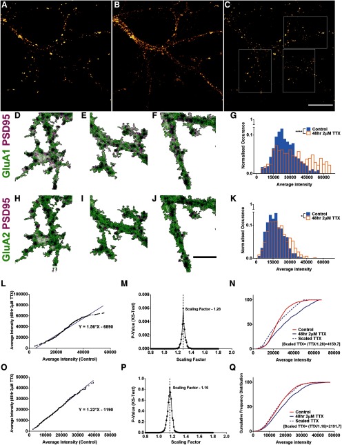
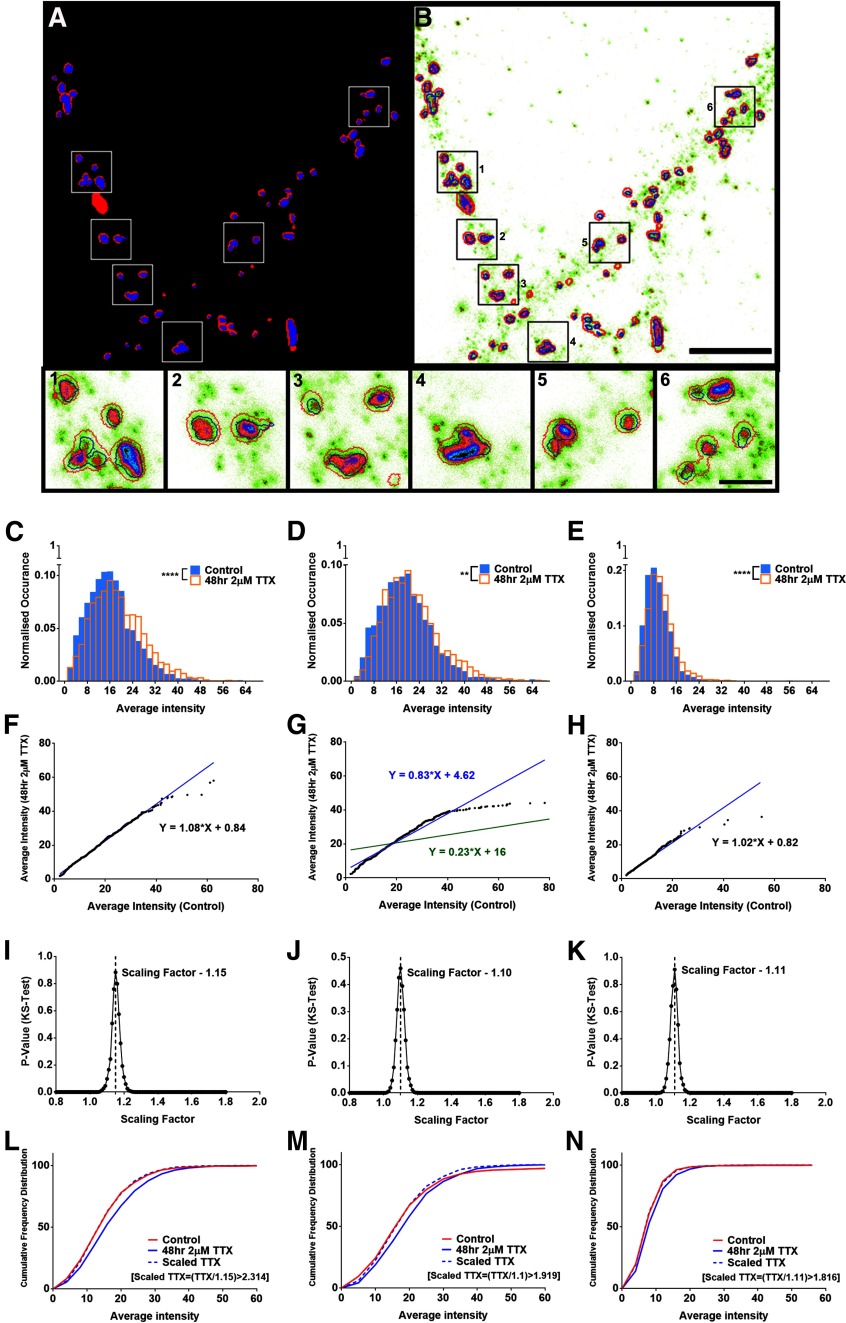
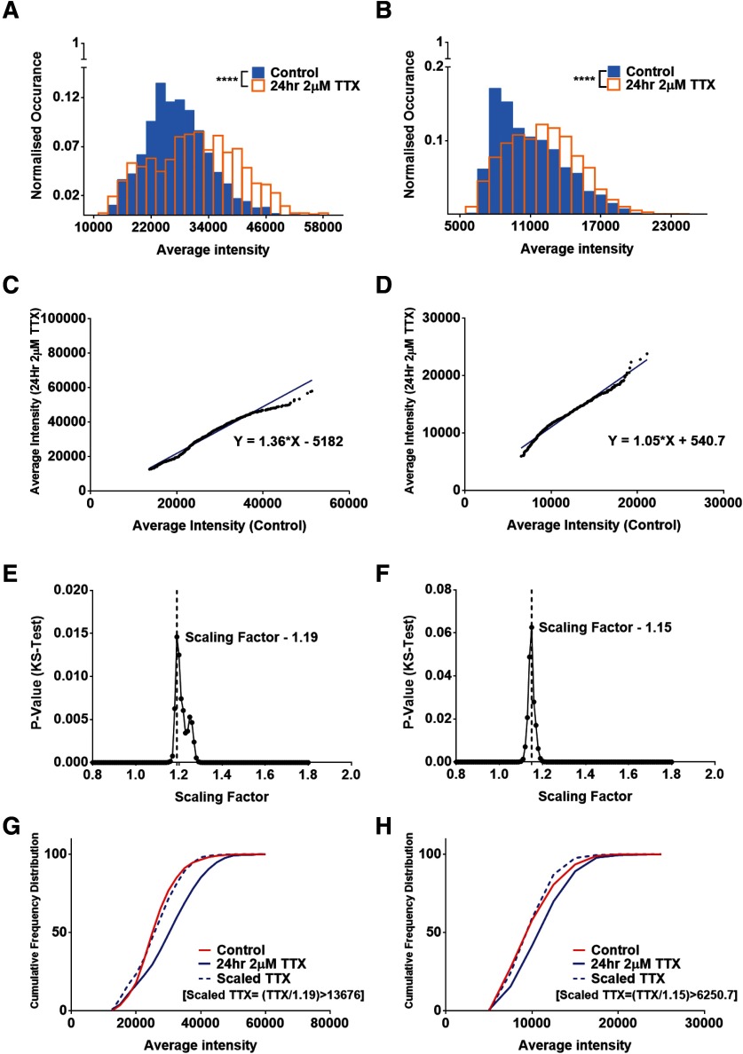
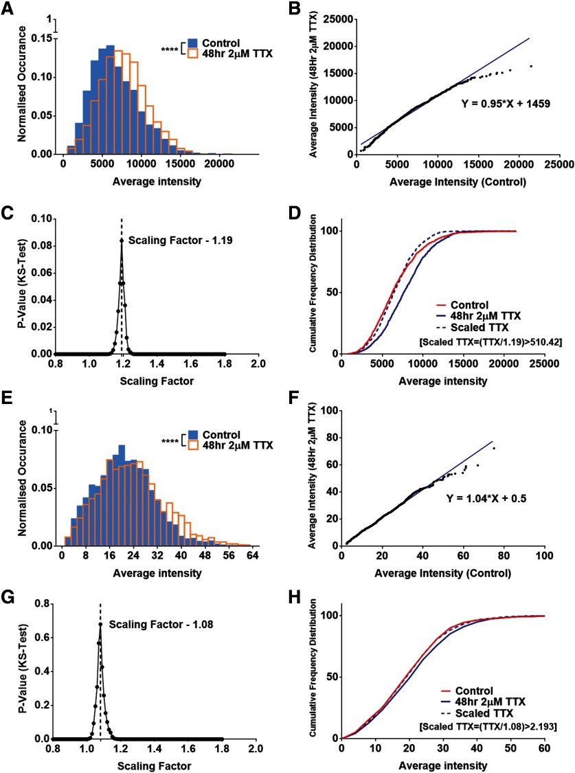
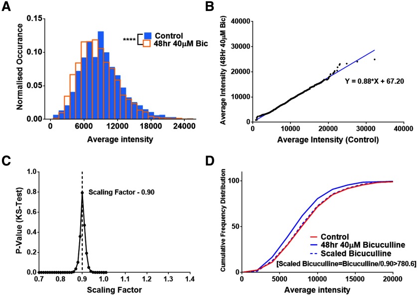
Homeostatic scaling is a form of synaptic plasticity where individual synapses scale their strengths to compensate for global suppression or elevation of neuronal activity. This process can be studied by measuring miniature EPSP (mEPSP) amplitudes and frequencies following the regulation of activity in neuronal cultures. Here, we demonstrate a quantitative approach to characterize multiplicative synaptic scaling using immunolabelling of hippocampal neuronal cultures treated with tetrodotoxin (TTX) or bicuculline to extract scaling factors for various synaptic proteins. This approach allowed us to directly examine the scaling of presynaptic and postsynaptic scaffolding molecules along with neurotransmitter receptors in primary cultures from mouse and rat hippocampal neurons. We show robust multiplicative scaling of synaptic scaffolding molecules namely, Shank2, PSD95, Bassoon, and AMPA receptor subunits and quantify their scaling factors. We use super-resolution microscopy to calculate scaling factors of surface expressed GluA2 within functional zones of the synapse and show that there is differential and correlated scaling of GluA2 levels within the spine, the postsynaptic density (PSD), and the perisynaptic regions. Our method opens a novel paradigm to quantify relative molecular changes of synaptic proteins within distinct subsynaptic compartments from a large number of synapses in response to alteration of neuronal activity, providing anatomic insights into the intricacies of variability in strength of individual synapses.
内稳态缩放是一种突触可塑性形式,其中单个突触会调整其强度以补偿神经元活动的全局抑制或升高。可以通过测量神经元培养物中活性调节后的微小 EPSP(mEPSP)幅度和频率来研究该过程。在这里,我们展示了一种使用海兔毒素(TTX)或印防己毒素处理的海马神经元培养物的免疫标记来定量表征乘法性突触缩放的方法,以提取各种突触蛋白的缩放因子。这种方法使我们能够直接检查突触前和突触后支架分子以及来自小鼠和大鼠海马神经元的原代培养物中的神经递质受体的缩放。我们显示了突触支架分子(Shank2、PSD95、Bassoon 和 AMPA 受体亚基)的强大乘法性缩放,并量化了它们的缩放因子。我们使用超分辨率显微镜计算了功能区内突触表面表达的 GluA2 的缩放因子,并显示 GluA2 水平在棘突、突触后密度(PSD)和突触周区域内存在差异和相关的缩放。我们的方法为定量测量神经元活动改变时不同亚突触隔室中突触蛋白的相对分子变化提供了一种新的范例,为单个突触强度变化的复杂性提供了解剖学见解。