Sun Shi-Yan, Li Xiao-Wei, Cao Ran, Zhao Yang, Sheng Nengyin, Tang Ai-Hui
Chinese Academy of Sciences (CAS) Key Laboratory of Brain Function and Disease, Ministry of Education Key Laboratory for Membrane-less Organelles and Cellular Dynamics, Division of Life Sciences and Medicine, University of Science and Technology of China, Hefei, China.
Institute of Artificial Intelligence, Hefei Comprehensive National Science Center, Hefei, China.
Front Synaptic Neurosci. 2022 May 24;14:748184. doi: 10.3389/fnsyn.2022.748184. eCollection 2022.
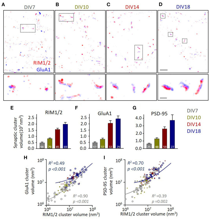
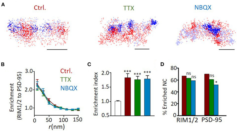
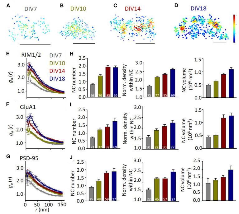
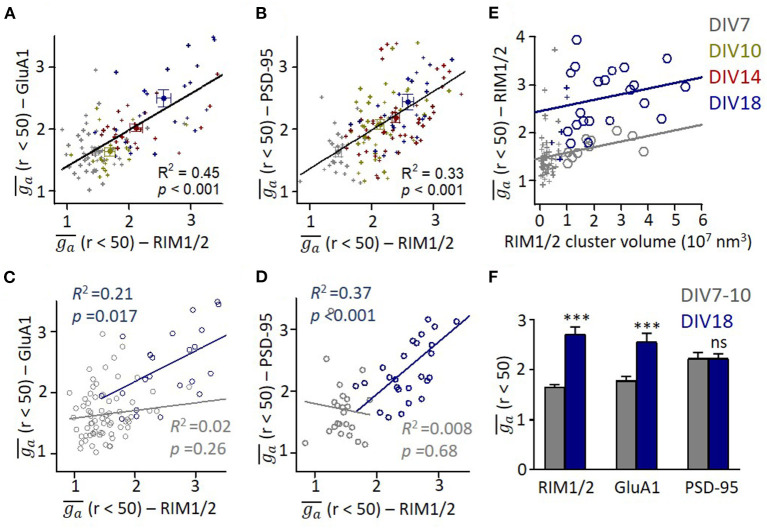
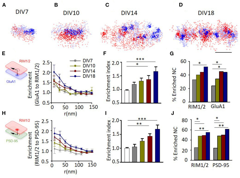
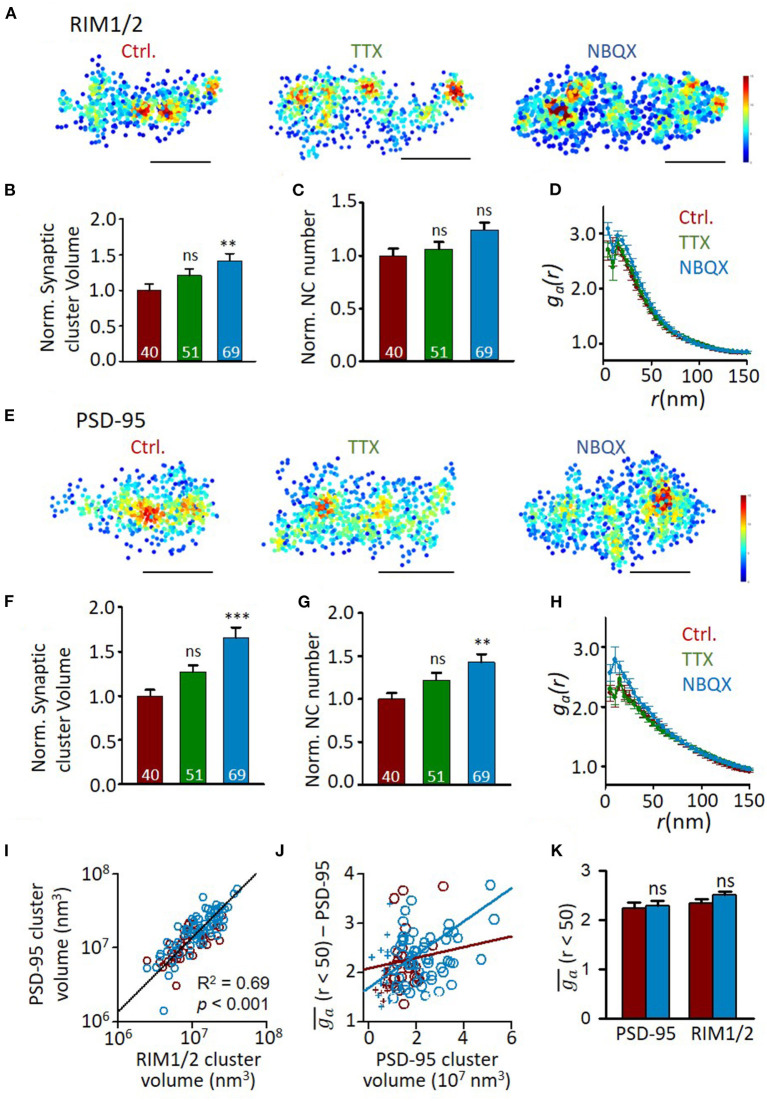
Nanoscale organization of presynaptic proteins determines the sites of transmitter release, and its alignment with assemblies of postsynaptic receptors through nanocolumns is suggested to optimize the efficiency of synaptic transmission. However, it remains unknown how these nano-organizations are formed during development. In this study, we used super-resolution stochastic optical reconstruction microscopy (STORM) imaging technique to systematically analyze the evolvement of subsynaptic organization of three key synaptic proteins, namely, RIM1/2, GluA1, and PSD-95, during synapse maturation in cultured hippocampal neurons. We found that volumes of synaptic clusters and their subsynaptic heterogeneity increase as synapses get matured. Synapse sizes of presynaptic and postsynaptic compartments correlated well at all stages, while only more mature synapses demonstrated a significant correlation between presynaptic and postsynaptic nano-organizations. After a long incubation with an inhibitor of action potentials or AMPA receptors, both presynaptic and postsynaptic compartments showed increased synaptic cluster volume and subsynaptic heterogeneity; however, the trans-synaptic alignment was intact. Together, our results characterize the evolvement of subsynaptic protein architectures during development and demonstrate that the nanocolumn is organized more likely by an intrinsic mechanism and independent of synaptic activities.
突触前蛋白的纳米级组织决定了神经递质释放的位点,并且通过纳米柱与突触后受体组件的对齐被认为可以优化突触传递的效率。然而,这些纳米组织在发育过程中是如何形成的仍然未知。在本研究中,我们使用超分辨率随机光学重建显微镜(STORM)成像技术,系统地分析了培养的海马神经元突触成熟过程中三种关键突触蛋白,即RIM1/2、GluA1和PSD-95的亚突触组织的演变。我们发现,随着突触成熟,突触簇的体积及其亚突触异质性增加。在所有阶段,突触前和突触后小室的突触大小都具有良好的相关性,而只有更成熟的突触在突触前和突触后纳米组织之间表现出显著的相关性。在用动作电位抑制剂或AMPA受体长时间孵育后,突触前和突触后小室的突触簇体积和亚突触异质性均增加;然而,跨突触对齐是完整的。总之,我们的结果描述了发育过程中亚突触蛋白结构的演变,并证明纳米柱更可能是由内在机制组织的,且独立于突触活动。