Division of Spine, Department of Orthopedics, Tongji Hospital Affiliated to Tongji University School of Medicine, School of Life Science and Technology, Tongji University, Shanghai, People's Republic of China.
Key Laboratory of Spine and Spinal Cord Injury Repair and Regeneration (Tongji University), Ministry of Education, Shanghai, People's Republic of China.
Int J Nanomedicine. 2020 Mar 3;15:1421-1435. doi: 10.2147/IJN.S225722. eCollection 2020.
In this study, we aim to explore the effects of graphene oxide (GO), a derivative of graphene, nanoparticles of four different sizes on the cellular fate of mouse neural stem cells (mNSCs).
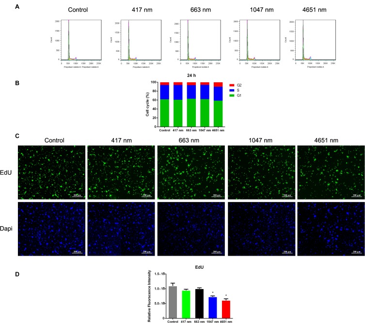
GO NPs were characterized with transmission electron microscopy (TEM), scanning electron micrography (SEM), atomic force microscopy (AFM) and Raman Spectra analysis. The cytotoxic effects of the GO NPs of different sizes on the mNSCs were determined using CCK-8 assay, Annexin V-APC/ 7-AAD staining and EdU staining assays. We investigated the biological and the mechanisms of GO NPs on cells using immunofluorescence analysis and quantitative real-time PCR (qPCR).
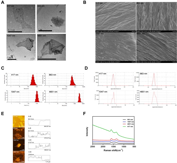
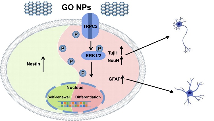
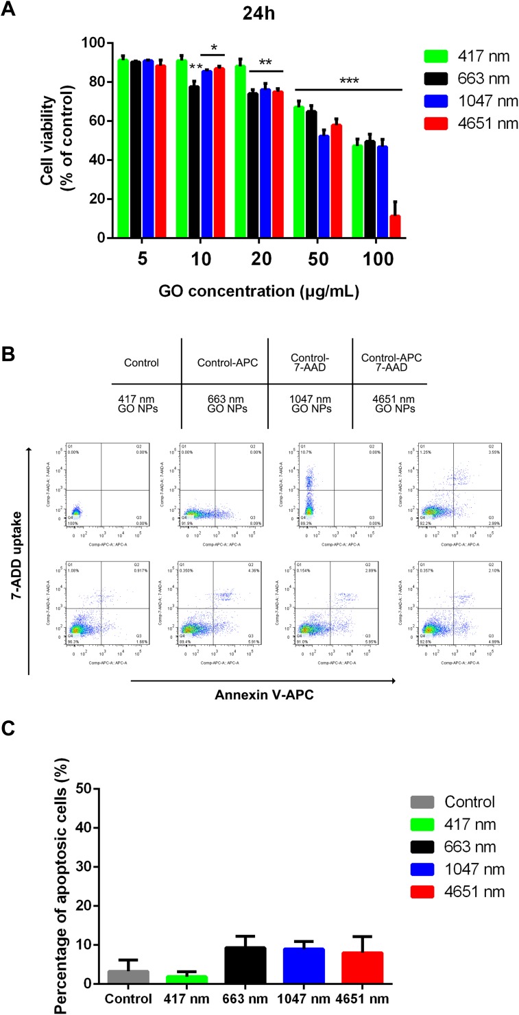
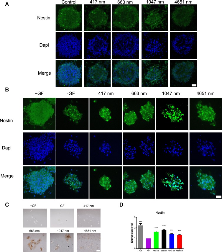
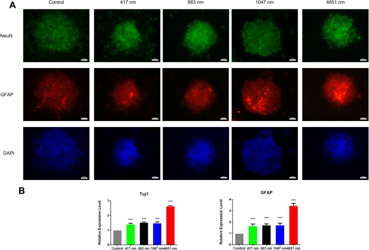
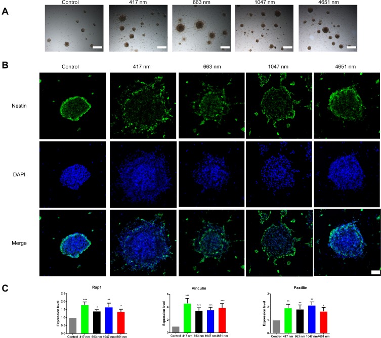
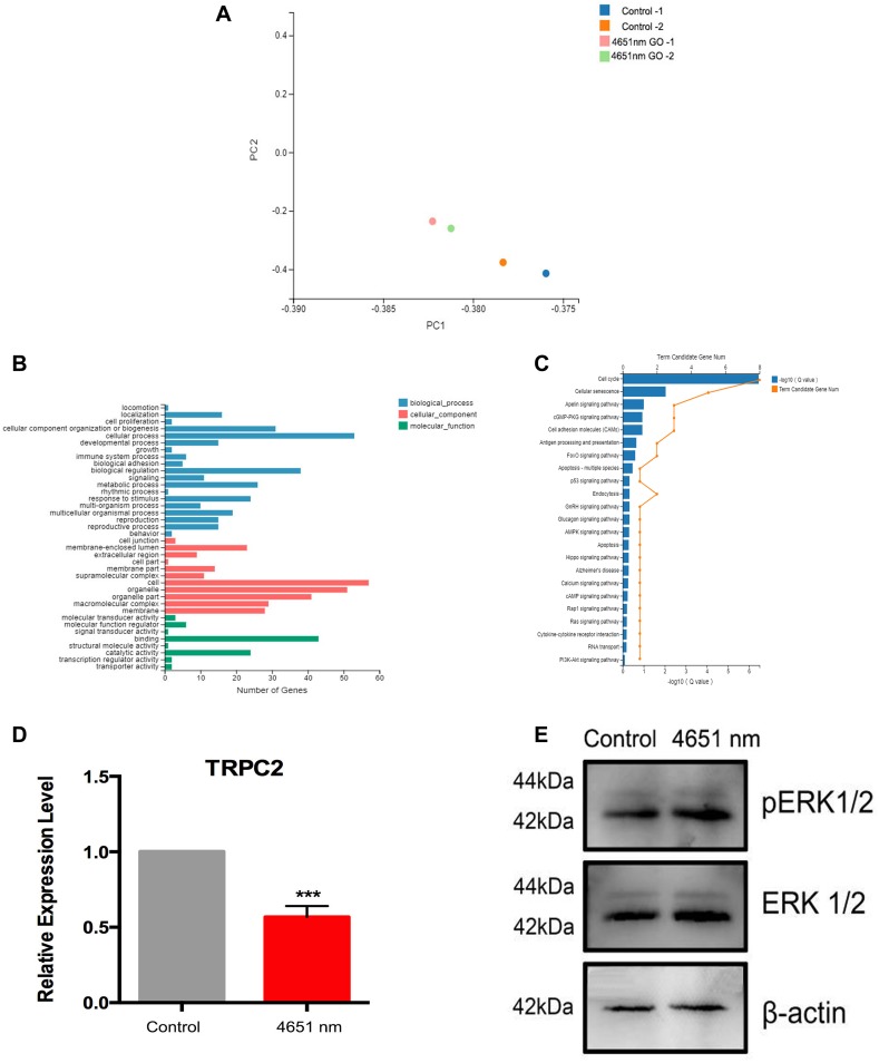
The average hydrodynamic sizes of the GO NPs were 417 nm, 663 nm, 1047 nm, and 4651 nm, with a thickness of approximately 22.5 nm, 17.7 nm, 22.4 nm, and 13.4 nm, respectively. GO NPs of all sizes showed low cytotoxicity at a concentration of 20 μg/mL on the mNSCs. Immunostaining demonstrated that treatment with GO NPs, especially the 663 nm ones, enhanced the self-renewal ability of mNSCs in the absence of EGF and bFGF. Under differentiation medium conditions that are free of mitogenic factors, all the GO NPs, particularly the 4651 nm ones, increased the expression level of Tuj1 and GFAP. With regards to the migration ability, we found that 417 nm GO-NP-treated mNSCs migrated over a longer distance than the control group obviously. In addition, higher expression of Rap1, Vinculin and Paxillin was observed in the GO NP-treated groups compared to the control group. mRNA-Sequence analysis and Western blotting results suggested that the 4651 nm GO NPs triggered positive neuronal differentiation through phosphorylation of ERK1/2 by the downregulating of TRPC2.
GO NPs play an important role in the applications of inducing self-renewal and differentiation of mNSC, and are promising in the future for further studies.
在本研究中,我们旨在探讨氧化石墨烯(GO),一种石墨烯的衍生物,四种不同大小的纳米颗粒对小鼠神经干细胞(mNSC)细胞命运的影响。
通过透射电子显微镜(TEM)、扫描电子显微镜(SEM)、原子力显微镜(AFM)和拉曼光谱分析对 GO NPs 进行了表征。使用 CCK-8 测定法、Annexin V-APC/7-AAD 染色和 EdU 染色试验测定不同大小的 GO NPs 对 mNSCs 的细胞毒性作用。我们使用免疫荧光分析和实时定量 PCR(qPCR)研究了 GO NPs 对细胞的生物学和机制。
GO NPs 的平均水动力粒径分别为 417nm、663nm、1047nm 和 4651nm,厚度分别约为 22.5nm、17.7nm、22.4nm 和 13.4nm。在浓度为 20μg/mL 时,所有大小的 GO NPs 对 mNSCs 的细胞毒性均较低。免疫染色表明,GO NPs 处理,特别是 663nm 的 GO NPs,增强了 mNSCs 在没有 EGF 和 bFGF 的情况下的自我更新能力。在没有有丝分裂原因子的分化培养基条件下,所有 GO NPs,特别是 4651nm 的 GO NPs,增加了 Tuj1 和 GFAP 的表达水平。关于迁移能力,我们发现 417nm GO-NP 处理的 mNSCs 比对照组明显迁移更长的距离。此外,与对照组相比,GO NP 处理组中 Rap1、Vinculin 和 Paxillin 的表达水平更高。mRNA-序列分析和 Western blotting 结果表明,4651nm GO NPs 通过下调 TRPC2 磷酸化 ERK1/2 触发阳性神经元分化。
GO NPs 在诱导 mNSC 的自我更新和分化的应用中起着重要的作用,在未来的研究中具有广阔的前景。