• 文献检索
  • 文档翻译
  • 深度研究
  • 学术资讯
  • Suppr Zotero 插件Zotero 插件
  • 邀请有礼
  • 套餐&价格
  • 历史记录
应用&插件
Suppr Zotero 插件Zotero 插件浏览器插件Mac 客户端Windows 客户端微信小程序
定价
高级版会员购买积分包购买API积分包
服务
文献检索文档翻译深度研究API 文档MCP 服务
关于我们
关于 Suppr公司介绍联系我们用户协议隐私条款
关注我们

Suppr 超能文献

核心技术专利:CN118964589B侵权必究
粤ICP备2023148730 号-1Suppr @ 2026

文献检索

告别复杂PubMed语法,用中文像聊天一样搜索,搜遍4000万医学文献。AI智能推荐,让科研检索更轻松。

立即免费搜索

文件翻译

保留排版,准确专业,支持PDF/Word/PPT等文件格式,支持 12+语言互译。

免费翻译文档

深度研究

AI帮你快速写综述,25分钟生成高质量综述,智能提取关键信息,辅助科研写作。

立即免费体验

CT 在 2019 年冠状病毒病(COVID-19)中的应用:对 4410 例成年患者胸部 CT 表现的系统综述。

CT in coronavirus disease 2019 (COVID-19): a systematic review of chest CT findings in 4410 adult patients.

机构信息

Department of Cardiovascular Radiology & Endovascular Interventions, All India Institute of Medical Sciences, New Delhi, 110029, India.

Department of Cardiology, Sri Chitra Tirunal Institute for Medical Sciences and Technology, Trivandrum, India.

出版信息

Eur Radiol. 2020 Nov;30(11):6129-6138. doi: 10.1007/s00330-020-06975-7. Epub 2020 May 30.

DOI:10.1007/s00330-020-06975-7
PMID:32474632
原文链接:https://pmc.ncbi.nlm.nih.gov/articles/PMC7261039/
Abstract

OBJECTIVE

The objective of this systematic review was to evaluate the key imaging manifestations of COVID-19 on chest CT in adult patients by providing a comprehensive review of the published literature.

METHODS

We performed a systematic literature search from the PubMed, Google Scholar, Embase, and WHO databases for studies mentioning the chest CT imaging findings of adult COVID-19 patients.

RESULTS

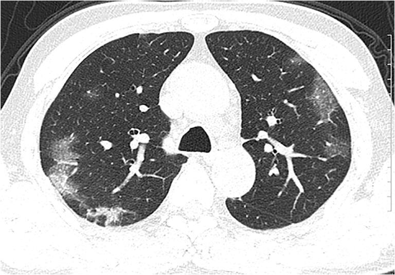
A total of 45 studies comprising 4410 patients were included. Ground glass opacities (GGO), in isolation (50.2%) or coexisting with consolidations (44.2%), were the most common lesions. Distribution of GGOs was most commonly bilateral, peripheral/subpleural, and posterior with predilection for lower lobes. Common ancillary findings included pulmonary vascular enlargement (64%), intralobular septal thickening (60%), adjacent pleural thickening (41.7%), air bronchograms (41.2%), subpleural lines, crazy paving, bronchus distortion, bronchiectasis, and interlobular septal thickening. CT in early follow-up period generally showed an increase in size, number, and density of GGOs, with progression into mixed areas of GGOs plus consolidations and crazy paving, peaking at 10-11 days, before gradually resolving or persisting as patchy fibrosis. While younger adults more commonly had GGOs, extensive/multilobar involvement with consolidations was prevalent in the older population and those with severe disease.

CONCLUSION

This review describes the imaging features for diagnosis, stratification, and follow-up of COVID-19 patients. The most common CT manifestations are bilateral, peripheral/subpleural, posterior GGOs with or without consolidations with a lower lobe predominance. It is pertinent to be familiar with the various imaging findings to positively impact the management of these patients.

KEY POINTS

• Ground glass opacities (GGOs), whether isolated or coexisting with consolidations, in bilateral and subpleural distribution, are the most prevalent chest CT findings in adult COVID-19 patients. • Follow-up CT shows a progression of GGOs into a mixed pattern, reaching a peak at 10-11 days, before gradually resolving or persisting as patchy fibrosis. • Younger people tend to have more GGOs. Older or sicker people tend to have more extensive involvement with consolidations.

摘要

目的

本系统评价旨在通过综合回顾已发表文献,评估成人 COVID-19 患者胸部 CT 的关键影像学表现。

方法

我们从 PubMed、Google Scholar、Embase 和世界卫生组织数据库中进行了系统文献检索,以查找提及成人 COVID-19 患者胸部 CT 影像学发现的研究。

结果

共纳入 45 项研究,共计 4410 例患者。磨玻璃影(GGO),无论是孤立存在(50.2%)还是与实变共存(44.2%),是最常见的病变。GGO 的分布最常见为双侧、外周/胸膜下和后背部,以下肺叶多见。常见的辅助发现包括肺血管增大(64%)、小叶间隔增厚(60%)、相邻胸膜增厚(41.7%)、空气支气管征(41.2%)、胸膜下线、铺路石征、支气管扭曲、支气管扩张和小叶间隔增厚。早期随访 CT 通常显示 GGO 数量、大小和密度增加,进展为 GGO 合并实变的混杂区和铺路石征,在第 10-11 天达到高峰,然后逐渐消退或持续存在为斑片状纤维化。年轻患者更常见 GGO,而老年患者和重症患者则更常见广泛/多叶累及实变。

结论

本综述描述了 COVID-19 患者的诊断、分层和随访的影像学特征。最常见的 CT 表现为双侧、外周/胸膜下、后背部 GGO 伴或不伴实变,以下肺叶为主。熟悉各种影像学表现对于积极管理这些患者至关重要。

关键要点

  • 磨玻璃影(GGOs),无论是孤立存在还是与实变共存,在双侧和胸膜下分布,是成人 COVID-19 患者胸部 CT 最常见的发现。

  • 随访 CT 显示 GGO 进展为混合模式,在第 10-11 天达到高峰,然后逐渐消退或持续存在为斑片状纤维化。

  • 年轻患者倾向于出现更多的 GGOs。年龄较大或病情较重的患者更倾向于出现广泛的实变累及。

https://cdn.ncbi.nlm.nih.gov/pmc/blobs/e06a/7261039/4d2107d1bc5e/330_2020_6975_Fig2_HTML.jpg
https://cdn.ncbi.nlm.nih.gov/pmc/blobs/e06a/7261039/df50a1479ee4/330_2020_6975_Fig1_HTML.jpg
https://cdn.ncbi.nlm.nih.gov/pmc/blobs/e06a/7261039/4d2107d1bc5e/330_2020_6975_Fig2_HTML.jpg
https://cdn.ncbi.nlm.nih.gov/pmc/blobs/e06a/7261039/df50a1479ee4/330_2020_6975_Fig1_HTML.jpg
https://cdn.ncbi.nlm.nih.gov/pmc/blobs/e06a/7261039/4d2107d1bc5e/330_2020_6975_Fig2_HTML.jpg

相似文献

1
CT in coronavirus disease 2019 (COVID-19): a systematic review of chest CT findings in 4410 adult patients.CT 在 2019 年冠状病毒病(COVID-19)中的应用:对 4410 例成年患者胸部 CT 表现的系统综述。
Eur Radiol. 2020 Nov;30(11):6129-6138. doi: 10.1007/s00330-020-06975-7. Epub 2020 May 30.
2
Coronavirus Disease 2019 (COVID-19): A Systematic Review of Imaging Findings in 919 Patients.新型冠状病毒病 2019(COVID-19):919 例患者影像学表现的系统评价。
AJR Am J Roentgenol. 2020 Jul;215(1):87-93. doi: 10.2214/AJR.20.23034. Epub 2020 Mar 14.
3
Coronavirus Disease 2019 (COVID-19) CT Findings: A Systematic Review and Meta-analysis.新型冠状病毒病 2019(COVID-19)的 CT 表现:系统评价和荟萃分析。
J Am Coll Radiol. 2020 Jun;17(6):701-709. doi: 10.1016/j.jacr.2020.03.006. Epub 2020 Mar 25.
4
CT image of novel coronavirus pneumonia: a case report.新型冠状病毒肺炎的CT图像:病例报告
Jpn J Radiol. 2020 May;38(5):407-408. doi: 10.1007/s11604-020-00945-1. Epub 2020 Mar 18.
5
A Comparison of Clinical and Chest CT Findings in Patients With Influenza A (H1N1) Virus Infection and Coronavirus Disease (COVID-19).甲型流感病毒(H1N1)感染与新型冠状病毒肺炎(COVID-19)患者的临床和胸部 CT 表现比较。
AJR Am J Roentgenol. 2020 Nov;215(5):1065-1071. doi: 10.2214/AJR.20.23214. Epub 2020 May 26.
6
Clinical and radiological features of novel coronavirus pneumonia.新型冠状病毒肺炎的临床和影像学特征。
J Xray Sci Technol. 2020;28(3):391-404. doi: 10.3233/XST-200687.
7
The chest CT features of coronavirus disease 2019 (COVID-19) in China: a meta-analysis of 19 retrospective studies.中国 2019 年冠状病毒病(COVID-19)的胸部 CT 特征:19 项回顾性研究的荟萃分析。
Virol J. 2020 Oct 21;17(1):159. doi: 10.1186/s12985-020-01432-9.
8
Chest CT-based differential diagnosis of 28 patients with suspected corona virus disease 2019 (COVID-19).基于胸部 CT 的 28 例疑似 2019 冠状病毒病(COVID-19)患者的鉴别诊断。
Br J Radiol. 2020 Aug;93(1112):20200243. doi: 10.1259/bjr.20200243. Epub 2020 Jun 2.
9
Analysis of clinical features and imaging signs of COVID-19 with the assistance of artificial intelligence.人工智能辅助分析 COVID-19 的临床特征和影像征象。
Eur Rev Med Pharmacol Sci. 2020 Aug;24(15):8210-8218. doi: 10.26355/eurrev_202008_22510.
10
CT findings of COVID-19 in follow-up: comparison between progression and recovery.新冠肺炎 CT 随访表现:进展与恢复的比较。
Diagn Interv Radiol. 2020 Jul;26(4):301-307. doi: 10.5152/dir.2019.20176.

引用本文的文献

1
The respiratory microbiome in patients with post-COVID-19 residual lung abnormalities resembles that of healthy individuals and is distinct from idiopathic pulmonary fibrosis.新冠后肺部残留异常患者的呼吸道微生物群与健康个体相似,且与特发性肺纤维化不同。
ERJ Open Res. 2025 May 27;11(3). doi: 10.1183/23120541.00826-2024. eCollection 2025 May.
2
Impact of the coronavirus disease 2019 pandemic on hospital admissions for idiopathic pulmonary fibrosis: a nationwide population-based study.2019 冠状病毒病大流行对特发性肺纤维化住院的影响:一项全国性基于人群的研究。
BMC Pulm Med. 2024 Aug 31;24(1):430. doi: 10.1186/s12890-024-03230-3.
3

本文引用的文献

1
HRCT imaging features in representative imported cases of 2019 novel coronavirus pneumonia.2019新型冠状病毒肺炎典型输入性病例的高分辨率CT成像特征
Precis Clin Med. 2020 Mar;3(1):9-13. doi: 10.1093/pcmedi/pbaa004. Epub 2020 Feb 11.
2
CT Features and Short-term Prognosis of COVID-19 Pneumonia: A Single-Center Study from Kashan, Iran.新型冠状病毒肺炎的CT特征及短期预后:来自伊朗卡尚的一项单中心研究
Radiol Cardiothorac Imaging. 2020 Apr 20;2(2):e200130. doi: 10.1148/ryct.2020200130. eCollection 2020 Apr.
3
Chest CT Findings in Cases from the Cruise Ship with Coronavirus Disease (COVID-19).
Exploring the prevalence and chest CT predictors of Long COVID in children: a comprehensive study from Shanghai and Linyi.
探索儿童长期新冠的患病率及胸部CT预测因素:来自上海和临沂的一项综合研究
Front Pediatr. 2024 Aug 7;12:1420196. doi: 10.3389/fped.2024.1420196. eCollection 2024.
4
Assessment of post-SARS-CoV-2 fatigue among physicians working in COVID-designated hospitals in Dhaka, Bangladesh.评估孟加拉国达卡的 COVID 指定医院工作的医生在新冠病毒后出现的疲劳症状。
Brain Behav. 2024 Jun;14(6):e3553. doi: 10.1002/brb3.3553.
5
Which sequence should be used in the thorax magnetic resonance imaging of COVID-19: a comparative study.COVID-19 胸部磁共振成像中应使用哪种序列:一项对比研究。
Turk J Med Sci. 2023 Aug 11;53(5):1214-1223. doi: 10.55730/1300-0144.5687. eCollection 2023.
6
Interest of Chest CT to Assess the Prognosis of SARS-CoV-2 Pneumonia: An In-Hospital-Based Experience in Sub-Saharan Africa.胸部CT对评估新型冠状病毒肺炎预后的价值:撒哈拉以南非洲地区的一项基于医院的经验
Pulm Med. 2024 Apr 25;2024:5520174. doi: 10.1155/2024/5520174. eCollection 2024.
7
Quantitative CT Texture Analysis of COVID-19 Hospitalized Patients during 3-24-Month Follow-Up and Correlation with Functional Parameters.COVID-19住院患者3至24个月随访期间的定量CT纹理分析及其与功能参数的相关性
Diagnostics (Basel). 2024 Mar 5;14(5):550. doi: 10.3390/diagnostics14050550.
8
The Multisystem Impact of Long COVID: A Comprehensive Review.长新冠的多系统影响:全面综述
Diagnostics (Basel). 2024 Jan 24;14(3):244. doi: 10.3390/diagnostics14030244.
9
Doubts and concerns about COVID-19 uncertainties on imaging data, clinical score, and outcomes.对 COVID-19 影像学数据、临床评分和结果的不确定性的疑虑和担忧。
BMC Pulm Med. 2023 Nov 25;23(1):472. doi: 10.1186/s12890-023-02763-3.
10
Cough and sputum in long COVID are associated with severe acute COVID-19: a Japanese cohort study.长新冠患者的咳嗽和咳痰与严重急性 COVID-19 相关:一项日本队列研究。
Respir Res. 2023 Nov 14;24(1):283. doi: 10.1186/s12931-023-02591-3.
邮轮上新型冠状病毒肺炎(COVID-19)病例的胸部CT表现
Radiol Cardiothorac Imaging. 2020 Mar 17;2(2):e200110. doi: 10.1148/ryct.2020200110. eCollection 2020 Apr.
4
Imaging Profile of the COVID-19 Infection: Radiologic Findings and Literature Review.新型冠状病毒肺炎感染的影像学表现:放射学发现与文献综述
Radiol Cardiothorac Imaging. 2020 Feb 13;2(1):e200034. doi: 10.1148/ryct.2020200034. eCollection 2020 Feb.
5
Pathogenesis of COVID-19 from a cell biology perspective.从细胞生物学角度看 COVID-19 的发病机制。
Eur Respir J. 2020 Apr 16;55(4). doi: 10.1183/13993003.00607-2020. Print 2020 Apr.
6
The Role of Chest Imaging in Patient Management during the COVID-19 Pandemic: A Multinational Consensus Statement from the Fleischner Society.《COVID-19 大流行期间患者管理中的胸部影像学作用:Fleischner 学会的一项多国共识声明》。
Radiology. 2020 Jul;296(1):172-180. doi: 10.1148/radiol.2020201365. Epub 2020 Apr 7.
7
CT imaging changes of corona virus disease 2019(COVID-19): a multi-center study in Southwest China.COVID-19 的 CT 影像学改变:中国西南部多中心研究。
J Transl Med. 2020 Apr 6;18(1):154. doi: 10.1186/s12967-020-02324-w.
8
Clinical characteristics of non-critically ill patients with novel coronavirus infection (COVID-19) in a Fangcang Hospital.方仓医院中新型冠状病毒感染(COVID-19)非危重症患者的临床特征。
Clin Microbiol Infect. 2020 Aug;26(8):1063-1068. doi: 10.1016/j.cmi.2020.03.032. Epub 2020 Apr 3.
9
Pulmonary High-Resolution Computed Tomography (HRCT) Findings of Patients with Early-Stage Coronavirus Disease 2019 (COVID-19) in Hangzhou, China.中国杭州早期 2019 冠状病毒病(COVID-19)患者的肺部高分辨率计算机断层扫描(HRCT)表现。
Med Sci Monit. 2020 Apr 4;26:e923885. doi: 10.12659/MSM.923885.
10
Chest CT Features of COVID-19 in Rome, Italy.意大利罗马地区 COVID-19 的胸部 CT 特征。
Radiology. 2020 Aug;296(2):E79-E85. doi: 10.1148/radiol.2020201237. Epub 2020 Apr 3.