Brozzetti Lorenzo, Sacchetto Luca, Cecchini Maria Paola, Avesani Anna, Perra Daniela, Bongianni Matilde, Portioli Corinne, Scupoli Maria, Ghetti Bernardino, Monaco Salvatore, Buffelli Mario, Zanusso Gianluigi
Neuropathology Section, Department of Neurosciences, Biomedicine, and Movement Sciences, University of Verona, Verona, Italy.
Otolaryngology Section, Department of Surgery, Dentistry, Paediatrics and Gynaecology, University of Verona, Verona, Italy.
Front Neurosci. 2020 Mar 5;14:145. doi: 10.3389/fnins.2020.00145. eCollection 2020.
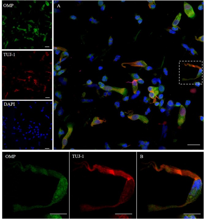
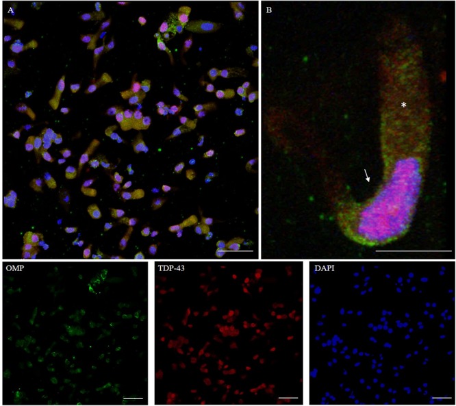
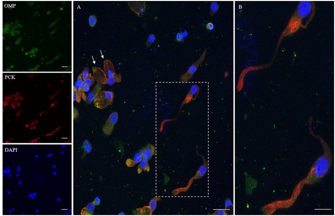
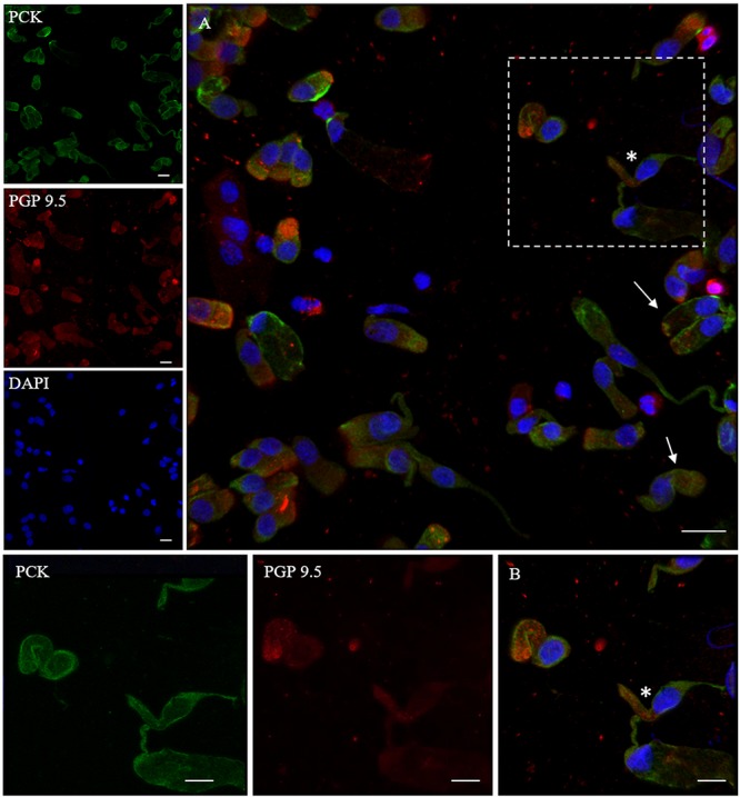
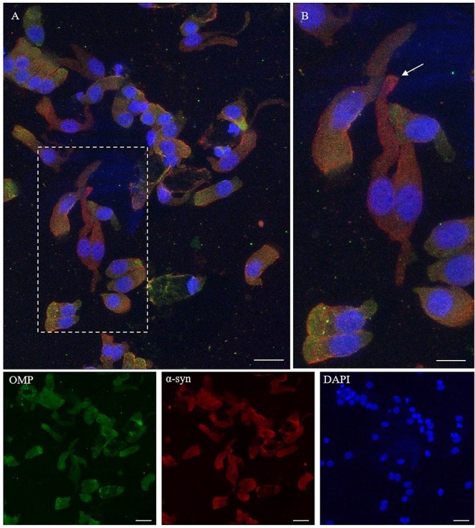
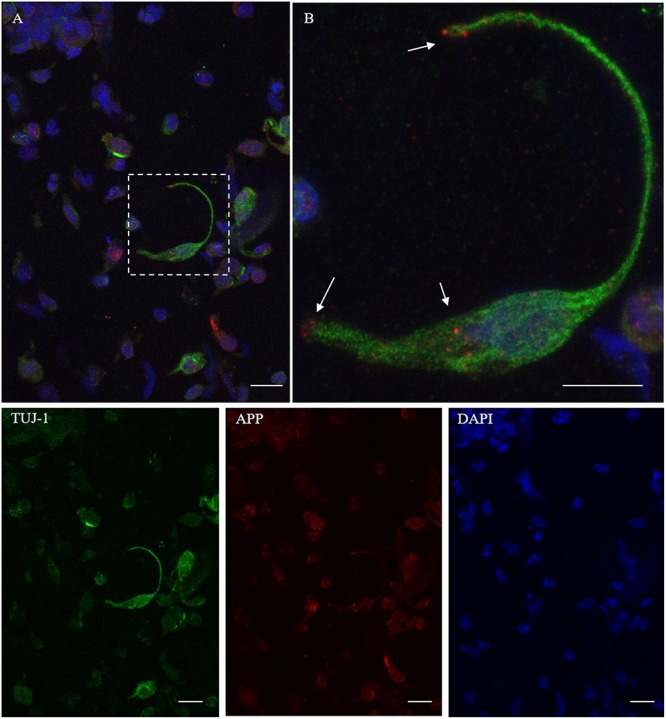
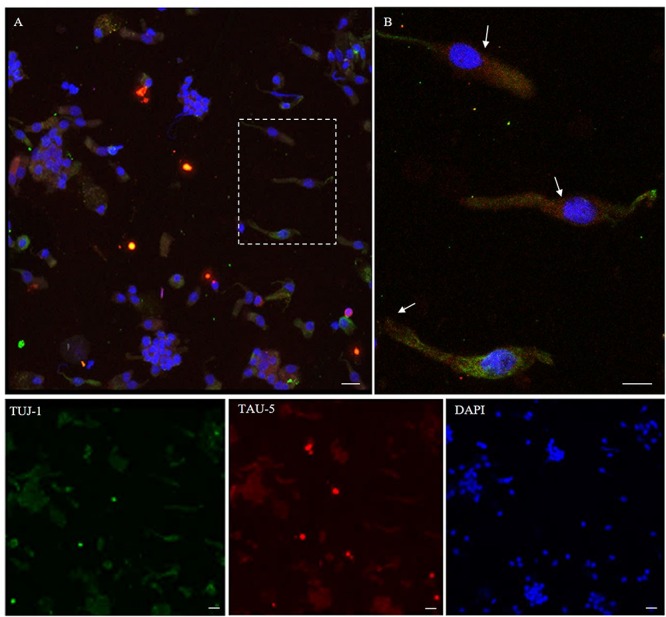
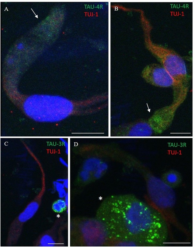
The olfactory neuroepithelium is located in the upper vault of the nasal cavity, lying on the olfactory cleft and projecting into the dorsal portion of the superior and middle turbinates beyond the mid-portion of the nasal septum. It is composed of a variety of cell types including olfactory sensory neurons, supporting glial-like cells, microvillar cells, and basal stem cells. The cells of the neuroepithelium are often intermingled with respiratory and metaplastic epithelial cells. Olfactory neurons undergo a constant self-renewal in the timespan of 2-3 months; they are directly exposed to the external environment, and thus they are vulnerable to physical and chemical injuries. The latter might induce metabolic perturbations and ultimately be the cause of cell death. However, the lifespan of olfactory neurons is biologically programmed, and for this reason, these cells have an accelerated metabolic cycle leading to an irreversible apoptosis. These characteristics make these cells suitable for research related to nerve cell degeneration and aging. Recent studies have shown that a non-invasive and painless olfactory brushing procedure allows an efficient sampling from the olfactory neuroepithelium. This approach allows to detect the pathologic prion protein in patients with sporadic Creutzfeldt-Jakob disease, using the real-time quaking-induced conversion assay. Investigating the expression of all the proteins associated to neurodegeneration in the cells of the olfactory mucosa is a novel approach toward understanding the pathogenesis of human neurodegenerative diseases. Our aim was to investigate the expression of α-synuclein, β-amyloid, tau, and TDP-43 in the olfactory neurons of normal subjects. We showed that these proteins that are involved in neurodegenerative diseases are expressed in olfactory neurons. These findings raise the question on whether a relationship exists between the mechanisms of protein aggregation that occur in the olfactory bulb during the early stage of the neurodegenerative process and the protein misfolding occurring in the olfactory neuroepithelium.
嗅神经上皮位于鼻腔顶部,位于嗅裂上,并伸入上鼻甲和中鼻甲的背侧部分,超出鼻中隔中部。它由多种细胞类型组成,包括嗅觉感觉神经元、支持性胶质样细胞、微绒毛细胞和基底干细胞。神经上皮细胞常与呼吸上皮细胞和化生上皮细胞相互交织。嗅觉神经元在2 - 3个月的时间跨度内不断自我更新;它们直接暴露于外部环境,因此容易受到物理和化学损伤。后者可能会引发代谢紊乱,最终导致细胞死亡。然而,嗅觉神经元的寿命是由生物程序设定的,因此,这些细胞具有加速的代谢周期,导致不可逆的细胞凋亡。这些特性使这些细胞适合用于与神经细胞退化和衰老相关的研究。最近的研究表明,一种非侵入性且无痛的嗅觉刷检程序能够从嗅神经上皮进行有效的采样。这种方法可以使用实时震颤诱导转化分析法检测散发性克雅氏病患者体内的病理性朊蛋白。研究嗅黏膜细胞中与神经退行性变相关的所有蛋白质的表达,是理解人类神经退行性疾病发病机制的一种新方法。我们的目的是研究正常受试者嗅神经元中α-突触核蛋白、β-淀粉样蛋白、tau蛋白和TDP-43的表达。我们发现这些与神经退行性疾病相关的蛋白质在嗅神经元中表达。这些发现引发了一个问题,即在神经退行性过程早期发生在嗅球中的蛋白质聚集机制与发生在嗅神经上皮中的蛋白质错误折叠之间是否存在关联。