Viterbi Family Department of Ophthalmology, Hamilton Glaucoma Center and Shiley Eye Institute, University of California San Diego, La Jolla, California, USA.
Viterbi Family Department of Ophthalmology, Hamilton Glaucoma Center and Shiley Eye Institute, University of California San Diego, La Jolla, California, USA.
Am J Ophthalmol. 2020 Sep;217:131-139. doi: 10.1016/j.ajo.2020.03.024. Epub 2020 Mar 25.
To compare gradient-boosting classifier (GBC) analysis of optical coherence tomography angiography (OCTA)-measured vessel density (VD) and OCT-measured tissue thickness to standard OCTA VD and OCT thickness parameters for classifying healthy eyes and eyes with early to moderate glaucoma.
Comparison of diagnostic tools.
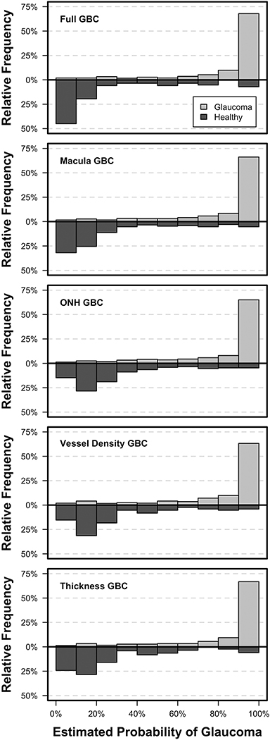
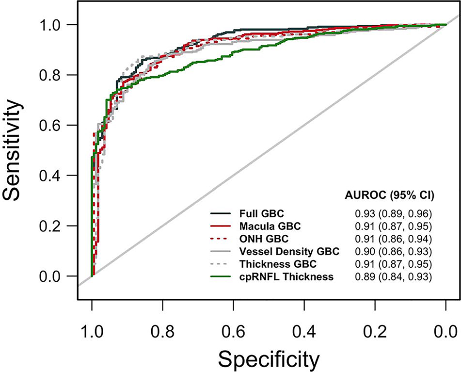
A total of 180 healthy eyes and 193 glaucomatous eyes with OCTA and OCT imaging of the macula and optic nerve head (ONH) were studied. Four GBCs were evaluated that combined 1) all macula VD and thickness measurements (Macula GBC), 2) all ONH VD and thickness measurements (ONH GBC), 3) all VD measurements from the macula and ONH (vessel density GBC), and 4) all thickness measurements from the macula and ONH (thickness GBC). ROC curve (AUROC) analyses compared the diagnostic accuracy of GBCs to that of standard instrument-provided parameters. A fifth GBC that combined all parameters (full GBC) also was investigated.
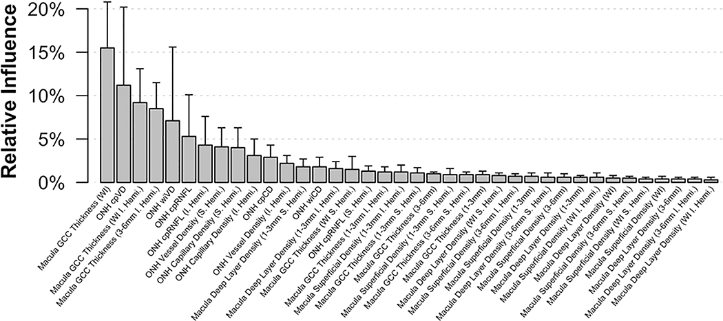
GBCs had better diagnostic accuracy than standard OCTA and OCT parameters with AUROCs ranging from 0.90 to 0.93 and 0.64 to 0.91, respectively. The full GBC (AUROC = 0.93) performed significantly better than the ONH GBC (AUROC = 0.91; P = .036) and the vessel density GBC (AUROC = 0.90; P = .010). All other GBCs performed similarly. The mean relative influence of each parameter included in the full GBC identified a combination of macular thickness and ONH VD measurements as the greatest contributors.
GBCs that combine OCTA and OCT macula and ONH measurements can improve diagnostic accuracy for glaucoma detection compared to most but not all instrument provided parameters.
比较梯度提升分类器(GBC)分析光学相干断层扫描血管造影(OCTA)测量的血管密度(VD)和 OCT 测量的组织厚度,以对健康眼和早期至中度青光眼眼进行分类。
诊断工具比较。
共研究了 180 只健康眼和 193 只患有 OCTA 和黄斑及视神经头(ONH)OCT 成像的青光眼眼。评估了 4 种 GBC,分别为:1)所有黄斑 VD 和厚度测量值(黄斑 GBC),2)所有 ONH VD 和厚度测量值(ONH GBC),3)所有来自黄斑和 ONH 的 VD 测量值(血管密度 GBC),4)所有来自黄斑和 ONH 的厚度测量值(厚度 GBC)。ROC 曲线(AUROC)分析比较了 GBC 与标准仪器提供的参数的诊断准确性。还研究了一种组合所有参数的第五个 GBC(全 GBC)。
GBC 的诊断准确性优于标准 OCTA 和 OCT 参数,AUROCs 范围分别为 0.90 至 0.93 和 0.64 至 0.91。全 GBC(AUROC = 0.93)的性能明显优于 ONH GBC(AUROC = 0.91;P =.036)和血管密度 GBC(AUROC = 0.90;P =.010)。其他所有 GBC 的表现相似。全 GBC 中包含的每个参数的平均相对影响确定了黄斑厚度和 ONH VD 测量值的组合是最大的贡献者。
与大多数但不是所有仪器提供的参数相比,组合 OCTA 和 OCT 黄斑和 ONH 测量值的 GBC 可以提高青光眼检测的诊断准确性。