Department of Pharmacy and Biotechnology, University of Bologna, 33-40126 Bologna, Italy.
Department of Electrical, Electronic and Information Engineering "Guglielmo Marconi" (DEI), University of Bologna 50, 47522 Cesena, Italy.
Int J Mol Sci. 2020 Mar 30;21(7):2368. doi: 10.3390/ijms21072368.
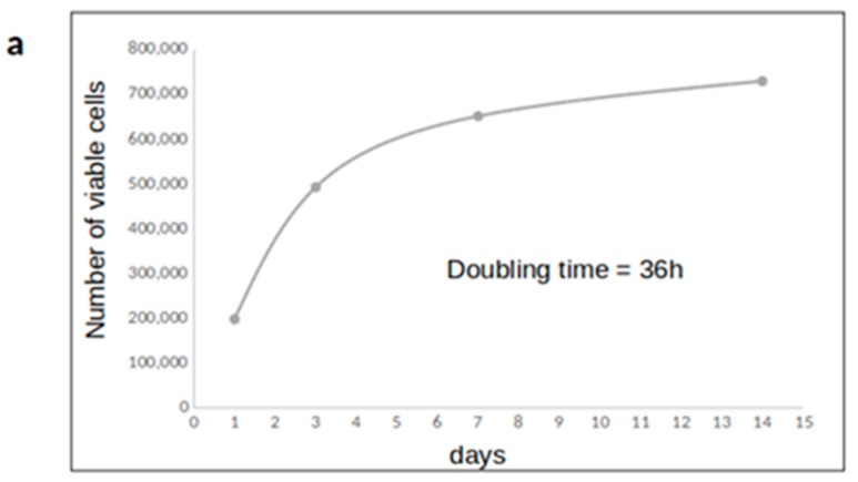
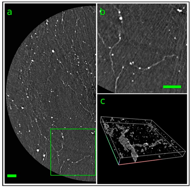
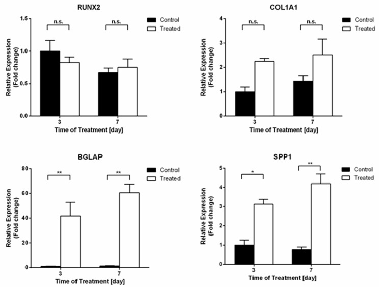
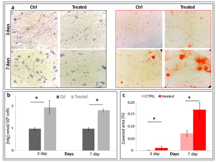
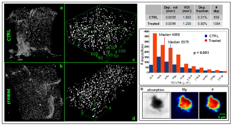
In this study, we explore the behaviour of intracellular magnesium during bone phenotype modulation in a 3D cell model built to mimic osteogenesis. In addition, we measured the amount of magnesium in the mineral depositions generated during osteogenic induction. A two-fold increase of intracellular magnesium content was found, both at three and seven days from the induction of differentiation. By X-ray microscopy, we characterized the morphology and chemical composition of the mineral depositions secreted by 3D cultured differentiated cells finding a marked co-localization of Mg with P at seven days of differentiation. This is the first experimental evidence on the presence of Mg in the mineral depositions generated during biomineralization, suggesting that Mg incorporation occurs during the bone forming process. In conclusion, this study on the one hand attests to an evident involvement of Mg in the process of cell differentiation, and, on the other hand, indicates that its multifaceted role needs further investigation.
在这项研究中,我们探索了在模拟成骨的 3D 细胞模型中,骨表型调节过程中细胞内镁的行为。此外,我们还测量了成骨诱导过程中生成的矿物质沉积中的镁含量。结果发现,在诱导分化后的第 3 天和第 7 天,细胞内镁含量增加了两倍。通过 X 射线显微镜,我们对 3D 培养分化细胞分泌的矿物质沉积的形态和化学成分进行了表征,发现分化第 7 天时 Mg 与 P 明显共定位。这是关于生物矿化过程中矿物质沉积中存在镁的第一个实验证据,表明镁的掺入发生在骨形成过程中。总之,这项研究一方面证明了镁在细胞分化过程中明显参与,另一方面表明其多方面的作用需要进一步研究。