Rasschaert Marlène, Weller Roy O, Schroeder Josef A, Brochhausen Christoph, Idée Jean-Marc
Guerbet, Research and Innovation Division, Aulnay-sous-Bois, France.
Neuropathology, Faculty of Medicine University of Southampton, Southampton General Hospital, Southampton, UK.
J Magn Reson Imaging. 2020 Nov;52(5):1293-1305. doi: 10.1002/jmri.27124. Epub 2020 Apr 4.
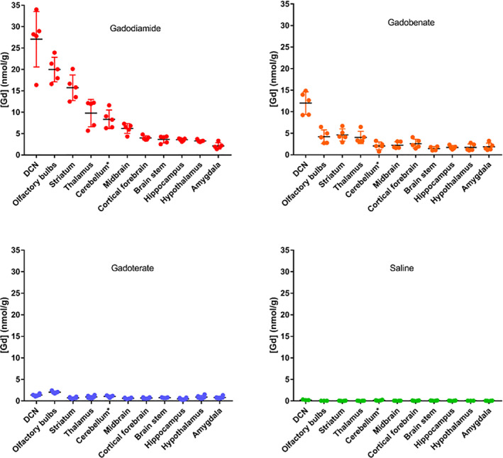
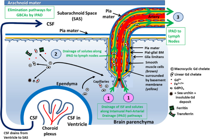
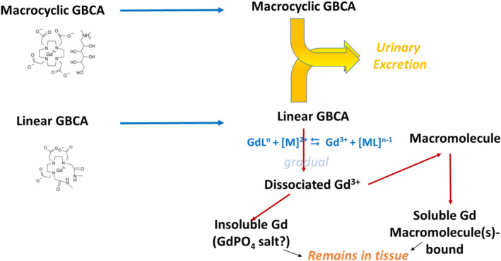
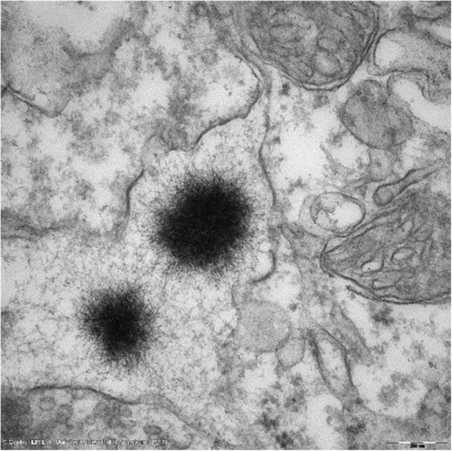
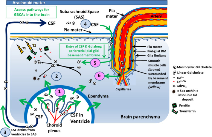
The unexpected appearance of T hyperintensities, mostly in the dentate nucleus and the globus pallidus, during nonenhanced MRI was reported in 2014. This effect is associated with prior repeated administrations of gadolinium (Gd)-based contrast agents (GBCAs) in patients with a functional blood-brain barrier (BBB). It is widely assumed that GBCAs do not cross the intact BBB, but the observation of these hypersignals raises questions regarding this assumption. This review critically discusses the mechanisms of Gd accumulation in the brain with regard to access pathways, Gd species, tissue distribution, and subcellular location. We propose the hypothesis that there is early access of Gd species to cerebrospinal fluid, followed by passive diffusion into the brain parenchyma close to the cerebral ventricles. When accessing areas rich in endogenous metals or phosphorus, the less kinetically stable GBCAs would dissociate, and Gd would bind to endogenous macromolecules, and/or precipitate within the brain tissue. It is also proposed that Gd species enter the brain parenchyma along penetrating cortical arteries in periarterial pial-glial basement membranes and leave the brain along intramural peri-arterial drainage (IPAD) pathways. Lastly, Gd/GBCAs may access the brain parenchyma directly from the blood through the BBB in the walls of capillaries. It is crucial to distinguish between the physiological distribution and drainage pathways for GBCAs and the possible dissociation of less thermodynamically/kinetically stable GBCAs that lead to long-term Gd deposition in the brain. LEVEL OF EVIDENCE: 5. TECHNICAL EFFICACY STAGE: 3.
2014年有报道称,在非增强磁共振成像(MRI)过程中意外出现了T2高信号,主要位于齿状核和苍白球。这种效应与先前在具有功能性血脑屏障(BBB)的患者中反复使用钆(Gd)基造影剂(GBCAs)有关。人们普遍认为GBCAs不会穿过完整的血脑屏障,但这些高信号的出现对这一假设提出了质疑。本综述批判性地讨论了Gd在脑内蓄积的机制,涉及进入途径、Gd种类、组织分布和亚细胞定位。我们提出一种假说,即Gd种类可早期进入脑脊液,随后被动扩散至靠近脑室的脑实质。当进入富含内源性金属或磷的区域时,动力学稳定性较差的GBCAs会解离,Gd会与内源性大分子结合,和/或在脑组织内沉淀。还提出Gd种类可沿动脉周围软膜-胶质基底膜中的穿透性皮质动脉进入脑实质,并沿壁内动脉周围引流(IPAD)途径离开大脑。最后,Gd/GBCAs可能通过毛细血管壁中的血脑屏障直接从血液进入脑实质。区分GBCAs的生理分布和引流途径以及热力学/动力学稳定性较差的GBCAs可能的解离导致Gd在脑内长期沉积至关重要。证据级别:5。技术疗效阶段:3。