• 文献检索
  • 文档翻译
  • 深度研究
  • 学术资讯
  • Suppr Zotero 插件Zotero 插件
  • 邀请有礼
  • 套餐&价格
  • 历史记录
应用&插件
Suppr Zotero 插件Zotero 插件浏览器插件Mac 客户端Windows 客户端微信小程序
定价
高级版会员购买积分包购买API积分包
服务
文献检索文档翻译深度研究API 文档MCP 服务
关于我们
关于 Suppr公司介绍联系我们用户协议隐私条款
关注我们

Suppr 超能文献

核心技术专利:CN118964589B侵权必究
粤ICP备2023148730 号-1Suppr @ 2026

文献检索

告别复杂PubMed语法,用中文像聊天一样搜索,搜遍4000万医学文献。AI智能推荐,让科研检索更轻松。

立即免费搜索

文件翻译

保留排版,准确专业,支持PDF/Word/PPT等文件格式,支持 12+语言互译。

免费翻译文档

深度研究

AI帮你快速写综述,25分钟生成高质量综述,智能提取关键信息,辅助科研写作。

立即免费体验

早期脑出血扩大的斑点征和非增强CT标记物的评分者间及评分者内一致性

Inter- and Intrarater Agreement of Spot Sign and Noncontrast CT Markers for Early Intracerebral Hemorrhage Expansion.

作者信息

Nawabi Jawed, Elsayed Sarah, Kniep Helge, Sporns Peter, Schlunk Frieder, McDonough Rosalie, Broocks Gabriel, Dührsen Lasse, Schön Gerhard, Götz Thomalla, Fiehler Jens, Hanning Uta

机构信息

Department of Radiology, Charité School of Medicine and University Hospital Berlin, 10117 Berlin, Germany.

Department of Diagnostic and Interventional Neuroradiology, University Medical Center Hamburg-Eppendorf, 20251 Hamburg, Germany.

出版信息

J Clin Med. 2020 Apr 4;9(4):1020. doi: 10.3390/jcm9041020.

DOI:10.3390/jcm9041020
PMID:32260409
原文链接:https://pmc.ncbi.nlm.nih.gov/articles/PMC7231301/
Abstract

BACKGROUND

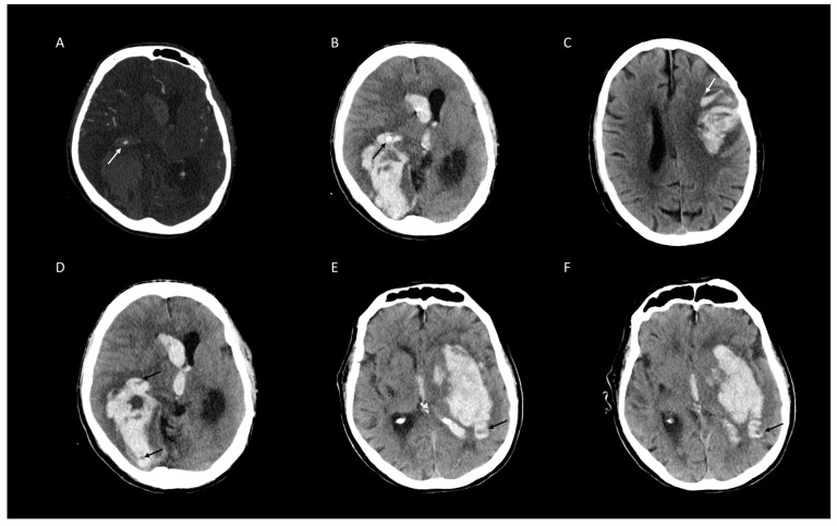
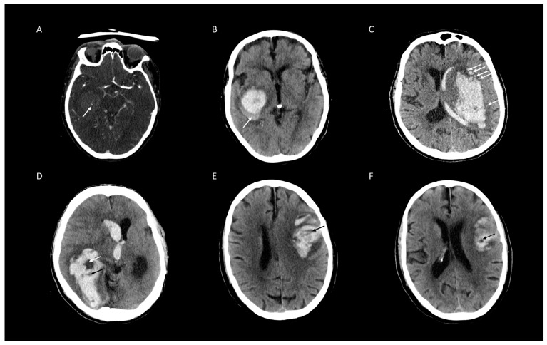
The aim of this study was to assess the inter- and intrarater reliability of noncontrast CT (NCCT) markers [Black Hole Sign (BH), Blend Sign (BS), Island Sign (IS), and Hypodensities (HD)] and Spot Sign (SS) on CTA in patients with spontaneous intracerebral hemorrhage (ICH).

METHODS

Patients with spontaneous ICH at three German tertiary stroke centers were retrospectively included. Each CT scan was rated for four NCCT markers and SS on CTA by two radiology residents. Raters were blind to all demographic and outcome data. Inter- and intrarater agreement was determined by Cohen's kappa (κ) coefficient and percentage of agreement.

RESULTS

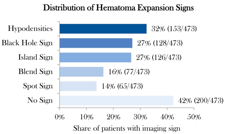
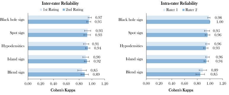
Interrater agreement was excellent in 473 included patients, ranging from 96% to 99%. Interrater κ ranged from 0.85 (95% CI [0.78-0.91]) to 0.97 (95% CI [0.94-0.99]) for NCCT markers and 0.93 (95% CI [0.88-0.98]) for SS, all -values < 0.001. Intrarrater agreement ranged from 96% to 100%, with κ ranging from 0.85 (95% CI [0.78-0.91]) to 1.00 (95% CI [0.10-0.85]) for NCCT markers and 0.96 (95% CI [0.92-1.00]) for SS, all -values < 0.001.

CONCLUSIONS

NCCT imaging findings and SS on CTA have good-to-excellent inter- and intrarater reliabilities, with the highest agreement for BH and SS.

摘要

背景

本研究旨在评估非增强CT(NCCT)标记物[黑洞征(BH)、融合征(BS)、岛征(IS)和低密度影(HD)]以及自发性脑出血(ICH)患者CT血管造影(CTA)上的斑点征(SS)的评分者间和评分者内可靠性。

方法

回顾性纳入德国三个三级卒中中心的自发性ICH患者。两名放射科住院医师对每次CT扫描的四个NCCT标记物和CTA上的SS进行评分。评分者对所有人口统计学和结局数据均不知情。评分者间和评分者内一致性通过科恩kappa(κ)系数和一致性百分比来确定。

结果

473例纳入患者的评分者间一致性极佳,范围为96%至99%。NCCT标记物的评分者间κ值范围为0.85(95%CI[0.78 - 0.91])至0.97(95%CI[0.94 - 0.99]),SS的评分者间κ值为0.93(95%CI[0.88 - 0.98]),所有P值均<0.001。评分者内一致性范围为96%至100%,NCCT标记物的κ值范围为0.85(95%CI[0.78 - 0.91])至1.00(95%CI[0.10 - 0.85]),SS的κ值为0.96(95%CI[0.92 - 1.00]),所有P值均<0.001。

结论

NCCT影像学表现和CTA上的SS具有良好至极佳的评分者间和评分者内可靠性,其中BH和SS的一致性最高。

https://cdn.ncbi.nlm.nih.gov/pmc/blobs/d78f/7231301/ad58889792cf/jcm-09-01020-g004.jpg
https://cdn.ncbi.nlm.nih.gov/pmc/blobs/d78f/7231301/8b9dc74fe4c3/jcm-09-01020-g001.jpg
https://cdn.ncbi.nlm.nih.gov/pmc/blobs/d78f/7231301/1d6dbc2ff91b/jcm-09-01020-g002.jpg
https://cdn.ncbi.nlm.nih.gov/pmc/blobs/d78f/7231301/bb13a41f2f97/jcm-09-01020-g003.jpg
https://cdn.ncbi.nlm.nih.gov/pmc/blobs/d78f/7231301/ad58889792cf/jcm-09-01020-g004.jpg

相似文献

1
Inter- and Intrarater Agreement of Spot Sign and Noncontrast CT Markers for Early Intracerebral Hemorrhage Expansion.早期脑出血扩大的斑点征和非增强CT标记物的评分者间及评分者内一致性
J Clin Med. 2020 Apr 4;9(4):1020. doi: 10.3390/jcm9041020.
2
Diagnostic Accuracy and Reliability of Noncontrast Computed Tomography Markers for Acute Hematoma Expansion among Radiologists.放射科医生对急性血肿扩大的非对比 CT 标志物的诊断准确性和可靠性。
Tomography. 2022 Dec 9;8(6):2893-2901. doi: 10.3390/tomography8060242.
3
Triage of 5 Noncontrast Computed Tomography Markers and Spot Sign for Outcome Prediction After Intracerebral Hemorrhage.颅内出血后结局预测的 5 项非对比计算机断层扫描标志物和斑点征分诊。
Stroke. 2018 Oct;49(10):2317-2322. doi: 10.1161/STROKEAHA.118.021625.
4
Prognostic Value of Non-Contrast CT Markers and Spot Sign for Outcome Prediction in Patients with Intracerebral Hemorrhage under Oral Anticoagulation.非增强CT标志物和斑点征对口服抗凝治疗的脑出血患者预后预测的价值
J Clin Med. 2020 Apr 10;9(4):1077. doi: 10.3390/jcm9041077.
5
Interrater and Intrarater Measurement Reliability of Noncontrast Computed Tomography Predictors of Intracerebral Hemorrhage Expansion.非对比计算机断层扫描预测脑出血扩大的观察者间和观察者内测量可靠性。
Stroke. 2019 May;50(5):1260-1262. doi: 10.1161/STROKEAHA.118.024050.
6
Comparison of Spot Sign, Blend Sign and Black Hole Sign for Outcome Prediction in Patients with Intracerebral Hemorrhage.斑点征、混合征和黑洞征对脑出血患者预后预测的比较
J Stroke. 2017 Sep;19(3):333-339. doi: 10.5853/jos.2016.02061. Epub 2017 Sep 29.
7
Accuracy of imaging markers on noncontrast computed tomography in predicting intracerebral hemorrhage expansion.非对比 CT 成像标志物预测脑出血扩大的准确性。
Neurol Res. 2020 Nov;42(11):973-979. doi: 10.1080/01616412.2020.1795577. Epub 2020 Jul 22.
8
Association Between Hypodensities Detected by Computed Tomography and Hematoma Expansion in Patients With Intracerebral Hemorrhage.计算机断层扫描检测到的低密度影与脑出血患者血肿扩大的关系。
JAMA Neurol. 2016 Aug 1;73(8):961-8. doi: 10.1001/jamaneurol.2016.1218.
9
The comprehensive comparison of imaging sign from CT angiography and noncontrast CT for predicting intracranial hemorrhage expansion: A comparative study.CT 血管造影与非对比 CT 成像征象在预测颅内出血扩展中的全面比较:一项对比研究。
Medicine (Baltimore). 2022 Dec 9;101(49):e31914. doi: 10.1097/MD.0000000000031914.
10
Multilesion Segmentations in Patients with Intracerebral Hemorrhage: Reliability of ICH, IVH and PHE Masks.脑出血患者的多病灶分割:ICH、IVH 和 PHE 掩码的可靠性。
Tomography. 2023 Jan 11;9(1):89-97. doi: 10.3390/tomography9010008.

引用本文的文献

1
Spot Sign in Intracerebral Hemorrhage: Critical Reappraisal and Future Clinical Implications.脑出血中的斑点征:批判性重新评估及未来临床意义
Stroke. 2025 Jun;56(6):1612-1624. doi: 10.1161/STROKEAHA.125.050637. Epub 2025 Apr 8.
2
Hematoma expansion prediction based on SMOTE and XGBoost algorithm.基于 SMOTE 和 XGBoost 算法的血肿扩张预测。
BMC Med Inform Decis Mak. 2024 Jun 19;24(1):172. doi: 10.1186/s12911-024-02561-9.
3
Uncertainty-aware deep-learning model for prediction of supratentorial hematoma expansion from admission non-contrast head computed tomography scan.

本文引用的文献

1
Standards for Detecting, Interpreting, and Reporting Noncontrast Computed Tomographic Markers of Intracerebral Hemorrhage Expansion.颅内出血扩展的非对比计算机断层扫描标志物的检测、解释和报告标准。
Ann Neurol. 2019 Oct;86(4):480-492. doi: 10.1002/ana.25563. Epub 2019 Aug 24.
2
Prediction of hematoma expansion in spontaneous intracerebral hemorrhage using support vector machine.基于支持向量机预测自发性脑出血的血肿扩大。
EBioMedicine. 2019 May;43:454-459. doi: 10.1016/j.ebiom.2019.04.040. Epub 2019 May 3.
3
Interrater and Intrarater Measurement Reliability of Noncontrast Computed Tomography Predictors of Intracerebral Hemorrhage Expansion.
用于从入院时的非增强头部计算机断层扫描预测幕上血肿扩大的不确定性感知深度学习模型。
NPJ Digit Med. 2024 Feb 6;7(1):26. doi: 10.1038/s41746-024-01007-w.
4
Non-contrast computed tomography features predict intraventricular hemorrhage growth.非对比计算机断层扫描特征可预测脑室内出血的进展。
Eur Radiol. 2023 Nov;33(11):7807-7817. doi: 10.1007/s00330-023-09707-9. Epub 2023 May 22.
5
Machine Learning for Onset Prediction of Patients with Intracerebral Hemorrhage.用于脑出血患者发病预测的机器学习
J Clin Med. 2023 Mar 31;12(7):2631. doi: 10.3390/jcm12072631.
6
Using Noncontrast Computed Tomography to Improve Prediction of Intracerebral Hemorrhage Expansion.利用非对比计算机断层扫描提高脑出血扩展的预测能力。
Stroke. 2023 Feb;54(2):567-574. doi: 10.1161/STROKEAHA.122.041302. Epub 2023 Jan 9.
7
Diagnostic Accuracy and Reliability of Noncontrast Computed Tomography Markers for Acute Hematoma Expansion among Radiologists.放射科医生对急性血肿扩大的非对比 CT 标志物的诊断准确性和可靠性。
Tomography. 2022 Dec 9;8(6):2893-2901. doi: 10.3390/tomography8060242.
8
Neuroimaging of Acute Intracerebral Hemorrhage.急性脑出血的神经影像学
J Clin Med. 2021 Mar 5;10(5):1086. doi: 10.3390/jcm10051086.
9
Associations Between Levels of High-Sensitivity C-Reactive Protein and Outcome After Intracerebral Hemorrhage.高敏C反应蛋白水平与脑出血后预后的关联
Front Neurol. 2020 Oct 6;11:535068. doi: 10.3389/fneur.2020.535068. eCollection 2020.
非对比计算机断层扫描预测脑出血扩大的观察者间和观察者内测量可靠性。
Stroke. 2019 May;50(5):1260-1262. doi: 10.1161/STROKEAHA.118.024050.
4
Triage of 5 Noncontrast Computed Tomography Markers and Spot Sign for Outcome Prediction After Intracerebral Hemorrhage.颅内出血后结局预测的 5 项非对比计算机断层扫描标志物和斑点征分诊。
Stroke. 2018 Oct;49(10):2317-2322. doi: 10.1161/STROKEAHA.118.021625.
5
Absolute risk and predictors of the growth of acute spontaneous intracerebral haemorrhage: a systematic review and meta-analysis of individual patient data.急性自发性脑出血增长的绝对风险和预测因素:一项个体患者数据的系统回顾和荟萃分析。
Lancet Neurol. 2018 Oct;17(10):885-894. doi: 10.1016/S1474-4422(18)30253-9. Epub 2018 Aug 14.
6
Predicting Intracerebral Hemorrhage Expansion With Noncontrast Computed Tomography: The BAT Score.基于非增强 CT 的脑出血扩大预测:BAT 评分。
Stroke. 2018 May;49(5):1163-1169. doi: 10.1161/STROKEAHA.117.020138. Epub 2018 Apr 18.
7
Comparison of Swirl Sign and Black Hole Sign in Predicting Early Hematoma Growth in Patients with Spontaneous Intracerebral Hemorrhage.比较自发性脑出血患者的漩涡征和黑洞征对预测早期血肿增长的作用。
Med Sci Monit. 2018 Jan 29;24:567-573. doi: 10.12659/msm.906708.
8
Predictors of hematoma expansion predictors after intracerebral hemorrhage.脑出血后血肿扩大的预测因素
Oncotarget. 2017 Jul 18;8(51):89348-89363. doi: 10.18632/oncotarget.19366. eCollection 2017 Oct 24.
9
Comparison of Spot Sign, Blend Sign and Black Hole Sign for Outcome Prediction in Patients with Intracerebral Hemorrhage.斑点征、混合征和黑洞征对脑出血患者预后预测的比较
J Stroke. 2017 Sep;19(3):333-339. doi: 10.5853/jos.2016.02061. Epub 2017 Sep 29.
10
Island Sign: An Imaging Predictor for Early Hematoma Expansion and Poor Outcome in Patients With Intracerebral Hemorrhage.岛征:脑出血患者早期血肿扩大及预后不良的影像学预测指标
Stroke. 2017 Nov;48(11):3019-3025. doi: 10.1161/STROKEAHA.117.017985. Epub 2017 Oct 10.