Vitreoretinal Service, Moorfields Eye Hospital, London, United Kingdom.
Institute of Ophthalmology, University College London, London, United Kingdom.
PLoS One. 2020 Apr 8;15(4):e0230713. doi: 10.1371/journal.pone.0230713. eCollection 2020.
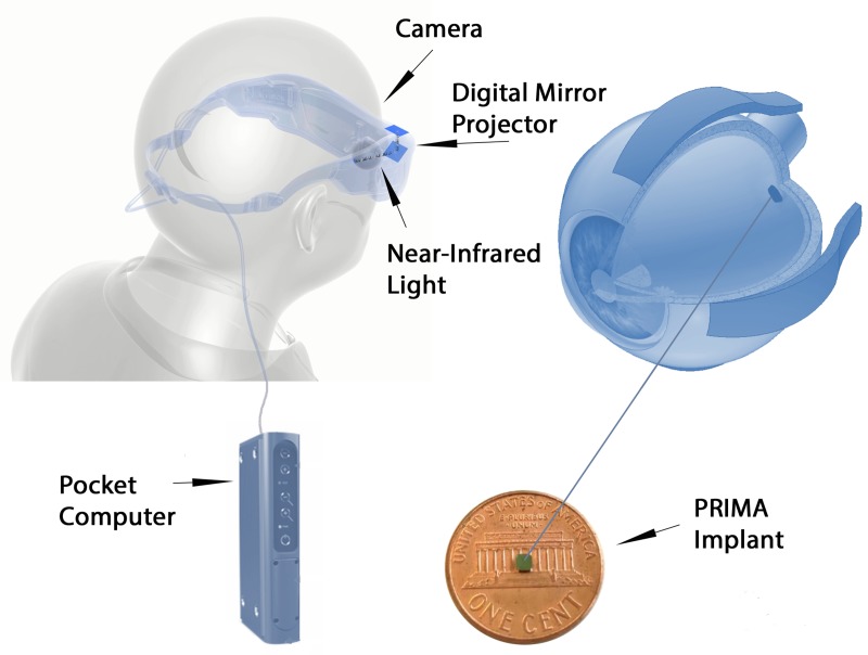
To evaluate the surgical technique for subretinal implantation of two sizes of PRIMA photovoltaic wireless microchip in two animal models, and refine these surgical procedures for human trials.
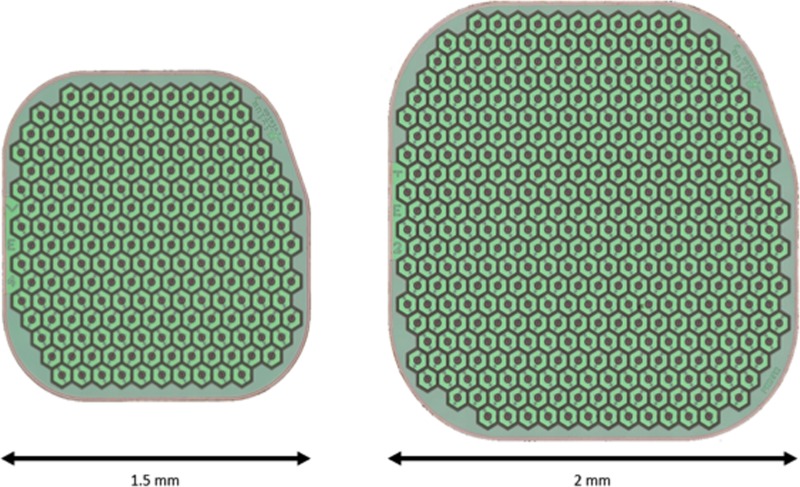
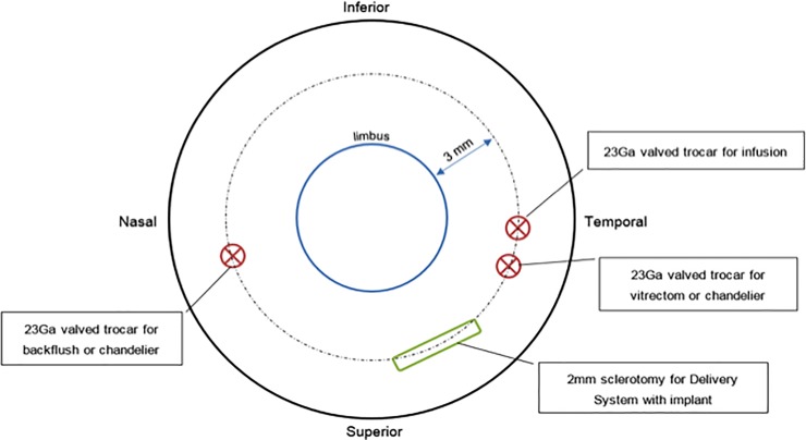
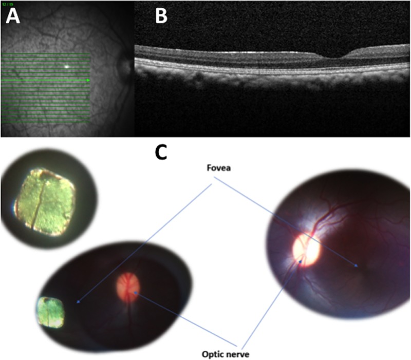
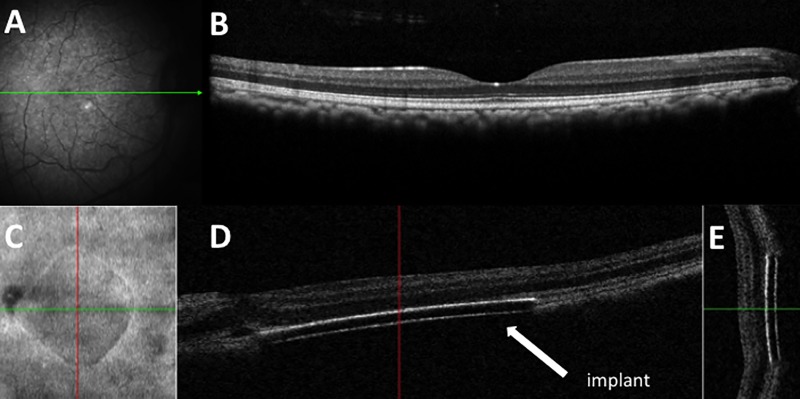
Cats and Macaca fascicularis primates with healthy retina underwent vitrectomy surgery and were implanted with subretinal wireless photovoltaic microchip at the macula/central retina. The 1.5mm PRIMA chip was initially studied in feline eyes. PRIMA implant (2mm,1.5mm sizes) arrays were studied in primates. Feasibility of subretinal chip implantation was evaluated with a newly-developed surgical technique, with surgical complications and adverse events recorded.
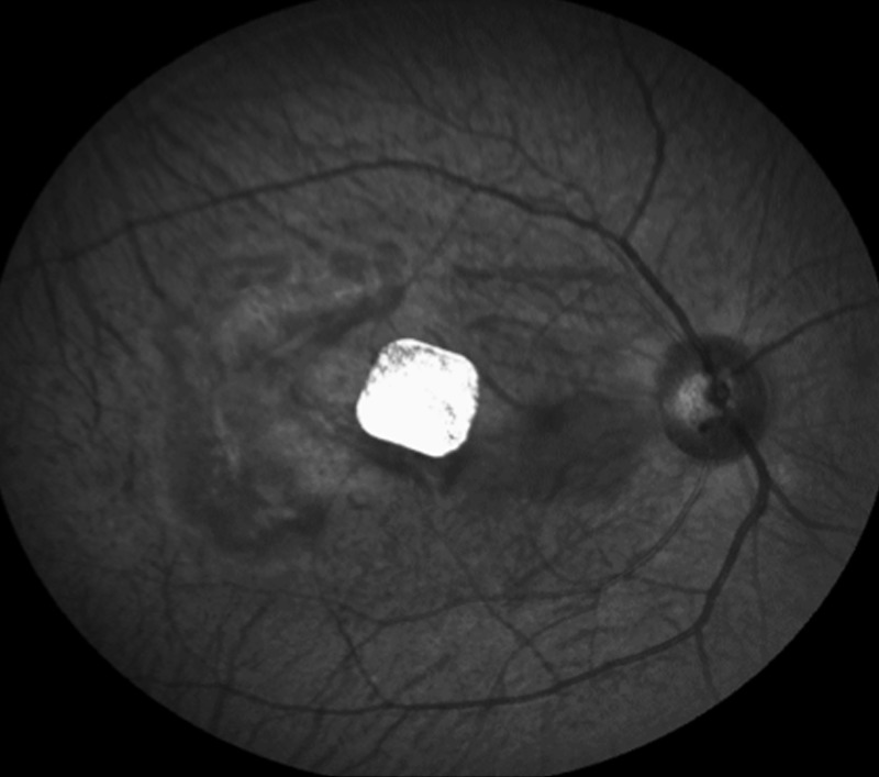
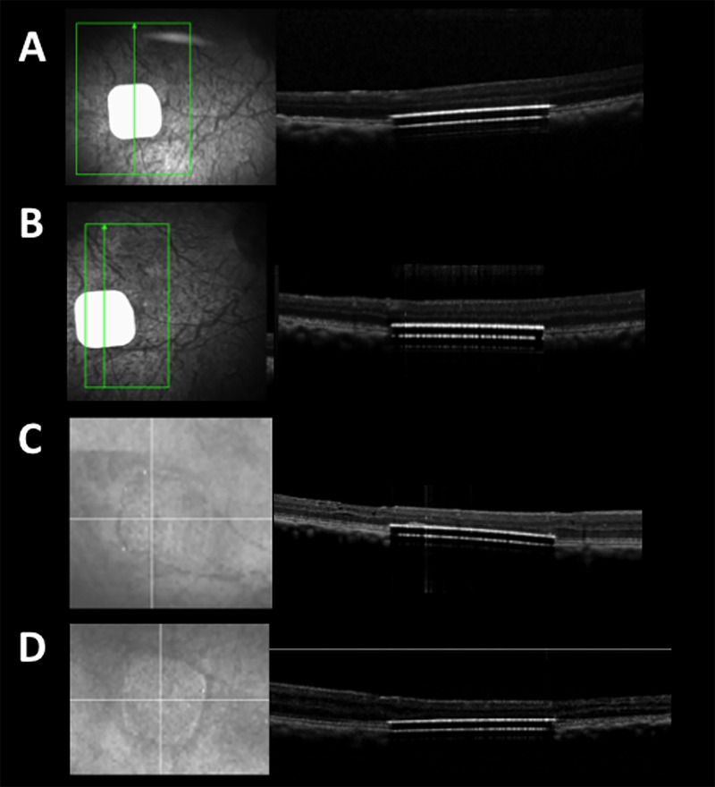
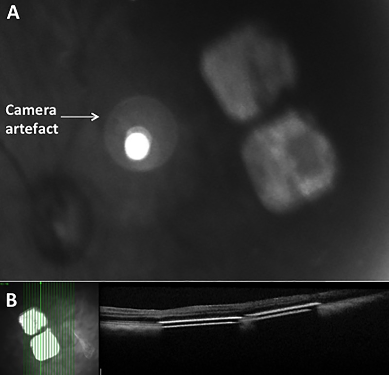
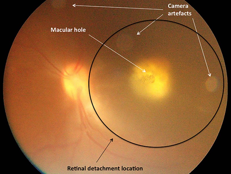
The 1.5mm implant was placed in the central retina of 11 feline eyes, with implantation duration 43-106 days. The 1.5mm implant was correctly positioned into central macula of 11 primate eyes, with follow-up periods of minimum 6 weeks (n = 11), 2 years (n = 2), and one eye for 3 years. One primate eye underwent multi-chip 1.5mm implantation using two 1.5mm chips. The 2mm implant was delivered to 4 primate eyes. Optical coherence tomography confirmed correct surgical placement of photovoltaic arrays in the subretinal space in all 26 eyes. Intraoperative complications in primate eyes included retinal tear, macular hole, retinal detachment, and vitreous hemorrhage that resolved spontaneously. Postoperatively, there was no case of significant ocular inflammation in the 1.5mm implant group.
We report subretinal implantation of 1.5mm and 2mm photovoltaic arrays in the central retina of feline and central macula of primate eyes with a low rate of device-related complications. The in vivo PRIMA implantation technique has been developed and refined for use for a 2mm PRIMA implant in ongoing human trials.
评估两种大小的 PRIMA 光伏无线微芯片在两种动物模型中经视网膜下植入的手术技术,并对这些手术程序进行改进,以用于人体试验。
健康视网膜的猫和猕猴接受玻璃体切除术,并在黄斑/中心视网膜下植入经视网膜下无线光伏微芯片。最初在猫眼中研究 1.5mm PRIMA 芯片。在灵长类动物中研究 PRIMA 植入物(2mm,1.5mm 尺寸)阵列。使用新开发的手术技术评估经视网膜下芯片植入的可行性,记录手术并发症和不良事件。
将 1.5mm 植入物放置在 11 只猫眼中的中心视网膜中,植入时间为 43-106 天。将 1.5mm 植入物正确定位在 11 只灵长类动物眼中的中央黄斑中,随访时间至少为 6 周(n=11)、2 年(n=2)和 1 只眼 3 年。一只灵长类动物眼接受了两个 1.5mm 芯片的多芯片 1.5mm 植入。将 2mm 植入物递送至 4 只灵长类动物眼。光学相干断层扫描证实了所有 26 只眼中光伏阵列在经视网膜下空间的正确手术位置。灵长类动物眼的术中并发症包括视网膜裂孔、黄斑裂孔、视网膜脱离和玻璃体出血,这些并发症均自发缓解。在 1.5mm 植入物组中,术后无一例出现明显的眼部炎症。
我们报告了在猫的中心视网膜和灵长类动物的中央黄斑下植入 1.5mm 和 2mm 光伏阵列,设备相关并发症的发生率较低。正在进行的人体试验中使用了一种改进的 2mm PRIMA 植入物的体内 PRIMA 植入技术。