• 文献检索
  • 文档翻译
  • 深度研究
  • 学术资讯
  • Suppr Zotero 插件Zotero 插件
  • 邀请有礼
  • 套餐&价格
  • 历史记录
应用&插件
Suppr Zotero 插件Zotero 插件浏览器插件Mac 客户端Windows 客户端微信小程序
定价
高级版会员购买积分包购买API积分包
服务
文献检索文档翻译深度研究API 文档MCP 服务
关于我们
关于 Suppr公司介绍联系我们用户协议隐私条款
关注我们

Suppr 超能文献

核心技术专利:CN118964589B侵权必究
粤ICP备2023148730 号-1Suppr @ 2026

文献检索

告别复杂PubMed语法,用中文像聊天一样搜索,搜遍4000万医学文献。AI智能推荐,让科研检索更轻松。

立即免费搜索

文件翻译

保留排版,准确专业,支持PDF/Word/PPT等文件格式,支持 12+语言互译。

免费翻译文档

深度研究

AI帮你快速写综述,25分钟生成高质量综述,智能提取关键信息,辅助科研写作。

立即免费体验

小型猪视网膜下植入三维植入物后视网膜的结构变化

Structural changes in the retina after implantation of subretinal three-dimensional implants in mini pigs.

作者信息

Vu Que Anh, Seo Hee Won, Choi Kwang-Eon, Kim Namju, Kang Yoo Na, Lee Jaemeun, Park Sun-Hyun, Kim Jee Taek, Kim Sohee, Kim Seong-Woo

机构信息

Department of Ophthalmology, Korea University School of Medicine, Seoul, South Korea.

Department of Ophthalmology, Hanoi Medical University, Hanoi, Vietnam.

出版信息

Front Neurosci. 2022 Sep 30;16:1010445. doi: 10.3389/fnins.2022.1010445. eCollection 2022.

DOI:10.3389/fnins.2022.1010445
PMID:36248640
原文链接:https://pmc.ncbi.nlm.nih.gov/articles/PMC9561346/
Abstract

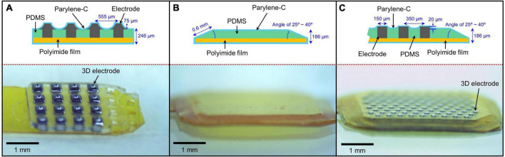
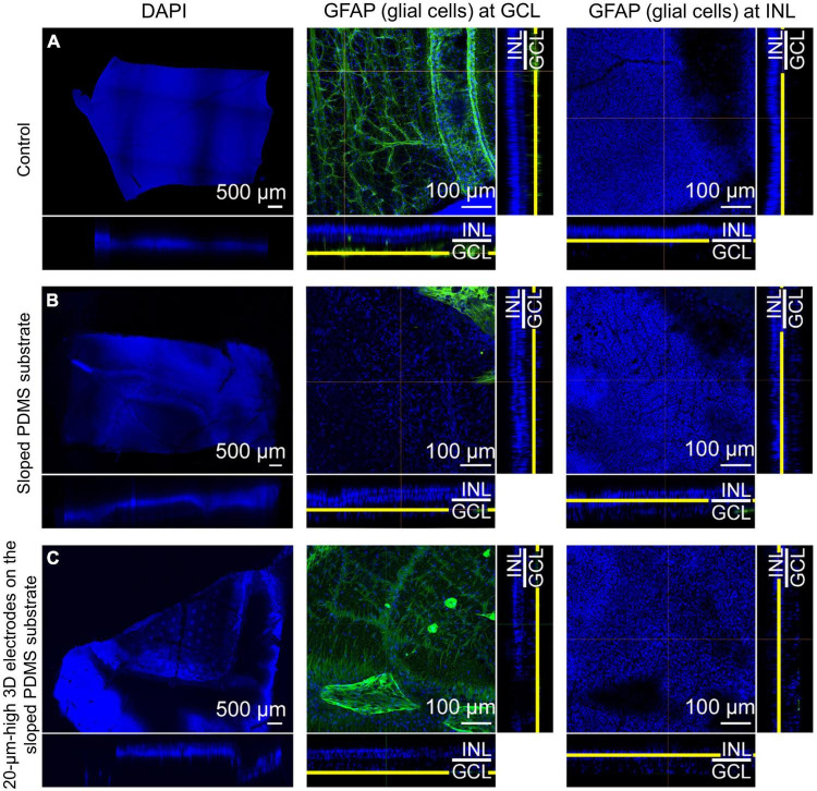
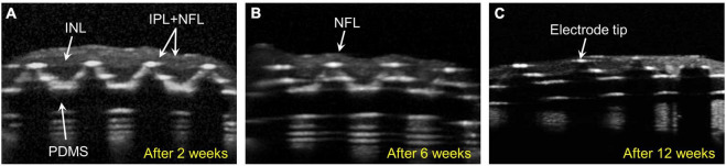
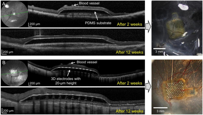
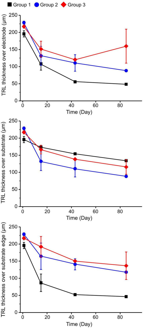
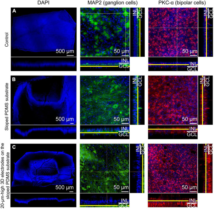
The retinal structural changes after subretinal implantation of three-dimensional (3D) microelectrodes were investigated in a mini pig. Three types of electrode were implanted into the subretinal spaces of nine mini pigs: 75-μm-high 3D electrodes on a 200-μm-thick right-angled polydimethylsiloxane (PDMS) substrate (group 1); a 140-μm-thick sloped PDMS substrate without electrodes (group 2); and a 140-μm-thick sloped PDMS substrate with 20-μm-high 3D electrodes (group 3). One mini pig was used as a control. Spectral domain-optical coherence tomography (SD-OCT) images were obtained at baseline and 2, 6, and 12 weeks post-surgery. Retinal specimens were immunostained using a tissue-clearing method 3 months post-implantation. The 75-μm-high 3D electrodes progressively penetrated the inner nuclear layer (INL) and touched the inner plexiform layer (IPL) 2 weeks post-surgery. At 6 weeks post-operatively, the electrodes were in contact with the nerve-fiber layer, accompanied by a severe fibrous reaction. In the other groups, the implants remained in place without subretinal migration. Immunostaining showed that retinal ganglion and bipolar cells were preserved without fibrosis over the retinal implants in groups 2 and 3 during the 12-week implantation period. In summary, SD-OCT and immunohistology results showed differences in the extent of reactions, such as fibrosis over the implants and penetration of the electrodes into the inner retinal layer depending on different types of electrodes. A sloped substrate performed better than a right-angled substrate in terms of retinal preservation over the implanted electrodes. The 20-μm-high electrodes showed better structural compatibility than the 75-μm-high 3D electrodes. There was no significant difference between the results of sloped implants without electrodes and 20-μm-high 3D electrodes, indicating that the latter had no adverse effects on retinal tissue.

摘要

在小型猪中研究了三维(3D)微电极视网膜下植入后的视网膜结构变化。将三种类型的电极植入9只小型猪的视网膜下间隙:在200μm厚的直角聚二甲基硅氧烷(PDMS)基底上的75μm高的3D电极(第1组);无电极的140μm厚的倾斜PDMS基底(第2组);以及有20μm高的3D电极的140μm厚的倾斜PDMS基底(第3组)。使用1只小型猪作为对照。在基线以及术后2、6和12周获取光谱域光学相干断层扫描(SD-OCT)图像。植入3个月后,使用组织透明法对视网膜标本进行免疫染色。术后2周,75μm高的3D电极逐渐穿透内核层(INL)并接触到内网状层(IPL)。术后6周,电极与神经纤维层接触,伴有严重的纤维反应。在其他组中,植入物保持在原位,没有视网膜下迁移。免疫染色显示,在12周的植入期内,第2组和第3组的视网膜植入物上的视网膜神经节细胞和双极细胞得以保留,没有纤维化。总之,SD-OCT和免疫组织学结果显示,根据不同类型的电极,在反应程度上存在差异,例如植入物上的纤维化以及电极向内视网膜层的穿透情况。就植入电极上方的视网膜保存而言,倾斜基底比直角基底表现更好。20μm高的电极比75μm高的3D电极显示出更好的结构兼容性。无电极的倾斜植入物和20μm高的3D电极的结果之间没有显著差异,表明后者对视网膜组织没有不良影响。

https://cdn.ncbi.nlm.nih.gov/pmc/blobs/7d91/9561346/243ab763848b/fnins-16-1010445-g006.jpg
https://cdn.ncbi.nlm.nih.gov/pmc/blobs/7d91/9561346/61b5449a5567/fnins-16-1010445-g001.jpg
https://cdn.ncbi.nlm.nih.gov/pmc/blobs/7d91/9561346/0c116234aa5e/fnins-16-1010445-g002.jpg
https://cdn.ncbi.nlm.nih.gov/pmc/blobs/7d91/9561346/f121b7ec62c1/fnins-16-1010445-g003.jpg
https://cdn.ncbi.nlm.nih.gov/pmc/blobs/7d91/9561346/212bca15a007/fnins-16-1010445-g004.jpg
https://cdn.ncbi.nlm.nih.gov/pmc/blobs/7d91/9561346/ed90ff0223d1/fnins-16-1010445-g005.jpg
https://cdn.ncbi.nlm.nih.gov/pmc/blobs/7d91/9561346/243ab763848b/fnins-16-1010445-g006.jpg

相似文献

1
Structural changes in the retina after implantation of subretinal three-dimensional implants in mini pigs.小型猪视网膜下植入三维植入物后视网膜的结构变化
Front Neurosci. 2022 Sep 30;16:1010445. doi: 10.3389/fnins.2022.1010445. eCollection 2022.
2
Optical Coherence Tomography in Patients With the Subretinal Implant Retina Implant Alpha IMS.视网膜下植入物Retina Implant Alpha IMS患者的光学相干断层扫描
Ophthalmic Surg Lasers Imaging Retina. 2017 Dec 1;48(12):993-999. doi: 10.3928/23258160-20171130-06.
3
Compound subretinal prostheses with extra-ocular parts designed for human trials: successful long-term implantation in pigs.用于人体试验的带有眼外部分的复合视网膜下假体:在猪体内成功长期植入。
Graefes Arch Clin Exp Ophthalmol. 2007 Feb;245(2):230-41. doi: 10.1007/s00417-006-0339-x.
4
Implantation and Extraction of Penetrating Electrode Arrays in Minipig Retinas.猪眼内穿透电极阵列的植入和取出。
Transl Vis Sci Technol. 2020 Apr 24;9(5):19. doi: 10.1167/tvst.9.5.19. eCollection 2020 Apr.
5
Integration of Perforated Subretinal Prostheses With Retinal Tissue.穿孔性视网膜下假体与视网膜组织的整合。
Transl Vis Sci Technol. 2015 Aug 14;4(4):5. doi: 10.1167/tvst.4.4.5. eCollection 2015 Aug.
6
Optimization of pillar electrodes in subretinal prosthesis for enhanced proximity to target neurons.优化视网膜下假体中的支柱电极,以增强与目标神经元的接近度。
J Neural Eng. 2018 Jun;15(3):036011. doi: 10.1088/1741-2552/aaac39. Epub 2018 Feb 1.
7
Three-dimensional electro-neural interfaces electroplated on subretinal prostheses.电镀在视网膜下假体上的三维电神经接口。
bioRxiv. 2023 Nov 13:2023.11.09.566003. doi: 10.1101/2023.11.09.566003.
8
Implantation and testing of subretinal film electrodes in domestic pigs.猪视网膜下膜电极的植入与测试
Exp Eye Res. 2006 Feb;82(2):332-40. doi: 10.1016/j.exer.2005.07.007. Epub 2005 Aug 25.
9
3D electronic implants in subretinal space: Long-term follow-up in rodents.3D 电子植入物在视网膜下腔:啮齿动物的长期随访。
Biomaterials. 2024 Dec;311:122674. doi: 10.1016/j.biomaterials.2024.122674. Epub 2024 Jun 17.
10
Effect of shape and coating of a subretinal prosthesis on its integration with the retina.视网膜下假体的形状和涂层对其与视网膜整合的影响。
Exp Eye Res. 2009 Jan;88(1):22-9. doi: 10.1016/j.exer.2008.09.018. Epub 2008 Oct 10.

引用本文的文献

1
Recent Progress in Photoreceptor Cell-Based Therapy for Degenerative Retinal Disease.基于光感受器细胞的退行性视网膜疾病治疗的最新进展
Stem Cells Transl Med. 2024 Apr 15;13(4):332-345. doi: 10.1093/stcltm/szae005.
2
Focal stimulation of retinal ganglion cells using subretinal 3D microelectrodes with peripheral electrodes of opposite current.使用带有反向电流外周电极的视网膜下3D微电极对视网膜神经节细胞进行局部刺激。
Biomed Eng Lett. 2023 Dec 26;14(2):355-365. doi: 10.1007/s13534-023-00342-3. eCollection 2024 Mar.
3
Retinal Prostheses: Engineering and Clinical Perspectives for Vision Restoration.

本文引用的文献

1
Normative Data of Axial Length, Retinal Thickness Measurements, Visual Evoked Potentials, and Full-Field Electroretinography in Female, Wild-Type Minipigs.女性野生型小型猪眼轴长度、视网膜厚度测量、视觉诱发电位和全视野视网膜电图的正常值。
Transl Vis Sci Technol. 2021 Oct 4;10(12):3. doi: 10.1167/tvst.10.12.3.
2
Ab-interno surgical technique for the implantation of a wireless subretinal prosthesis in mini-pigs.经瞳孔内外科手术在小型猪中植入无线式视网膜下假体。
Sci Rep. 2020 Oct 28;10(1):18507. doi: 10.1038/s41598-020-75579-4.
3
Implantation and Extraction of Penetrating Electrode Arrays in Minipig Retinas.
视网膜假体:视觉恢复的工程与临床视角。
Sensors (Basel). 2023 Jun 21;23(13):5782. doi: 10.3390/s23135782.
猪眼内穿透电极阵列的植入和取出。
Transl Vis Sci Technol. 2020 Apr 24;9(5):19. doi: 10.1167/tvst.9.5.19. eCollection 2020 Apr.
4
Fabrication of Subretinal 3D Microelectrodes with Hexagonal Arrangement.具有六边形排列的视网膜下3D微电极的制造
Micromachines (Basel). 2020 Apr 29;11(5):467. doi: 10.3390/mi11050467.
5
In vitro reliability testing and in vivo lifespan estimation of wireless Pixium Vision PRIMA photovoltaic subretinal prostheses suggest prolonged durability and functionality in clinical practice.体外可靠性测试和体内寿命估计表明,Pixium Vision PRIMA 型光感无线式视网膜假体在临床应用中具有较长的耐用性和功能性。
J Neural Eng. 2020 Jun 12;17(3):035005. doi: 10.1088/1741-2552/ab8f70.
6
PRIMA subretinal wireless photovoltaic microchip implantation in non-human primate and feline models.在非人类灵长类动物和猫科动物模型中进行 PRIMA 视网膜下无线光电池微芯片植入。
PLoS One. 2020 Apr 8;15(4):e0230713. doi: 10.1371/journal.pone.0230713. eCollection 2020.
7
Photovoltaic Restoration of Central Vision in Atrophic Age-Related Macular Degeneration.光感受器细胞移植治疗萎缩型年龄相关性黄斑变性的中心视力恢复。
Ophthalmology. 2020 Aug;127(8):1097-1104. doi: 10.1016/j.ophtha.2020.02.024. Epub 2020 Feb 25.
8
Protuberant Electrode Structures for Subretinal Electrical Stimulation: Modeling, Fabrication and Evaluation.用于视网膜下电刺激的突出电极结构:建模、制造与评估
Front Neurosci. 2019 Aug 27;13:885. doi: 10.3389/fnins.2019.00885. eCollection 2019.
9
A 3D flexible microelectrode array for subretinal stimulation.用于视网膜下刺激的三维柔性微电极阵列。
J Neural Eng. 2019 Aug 21;16(5):056016. doi: 10.1088/1741-2552/ab36ab.
10
Characteristics of prosthetic vision in rats with subretinal flat and pillar electrode arrays.视网膜下平板和柱状电极阵列大鼠的假体视觉特征。
J Neural Eng. 2019 Oct 30;16(6):066027. doi: 10.1088/1741-2552/ab34b3.