• 文献检索
  • 文档翻译
  • 深度研究
  • 学术资讯
  • Suppr Zotero 插件Zotero 插件
  • 邀请有礼
  • 套餐&价格
  • 历史记录
应用&插件
Suppr Zotero 插件Zotero 插件浏览器插件Mac 客户端Windows 客户端微信小程序
定价
高级版会员购买积分包购买API积分包
服务
文献检索文档翻译深度研究API 文档MCP 服务
关于我们
关于 Suppr公司介绍联系我们用户协议隐私条款
关注我们

Suppr 超能文献

核心技术专利:CN118964589B侵权必究
粤ICP备2023148730 号-1Suppr @ 2026

文献检索

告别复杂PubMed语法,用中文像聊天一样搜索,搜遍4000万医学文献。AI智能推荐,让科研检索更轻松。

立即免费搜索

文件翻译

保留排版,准确专业,支持PDF/Word/PPT等文件格式,支持 12+语言互译。

免费翻译文档

深度研究

AI帮你快速写综述,25分钟生成高质量综述,智能提取关键信息,辅助科研写作。

立即免费体验

动脉炎性和非动脉炎性前部缺血性视神经病变的血管密度和血管迂曲定量分析:光学相干断层扫描血管造影研究

Vessel Density and Vessel Tortuosity Quantitative Analysis of Arteritic and Non-arteritic Anterior Ischemic Optic Neuropathies: An Optical Coherence Tomography Angiography Study.

作者信息

Pierro Luisa, Arrigo Alessandro, Aragona Emanuela, Cavalleri Michele, Bandello Francesco

机构信息

Department of Ophthalmology, Scientific Institute San Raffaele, Vita-Salute University, 20132 Milan, Italy.

出版信息

J Clin Med. 2020 Apr 12;9(4):1094. doi: 10.3390/jcm9041094.

DOI:10.3390/jcm9041094
PMID:32290548
原文链接:https://pmc.ncbi.nlm.nih.gov/articles/PMC7230318/
Abstract

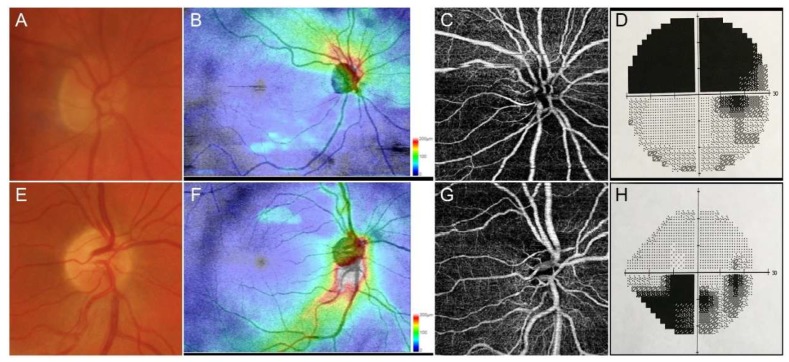
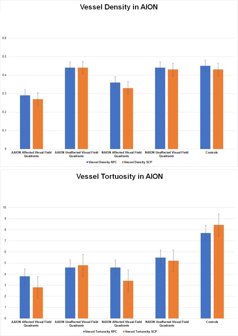
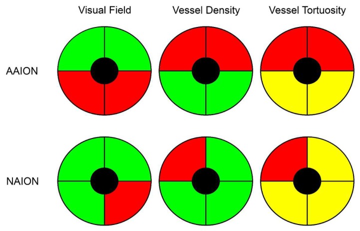
The aim of this study was to perform quantitative optical coherence tomography angiography (OCTA) assessment of arteritic and non-arteritic anterior ischemic optic neuropathies (AION; NAION). The study was designed as an observational, cross-sectional case series. All patients underwent complete ophthalmologic evaluation including LogMAR best-corrected visual acuity (BCVA), structural optical coherence tomography (OCT) and OCTA images, and dye-based angiography. Retinal nerve fiber layer (RNFL) thickness was obtained from structural OCT, and vessel density (VD) and vessel tortuosity (VT) were measured for each optic nerve head vascular plexus. After selecting the quadrants showing visual field defects, measured by Humphrey 30.2 perimetry (Zeiss Meditec, Dublin, CA, USA), we assessed the correlation between the localization of visual field defects and the quadrants showing impairments of RNFL, VD, and VT. Thirty naïve AION patients (15 arteritic AION (AAION) and 15 non-arteritic AION (NAION)) were included. LogMAR BCVA was 0.6 ± 0.2 for AAION and 0.3 ± 0.3 for NAION ( < 0.01). AAION and NAION eyes showed significant differences in terms of visual field involvement as well as VD and VT values, with remarkably worse alterations affecting AAION eyes. VD values perfectly matched with the quadrants showing RNFL and visual field defects. On the contrary, VT resulted remarkably decreased in all the quadrants, with even worse values in the quadrants showing RNFL and visual field alterations. The present study showed that AAION eyes are more injured than NAION ones. VD represents a good parameter for the detection of the main site on vascular impairment. Remarkably, VT resulted in a more sensitive parameter for the quantitative detection of blood flow impairment in AION disease.

摘要

本研究旨在对动脉炎性和非动脉炎性前部缺血性视神经病变(AION;NAION)进行定量光学相干断层扫描血管造影(OCTA)评估。该研究设计为一项观察性横断面病例系列研究。所有患者均接受了全面的眼科评估,包括LogMAR最佳矫正视力(BCVA)、结构性光学相干断层扫描(OCT)和OCTA图像以及染料血管造影。从结构性OCT获取视网膜神经纤维层(RNFL)厚度,并测量每个视神经乳头血管丛的血管密度(VD)和血管迂曲度(VT)。在选择通过Humphrey 30.2视野计(蔡司医疗技术公司,美国加利福尼亚州都柏林)测量显示视野缺损的象限后,我们评估了视野缺损的定位与显示RNFL、VD和VT受损的象限之间的相关性。纳入了30例初发AION患者(15例动脉炎性AION(AAION)和15例非动脉炎性AION(NAION))。AAION的LogMAR BCVA为0.6±0.2,NAION为0.3±0.3(<0.01)。AAION和NAION眼在视野受累以及VD和VT值方面存在显著差异,AAION眼的改变明显更严重。VD值与显示RNFL和视野缺损的象限完全匹配。相反,所有象限的VT均显著降低,在显示RNFL和视野改变的象限中值甚至更差。本研究表明,AAION眼比NAION眼损伤更严重。VD是检测血管损伤主要部位的一个良好参数。值得注意的是,VT是AION疾病中血流损伤定量检测的一个更敏感参数。

https://cdn.ncbi.nlm.nih.gov/pmc/blobs/14d1/7230318/886b9612cf60/jcm-09-01094-g003.jpg
https://cdn.ncbi.nlm.nih.gov/pmc/blobs/14d1/7230318/5370dc480f67/jcm-09-01094-g001.jpg
https://cdn.ncbi.nlm.nih.gov/pmc/blobs/14d1/7230318/52824f85d16c/jcm-09-01094-g002.jpg
https://cdn.ncbi.nlm.nih.gov/pmc/blobs/14d1/7230318/886b9612cf60/jcm-09-01094-g003.jpg
https://cdn.ncbi.nlm.nih.gov/pmc/blobs/14d1/7230318/5370dc480f67/jcm-09-01094-g001.jpg
https://cdn.ncbi.nlm.nih.gov/pmc/blobs/14d1/7230318/52824f85d16c/jcm-09-01094-g002.jpg
https://cdn.ncbi.nlm.nih.gov/pmc/blobs/14d1/7230318/886b9612cf60/jcm-09-01094-g003.jpg

相似文献

1
Vessel Density and Vessel Tortuosity Quantitative Analysis of Arteritic and Non-arteritic Anterior Ischemic Optic Neuropathies: An Optical Coherence Tomography Angiography Study.动脉炎性和非动脉炎性前部缺血性视神经病变的血管密度和血管迂曲定量分析:光学相干断层扫描血管造影研究
J Clin Med. 2020 Apr 12;9(4):1094. doi: 10.3390/jcm9041094.
2
Microvascular and structural alterations in the optic nerve head of advanced primary open-angle glaucoma compared with atrophic non-arteritic anterior ischemic optic neuropathy.与非动脉炎性前部缺血性视神经病变相比,晚期原发性开角型青光眼的视神经头微血管和结构改变。
Graefes Arch Clin Exp Ophthalmol. 2021 Jul;259(7):1945-1953. doi: 10.1007/s00417-021-05122-2. Epub 2021 Mar 4.
3
Peripapillary and macular microvasculature features of non-arteritic anterior ischemic optic neuropathy.非动脉炎性前部缺血性视神经病变的视乳头周围和黄斑部微血管特征
Front Med (Lausanne). 2023 Jan 12;9:1033838. doi: 10.3389/fmed.2022.1033838. eCollection 2022.
4
[Optic nerve morphology and vessel density in eyes with different phases of non-arteritic anterior ischemic optic neuropathy].[非动脉炎性前部缺血性视神经病变不同阶段眼睛的视神经形态与血管密度]
Zhonghua Yan Ke Za Zhi. 2019 Sep 11;55(9):677-686. doi: 10.3760/cma.j.issn.0412-4081.2019.09.010.
5
Optic disc morphology in open-angle glaucoma compared with anterior ischemic optic neuropathies.开角型青光眼与前部缺血性视神经病变的视盘形态比较。
Invest Ophthalmol Vis Sci. 2010 Apr;51(4):2003-10. doi: 10.1167/iovs.09-3492. Epub 2009 Sep 8.
6
Comparison of optic nerve head topography findings in eyes with non-arteritic anterior ischemic optic neuropathy and eyes with glaucoma.非动脉炎性前部缺血性视神经病变眼与青光眼眼中视神经头地形学发现的比较。
Graefes Arch Clin Exp Ophthalmol. 2010 Jun;248(6):845-51. doi: 10.1007/s00417-010-1323-z. Epub 2010 Mar 6.
7
Comparison of peripapillary retinal nerve fiber layer loss and visual outcome in fellow eyes following sequential bilateral non-arteritic anterior ischemic optic neuropathy.双侧非动脉性前部缺血性视神经病变先后发病后,对侧眼视乳头周围视网膜神经纤维层损失与视觉预后的比较。
Curr Eye Res. 2015 May;40(6):632-7. doi: 10.3109/02713683.2014.952829. Epub 2014 Aug 25.
8
Optical coherence tomography angiography in acute arteritic and non-arteritic anterior ischemic optic neuropathy.光学相干断层扫描血管造影在急性动脉炎性和非动脉炎性前部缺血性视神经病变中的应用
Graefes Arch Clin Exp Ophthalmol. 2017 Nov;255(11):2255-2261. doi: 10.1007/s00417-017-3774-y. Epub 2017 Aug 31.
9
Detecting changes in the blood flow of the optic disk in patients with nonarteritic anterior ischemic optic neuropathy optical coherence tomography-angiography.检测非动脉炎性前部缺血性视神经病变患者视盘血流变化的光学相干断层扫描血管造影术。
Front Neurol. 2023 Mar 24;14:1140770. doi: 10.3389/fneur.2023.1140770. eCollection 2023.
10
Quantification of peripapillary vessel density in non-arteritic anterior ischemic optic neuropathy patients with optical coherence tomography angiography.光学相干断层扫描血管造影术对非动脉炎性前部缺血性视神经病变患者视乳头周围血管密度的定量分析
Quant Imaging Med Surg. 2022 Feb;12(2):1549-1557. doi: 10.21037/qims-21-800.

引用本文的文献

1
Peripapillary Microvascular and Structural Parameters in Atrophic Nonarteritic Anterior Ischemic Optic Neuropathy, Unaffected Fellow Eyes and Controls in an Indian Population.印度人群中萎缩性非动脉炎性前部缺血性视神经病变患者、未受影响的对侧眼及对照组的视乳头周围微血管和结构参数
Neuroophthalmology. 2024 Jun 27;49(1):35-42. doi: 10.1080/01658107.2024.2367055. eCollection 2025.
2
Swept-Source OCT Angiography-Derived Regional Normative Data of Peripapillary Vessel Density in Healthy Populations.扫频源光学相干断层扫描血管造影术得出的健康人群视乳头周围血管密度的区域规范数据。
Transl Vis Sci Technol. 2025 Aug 1;14(8):5. doi: 10.1167/tvst.14.8.5.
3

本文引用的文献

1
OPTICAL COHERENCE TOMOGRAPHY ANGIOGRAPHY CAN CATEGORIZE DIFFERENT SUBGROUPS OF CHOROIDAL NEOVASCULARIZATION SECONDARY TO AGE-RELATED MACULAR DEGENERATION.光学相干断层扫描血管造影术可以对与年龄相关性黄斑变性相关的脉络膜新生血管进行分类。
Retina. 2020 Dec;40(12):2263-2269. doi: 10.1097/IAE.0000000000002775.
2
Advanced Optical Coherence Tomography Angiography Analysis of Age-related Macular Degeneration Complicated by Onset of Unilateral Choroidal Neovascularization.基于先进光学相干断层扫描血管造影分析单侧脉络膜新生血管性年龄相关性黄斑变性发病情况。
Am J Ophthalmol. 2018 Nov;195:233-242. doi: 10.1016/j.ajo.2018.08.001. Epub 2018 Aug 8.
3
Correlation of Structure With Function: Future Utilities for Optical Coherence Tomography Angiography in Neuro-Ophthalmology.
结构与功能的相关性:光学相干断层扫描血管造影术在神经眼科中的未来应用
Clin Exp Ophthalmol. 2025 Aug;53(6):682-697. doi: 10.1111/ceo.14544. Epub 2025 Apr 23.
4
Evaluation of blood flow in arteritic anterior ischemic optic neuropathy using laser speckle flowgraphy: A case series.使用激光散斑血流图评估动脉炎性前部缺血性视神经病变中的血流:病例系列
Am J Ophthalmol Case Rep. 2025 Mar 24;38:102316. doi: 10.1016/j.ajoc.2025.102316. eCollection 2025 Jun.
5
Optic disc characteristics evaluated with swept source optical coherence tomography angiography (SS-OCTA) in nonarteritic anterior ischemic optic neuropathy (NAION): a cross-sectional study.扫频源光学相干断层扫描血管造影(SS-OCTA)评估非动脉炎性前部缺血性视神经病变(NAION)的视盘特征:一项横断面研究。
BMC Ophthalmol. 2025 Apr 2;25(1):166. doi: 10.1186/s12886-025-04002-1.
6
Effect of modified surgical conditions on the peripapillary microvasculature and RNFL in patients receiving vitrectomy: an OCTA study.改良手术条件对接受玻璃体切除术患者视乳头周围微血管系统和视网膜神经纤维层的影响:一项光学相干断层扫描血管造影研究
BMC Ophthalmol. 2025 Feb 11;25(1):74. doi: 10.1186/s12886-024-03832-9.
7
A case-control study of peripapillary microvascular structure by OCT-angiography in non-arteritic ischaemic optic neuropathy at early and resolutive stages.一项通过光学相干断层扫描血管造影术对非动脉炎性缺血性视神经病变早期和恢复期视乳头周围微血管结构进行的病例对照研究。
Eye (Lond). 2025 Mar;39(4):771-778. doi: 10.1038/s41433-024-03439-w. Epub 2024 Nov 22.
8
Optical coherence tomography angiography findings in patients affected by giant cell arteritis, with and without ocular involvement: a pilot study.巨细胞动脉炎患者伴或不伴眼部受累的光学相干断层扫描血管造影结果:一项初步研究。
Front Med (Lausanne). 2024 Aug 12;11:1408821. doi: 10.3389/fmed.2024.1408821. eCollection 2024.
9
Microvascular Changes in Pituitary Adenoma Correlate with Structural Measurements and Visual Field Loss.垂体腺瘤的微血管变化与结构测量及视野缺损相关。
Neuropsychiatr Dis Treat. 2023 Dec 6;19:2745-2754. doi: 10.2147/NDT.S425454. eCollection 2023.
10
Vascular Changes in the Macula of Patients after Previous Episodes of Vision Loss Due to Leber Hereditary Optic Neuropathy and Non-Arteritic Ischemic Optic Neuropathy.既往因Leber遗传性视神经病变和非动脉炎性缺血性视神经病变导致视力丧失的患者黄斑区血管变化。
Diagnostics (Basel). 2023 May 12;13(10):1726. doi: 10.3390/diagnostics13101726.
Quantitative analysis of optical coherence tomographic angiography (OCT-A) in patients with non-arteritic anterior ischemic optic neuropathy (NAION) corresponds to visual function.
非动脉炎性前部缺血性视神经病变(NAION)患者的光学相干断层扫描血管造影(OCT-A)定量分析与视觉功能相对应。
PLoS One. 2018 Jun 28;13(6):e0199793. doi: 10.1371/journal.pone.0199793. eCollection 2018.
4
Reduction of Peripapillary Vessel Density by Optical Coherence Tomography Angiography from the Acute to the Atrophic Stage in Non-Arteritic Anterior Ischaemic Optic Neuropathy.光学相干断层扫描血管造影术观察非动脉炎性前部缺血性视神经病变从急性期到萎缩期视乳头周围血管密度的降低情况
Ophthalmologica. 2018;240(4):191-199. doi: 10.1159/000489226. Epub 2018 Jun 26.
5
Retinal and Choroidal Microvasculature in Nonarteritic Anterior Ischemic Optic Neuropathy: An Optical Coherence Tomography Angiography Study.非动脉炎性前部缺血性视神经病变的视网膜和脉络膜微血管:一项光相干断层扫描血管造影研究。
Invest Ophthalmol Vis Sci. 2018 Feb 1;59(2):870-877. doi: 10.1167/iovs.17-22996.
6
Optical Coherence Tomography Angiography of the Peripapillary Retina in Normal-Tension Glaucoma and Chronic Nonarteritic Anterior Ischemic Optic Neuropathy.正常眼压性青光眼和慢性非动脉性前部缺血性视神经病变患者视乳头周围视网膜的光学相干断层扫描血管造影
Curr Eye Res. 2018 Jun;43(6):778-784. doi: 10.1080/02713683.2018.1438630. Epub 2018 Feb 16.
7
Optical coherence tomography angiography.光学相干断层扫描血管造影术。
Prog Retin Eye Res. 2018 May;64:1-55. doi: 10.1016/j.preteyeres.2017.11.003. Epub 2017 Dec 8.
8
Optical coherence tomography angiography in acute arteritic and non-arteritic anterior ischemic optic neuropathy.光学相干断层扫描血管造影在急性动脉炎性和非动脉炎性前部缺血性视神经病变中的应用
Graefes Arch Clin Exp Ophthalmol. 2017 Nov;255(11):2255-2261. doi: 10.1007/s00417-017-3774-y. Epub 2017 Aug 31.
9
Impact of image quality on OCT angiography based quantitative measurements.图像质量对基于光学相干断层扫描血管造影的定量测量的影响。
Int J Retina Vitreous. 2017 May 15;3:13. doi: 10.1186/s40942-017-0068-9. eCollection 2017.
10
Optical Coherence Tomography Angiography in Nonarteritic Anterior Ischemic Optic Neuropathy.光学相干断层扫描血管造影术在非动脉炎性前部缺血性视神经病变中的应用
J Neuroophthalmol. 2017 Dec;37(4):358-364. doi: 10.1097/WNO.0000000000000493.