• 文献检索
  • 文档翻译
  • 深度研究
  • 学术资讯
  • Suppr Zotero 插件Zotero 插件
  • 邀请有礼
  • 套餐&价格
  • 历史记录
应用&插件
Suppr Zotero 插件Zotero 插件浏览器插件Mac 客户端Windows 客户端微信小程序
定价
高级版会员购买积分包购买API积分包
服务
文献检索文档翻译深度研究API 文档MCP 服务
关于我们
关于 Suppr公司介绍联系我们用户协议隐私条款
关注我们

Suppr 超能文献

核心技术专利:CN118964589B侵权必究
粤ICP备2023148730 号-1Suppr @ 2026

文献检索

告别复杂PubMed语法,用中文像聊天一样搜索,搜遍4000万医学文献。AI智能推荐,让科研检索更轻松。

立即免费搜索

文件翻译

保留排版,准确专业,支持PDF/Word/PPT等文件格式,支持 12+语言互译。

免费翻译文档

深度研究

AI帮你快速写综述,25分钟生成高质量综述,智能提取关键信息,辅助科研写作。

立即免费体验

图像质量对基于光学相干断层扫描血管造影的定量测量的影响。

Impact of image quality on OCT angiography based quantitative measurements.

作者信息

Al-Sheikh Mayss, Ghasemi Falavarjani Khalil, Akil Handan, Sadda SriniVas R

机构信息

Doheny Image Reading Center, Doheny Eye Institute, Los Angeles, CA USA.

Department of Ophthalmology, David Geffen School of Medicine, University of California - Los Angeles, Los Angeles, CA USA.

出版信息

Int J Retina Vitreous. 2017 May 15;3:13. doi: 10.1186/s40942-017-0068-9. eCollection 2017.

DOI:10.1186/s40942-017-0068-9
PMID:28515959
原文链接:https://pmc.ncbi.nlm.nih.gov/articles/PMC5430594/
Abstract

BACKGROUND

To study the impact of image quality on quantitative measurements and the frequency of segmentation error with optical coherence tomography angiography (OCTA).

METHODS

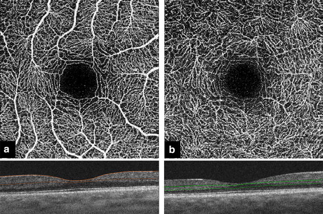
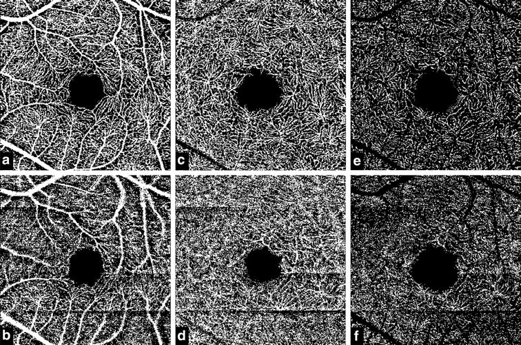
Seventeen eyes of 10 healthy individuals were included in this study. OCTA was performed using a swept-source device (Triton, Topcon). Each subject underwent three scanning sessions 1-2 min apart; the first two scans were obtained under standard conditions and for the third session, the image quality index was reduced using application of a topical ointment. En face OCTA images of the retinal vasculature were generated using the default segmentation for the superficial and deep retinal layer (SRL, DRL). Intraclass correlation coefficient (ICC) was used as a measure for repeatability. The frequency of segmentation error, motion artifact, banding artifact and projection artifact was also compared among the three sessions.

RESULTS

The frequency of segmentation error, and motion artifact was statistically similar between high and low image quality sessions (P = 0.707, and P = 1 respectively). However, the frequency of projection and banding artifact was higher with a lower image quality. The vessel density in the SRL was highly repeatable in the high image quality sessions (ICC = 0.8), however, the repeatability was low, comparing the high and low image quality measurements (ICC = 0.3). In the DRL, the repeatability of the vessel density measurements was fair in the high quality sessions (ICC = 0.6 and ICC = 0.5, with and without automatic artifact removal, respectively) and poor comparing high and low image quality sessions (ICC = 0.3 and ICC = 0.06, with and without automatic artifact removal, respectively).

CONCLUSIONS

The frequency of artifacts is higher and the repeatability of the measurements is lower with lower image quality. The impact of image quality index should be always considered in OCTA based quantitative measurements.

摘要

背景

研究图像质量对光学相干断层扫描血管造影(OCTA)定量测量及分割误差频率的影响。

方法

本研究纳入10名健康个体的17只眼睛。使用扫频源设备(Triton,拓普康)进行OCTA检查。每位受试者间隔1 - 2分钟进行三次扫描;前两次扫描在标准条件下进行,第三次扫描时,通过应用局部药膏降低图像质量指数。使用视网膜浅层和深层(SRL、DRL)的默认分割方法生成视网膜血管的OCTA正面图像。组内相关系数(ICC)用作重复性的度量指标。还比较了三次扫描中分割误差、运动伪影、条带伪影和投影伪影的频率。

结果

高图像质量和低图像质量扫描之间,分割误差和运动伪影的频率在统计学上相似(P分别为0.707和1)。然而,投影和条带伪影的频率在图像质量较低时更高。SRL中的血管密度在高图像质量扫描中具有高度重复性(ICC = 0.8),但是,比较高图像质量和低图像质量测量时,重复性较低(ICC = 0.3)。在DRL中,血管密度测量的重复性在高质量扫描中尚可(有和无自动伪影去除时,ICC分别为0.6和0.5),而比较高图像质量和低图像质量扫描时较差(有和无自动伪影去除时,ICC分别为0.3和0.06)。

结论

图像质量较低时,伪影频率更高,测量的重复性更低。在基于OCTA的定量测量中应始终考虑图像质量指数的影响。

https://cdn.ncbi.nlm.nih.gov/pmc/blobs/00a7/5430594/b12a40107375/40942_2017_68_Fig2_HTML.jpg
https://cdn.ncbi.nlm.nih.gov/pmc/blobs/00a7/5430594/5ea487a500ba/40942_2017_68_Fig1_HTML.jpg
https://cdn.ncbi.nlm.nih.gov/pmc/blobs/00a7/5430594/b12a40107375/40942_2017_68_Fig2_HTML.jpg
https://cdn.ncbi.nlm.nih.gov/pmc/blobs/00a7/5430594/5ea487a500ba/40942_2017_68_Fig1_HTML.jpg
https://cdn.ncbi.nlm.nih.gov/pmc/blobs/00a7/5430594/b12a40107375/40942_2017_68_Fig2_HTML.jpg

相似文献

1
Impact of image quality on OCT angiography based quantitative measurements.图像质量对基于光学相干断层扫描血管造影的定量测量的影响。
Int J Retina Vitreous. 2017 May 15;3:13. doi: 10.1186/s40942-017-0068-9. eCollection 2017.
2
Repeatability of automated vessel density measurements using optical coherence tomography angiography.使用光学相干断层扫描血管造影术进行自动血管密度测量的可重复性
Br J Ophthalmol. 2017 Apr;101(4):449-452. doi: 10.1136/bjophthalmol-2016-308764. Epub 2016 Jul 22.
3
Impact of Multiple En Face Image Averaging on Quantitative Assessment from Optical Coherence Tomography Angiography Images.多层面平均对视盘 OCT 血管成像定量评估的影响。
Ophthalmology. 2017 Jul;124(7):944-952. doi: 10.1016/j.ophtha.2017.02.006. Epub 2017 Mar 17.
4
Image artefacts in swept-source optical coherence tomography angiography.扫频源光学相干断层扫描血管造影中的图像伪影。
Br J Ophthalmol. 2017 May;101(5):564-568. doi: 10.1136/bjophthalmol-2016-309104. Epub 2016 Jul 20.
5
CHOROIDAL NEOVASCULAR AREA AND VESSEL DENSITY COMPARISON BETWEEN TWO SWEPT-SOURCE OPTICAL COHERENCE TOMOGRAPHY ANGIOGRAPHY DEVICES.两种扫频源光学相干断层血管成像设备的脉络膜新生血管区和血管密度比较。
Retina. 2020 Mar;40(3):521-528. doi: 10.1097/IAE.0000000000002430.
6
Repeatability and Reproducibility of Superficial Macular Retinal Vessel Density Measurements Using Optical Coherence Tomography Angiography En Face Images.使用光学相干断层扫描血管造影正面图像测量浅表黄斑视网膜血管密度的可重复性和再现性
JAMA Ophthalmol. 2017 Oct 1;135(10):1092-1098. doi: 10.1001/jamaophthalmol.2017.3431.
7
Optimizing the Repeatability of Choriocapillaris Flow Deficit Measurement From Optical Coherence Tomography Angiography.优化光学相干断层扫描血管造影中脉络膜血流缺损测量的重复性。
Am J Ophthalmol. 2020 Nov;219:21-32. doi: 10.1016/j.ajo.2020.05.027. Epub 2020 May 23.
8
Optical Coherence Tomography Angiography in Healthy Adult Subjects: Normative Values, Frequency, and Impact of Artifacts.健康成年人的光学相干断层扫描血管造影:正常值、频率和伪影的影响。
Biomed Res Int. 2022 Mar 7;2022:7286252. doi: 10.1155/2022/7286252. eCollection 2022.
9
Effect of segmentation error correction on optical coherence tomography angiography measurements in healthy subjects and diabetic macular oedema.分段误差校正对健康受试者和糖尿病性黄斑水肿的光学相干断层扫描血管造影测量的影响。
Br J Ophthalmol. 2020 Feb;104(2):162-166. doi: 10.1136/bjophthalmol-2019-314018. Epub 2019 Apr 29.
10
Identification of imaging features that determine quality and repeatability of retinal capillary plexus density measurements in OCT angiography.确定影响 OCT 血管造影中视网膜毛细血管丛密度测量质量和可重复性的影像学特征。
Br J Ophthalmol. 2018 Apr;102(4):509-514. doi: 10.1136/bjophthalmol-2017-310700. Epub 2017 Aug 16.

引用本文的文献

1
Assessment of retinal blood flow changes in early Parkinson's disease: An optical coherence tomography angiography study.早期帕金森病视网膜血流变化的评估:一项光学相干断层扫描血管造影研究。
J Res Med Sci. 2025 Jul 15;30:35. doi: 10.4103/jrms.jrms_250_24. eCollection 2025.
2
Changes in Retinal Nerve Fiber and Ganglion Cell Layers After Chemical Injury: A Prospective Study.化学损伤后视网膜神经纤维层和神经节细胞层的变化:一项前瞻性研究。
J Clin Med. 2025 Aug 7;14(15):5601. doi: 10.3390/jcm14155601.
3
Open ultrawidefield fundus image dataset with disease diagnosis and clinical image quality assessment.

本文引用的文献

1
SWEPT-SOURCE OPTICAL COHERENCE TOMOGRAPHY ANGIOGRAPHY OF THE OPTIC DISK IN OPTIC NEUROPATHY.视神经病变中视盘的扫频源光学相干断层扫描血管造影
Retina. 2016 Dec;36 Suppl 1:S168-S177. doi: 10.1097/IAE.0000000000001259.
2
Retinal vessel calibre measurements by optical coherence tomography angiography.通过光学相干断层扫描血管造影术测量视网膜血管管径
Br J Ophthalmol. 2017 Jul;101(7):989-992. doi: 10.1136/bjophthalmol-2016-309678. Epub 2016 Nov 16.
3
Retinal Microvasculature Alteration in High Myopia.高度近视患者视网膜微血管改变
带有疾病诊断和临床图像质量评估的开放超广角眼底图像数据集。
Sci Data. 2024 Nov 20;11(1):1251. doi: 10.1038/s41597-024-04113-2.
4
Optical coherence tomography angiography in cardiovascular disease.光学相干断层扫描血管造影术在心血管疾病中的应用
Prog Cardiovasc Dis. 2024 Nov-Dec;87:60-72. doi: 10.1016/j.pcad.2024.10.011. Epub 2024 Oct 21.
5
Comparative of OCT and OCTA parameters in patients with early chronic angle-closure glaucoma and early pituitary adenoma.比较早期慢性闭角型青光眼和早期垂体腺瘤患者的 OCT 和 OCTA 参数。
Sci Rep. 2024 Sep 13;14(1):21448. doi: 10.1038/s41598-024-71103-0.
6
High Prevalence of Artifacts in Optical Coherence Tomography With Adequate Signal Strength.光学相干断层扫描在信号强度足够的情况下存在较高的伪影发生率。
Transl Vis Sci Technol. 2024 Aug 1;13(8):43. doi: 10.1167/tvst.13.8.43.
7
Visual Pathway Recovery Post Pituitary Adenoma Surgery: Insights from Retinal Structure, Vascular Density, and Neural Conduction Analysis.垂体腺瘤手术后视觉通路的恢复:来自视网膜结构、血管密度和神经传导分析的见解
Ophthalmol Ther. 2024 Jul;13(7):1993-2008. doi: 10.1007/s40123-024-00966-3. Epub 2024 May 31.
8
Non-Invasive Tools in Perioperative Stroke Risk Assessment for Asymptomatic Carotid Artery Stenosis with a Focus on the Circle of Willis.围手术期无症状性颈动脉狭窄卒中风险评估中的非侵入性工具,重点关注 Willis 环
J Clin Med. 2024 Apr 24;13(9):2487. doi: 10.3390/jcm13092487.
9
Reduced Retinal Blood Vessel Densities Measured by Optical Coherence Tomography Angiography in Keratoconus Patients Are Negatively Correlated with Keratoconus Severity.圆锥角膜患者中通过光学相干断层扫描血管造影测量的视网膜血管密度降低与圆锥角膜严重程度呈负相关。
Diagnostics (Basel). 2024 Mar 27;14(7):707. doi: 10.3390/diagnostics14070707.
10
OCTA Signal Quality Augmentation Using the Isometric Handgrip Test to Maximize Vascular Flow (SQUEEZE): A Randomized Crossover Trial.使用等长握力测试增强 OCTA 信号质量以最大限度增加血管流量(挤压):一项随机交叉试验。
Transl Vis Sci Technol. 2024 Mar 1;13(3):22. doi: 10.1167/tvst.13.3.22.
Invest Ophthalmol Vis Sci. 2016 Nov 1;57(14):6020-6030. doi: 10.1167/iovs.16-19542.
4
Relationship between Optical Coherence Tomography Angiography Vessel Density and Severity of Visual Field Loss in Glaucoma.光学相干断层扫描血管造影血管密度与青光眼视野缺损严重程度之间的关系
Ophthalmology. 2016 Dec;123(12):2498-2508. doi: 10.1016/j.ophtha.2016.08.041. Epub 2016 Oct 7.
5
Compensation for Reflectance Variation in Vessel Density Quantification by Optical Coherence Tomography Angiography.光学相干断层扫描血管造影术在血管密度定量分析中对反射率变化的补偿
Invest Ophthalmol Vis Sci. 2016 Aug 1;57(10):4485-92. doi: 10.1167/iovs.16-20080.
6
Swept-Source OCT Angiography Imaging of the Foveal Avascular Zone and Macular Capillary Network Density in Diabetic Retinopathy.糖尿病视网膜病变中黄斑无血管区和黄斑毛细血管网络密度的扫频源光学相干断层扫描血管造影成像
Invest Ophthalmol Vis Sci. 2016 Jul 1;57(8):3907-13. doi: 10.1167/iovs.16-19570.
7
The Characteristics of Peripapillary Retinal Perfusion by Optical Coherence Tomography Angiography in Tessellated Fundus Eyes.视网膜呈镶嵌状眼底眼中光学相干断层扫描血管造影显示的视乳头周围视网膜灌注特征
PLoS One. 2016 Jul 27;11(7):e0159911. doi: 10.1371/journal.pone.0159911. eCollection 2016.
8
Repeatability of automated vessel density measurements using optical coherence tomography angiography.使用光学相干断层扫描血管造影术进行自动血管密度测量的可重复性
Br J Ophthalmol. 2017 Apr;101(4):449-452. doi: 10.1136/bjophthalmol-2016-308764. Epub 2016 Jul 22.
9
Image artefacts in swept-source optical coherence tomography angiography.扫频源光学相干断层扫描血管造影中的图像伪影。
Br J Ophthalmol. 2017 May;101(5):564-568. doi: 10.1136/bjophthalmol-2016-309104. Epub 2016 Jul 20.
10
Optical Coherence Tomography Angiography Vessel Density in Healthy, Glaucoma Suspect, and Glaucoma Eyes.健康眼、青光眼可疑眼和青光眼眼中光学相干断层扫描血管造影的血管密度
Invest Ophthalmol Vis Sci. 2016 Jul 1;57(9):OCT451-9. doi: 10.1167/iovs.15-18944.