Center for Molecular Recognition and Biosensing, School of Life Sciences, Shanghai University, Shanghai 200444, P. R. China.
Plant Science Center, School of Life Sciences, Shanghai University, Shanghai 200444, P. R. China.
Theranostics. 2020 Mar 15;10(10):4410-4421. doi: 10.7150/thno.42951. eCollection 2020.
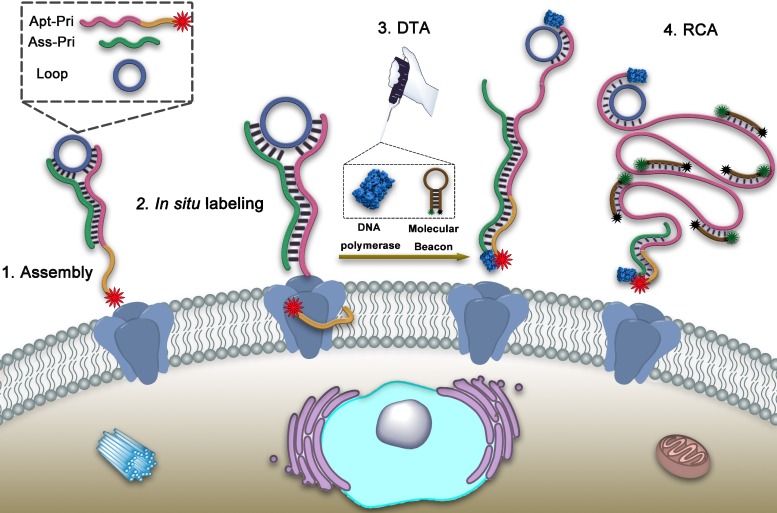
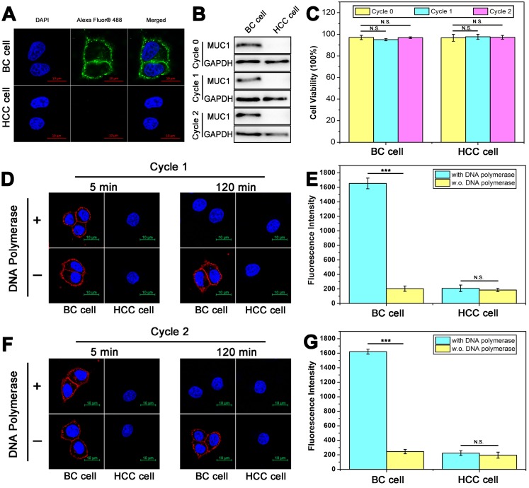
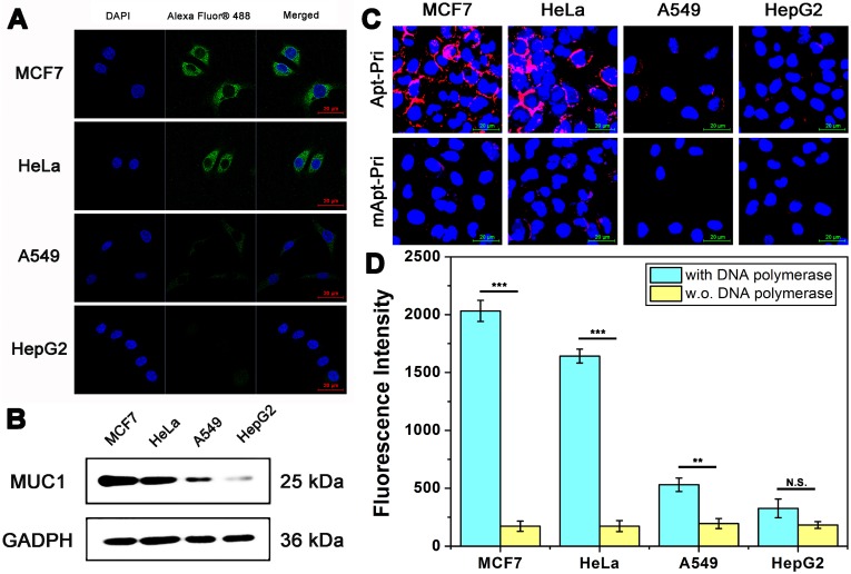
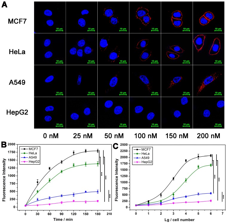
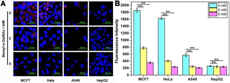
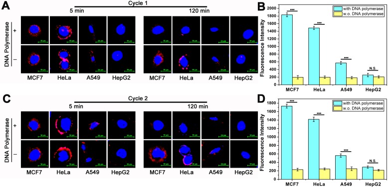
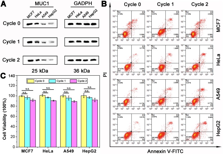
Non-destructive analysis of cells at the molecular level is of critical importance for cell research. At present, immunoassay-based and aptamer-based methods can achieve non-structural destructive cell analysis, but still lead to changes in cells at the molecular level. Here, we have proposed a dual-terminal amplification (DTA) strategy, which enables nondestructive analysis of membrane protein MUC1 without the effect on protein expression and cell viability in living cells. : A fluorophore (Cy5)-labeled DNA ternary complex consisting of three oligonucleotides is designed. It can recognize MUC1 through its aptamer region, and thus make the MUC1 of cells visible under a fluorescence microscope. When DNA polymerase is added, dual-terminal amplification is performed. One direction dissociates aptamer from MUC1, and the other direction, also known as rolling circle amplification (RCA), produces long linear DNA strands, which can be further adopted for quantitative analysis of MUC1. In this way, all reagents are removed from the surface of the cells after the analysis, which allows nondestructive analysis. We named this strategy dual-terminal amplification (DTA) analysis. : By using the DTA analysis, both fluorescence imaging analysis and fluorescence quantitative analysis of MUC1 were achieved. In addition, the aptamer-containing DNA ternary complex stays on cell surface only during the analysis and leaves the cell after the analysis is complete. The cells can be maintained in a non-interfering state for the rest of the time. So after the analysis, it is found that there are no effect on the physiological activity of cells and the expression of target protein even after two rounds of repeatable imaging and quantitative analysis. : In summary, we have successfully constructed a strategy for nondestructive analysis of membrane protein in living cells. We believe that this method provides a promising way for the analysis of the key membrane proteins of cells and the versatile utilization of precious cell samples.
对活细胞中的膜蛋白进行非破坏性分析。目前,基于免疫测定和适体的方法可以实现对非结构破坏性细胞的分析,但仍会导致细胞在分子水平上发生变化。在这里,我们提出了一种双端扩增(DTA)策略,该策略能够实现对活细胞中膜蛋白 MUC1 的非破坏性分析,而不会影响蛋白质表达和细胞活力。 :设计了一种由三个寡核苷酸组成的荧光素(Cy5)标记的 DNA 三元复合物。它可以通过其适体区域识别 MUC1,从而使细胞中的 MUC1在荧光显微镜下可见。当加入 DNA 聚合酶时,进行双端扩增。一个方向使适体从 MUC1 上解离,另一个方向,也称为滚环扩增(RCA),产生长线性 DNA 链,可进一步用于 MUC1 的定量分析。通过这种方式,在分析后从细胞表面去除所有试剂,从而实现非破坏性分析。我们将这种策略命名为双端扩增(DTA)分析。 :通过使用 DTA 分析,实现了 MUC1 的荧光成像分析和荧光定量分析。此外,含有适体的 DNA 三元复合物仅在分析期间留在细胞表面,并且在分析完成后离开细胞。在其余时间,细胞可以保持在非干扰状态。因此,在分析后,即使经过两轮可重复的成像和定量分析,也不会对细胞的生理活性和靶蛋白的表达产生影响。 :总之,我们成功构建了一种用于活细胞中膜蛋白非破坏性分析的策略。我们相信,这种方法为细胞关键膜蛋白的分析和宝贵细胞样本的多功能利用提供了一种有前途的方法。