• 文献检索
  • 文档翻译
  • 深度研究
  • 学术资讯
  • Suppr Zotero 插件Zotero 插件
  • 邀请有礼
  • 套餐&价格
  • 历史记录
应用&插件
Suppr Zotero 插件Zotero 插件浏览器插件Mac 客户端Windows 客户端微信小程序
定价
高级版会员购买积分包购买API积分包
服务
文献检索文档翻译深度研究API 文档MCP 服务
关于我们
关于 Suppr公司介绍联系我们用户协议隐私条款
关注我们

Suppr 超能文献

核心技术专利:CN118964589B侵权必究
粤ICP备2023148730 号-1Suppr @ 2026

文献检索

告别复杂PubMed语法,用中文像聊天一样搜索,搜遍4000万医学文献。AI智能推荐,让科研检索更轻松。

立即免费搜索

文件翻译

保留排版,准确专业,支持PDF/Word/PPT等文件格式,支持 12+语言互译。

免费翻译文档

深度研究

AI帮你快速写综述,25分钟生成高质量综述,智能提取关键信息,辅助科研写作。

立即免费体验

新型冠状病毒肺炎影像学诊断的研究进展与展望

Progress and prospect on imaging diagnosis of COVID-19.

作者信息

Fan Li, Li Dong, Xue Huadan, Zhang Longjiang, Liu Zaiyi, Zhang Bing, Zhang Lina, Yang Wenjie, Xie Baojun, Duan Xiaoyi, Hu Xiuhua, Cheng Kailiang, Peng Liqing, Yu Nan, Song Lan, Chen Huai, Sui Xin, Zheng Nannan, Liu Shiyuan, Jin Zhengyu

机构信息

Department of Radiology, Changzheng Hospital, Second Military Medical University, No. 415 Fengyang Road, Shanghai, 200003 China.

Department of Radiology, Tianjin Medical University General Hospital, No. 154 Anshan Road, Heping District, Tianjin, 300052 China.

出版信息

Chin J Acad Radiol. 2020;3(1):4-13. doi: 10.1007/s42058-020-00031-5. Epub 2020 Mar 18.

DOI:10.1007/s42058-020-00031-5
PMID:32292880
原文链接:https://pmc.ncbi.nlm.nih.gov/articles/PMC7149076/
Abstract

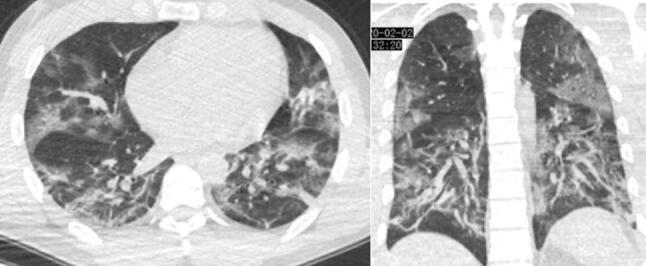
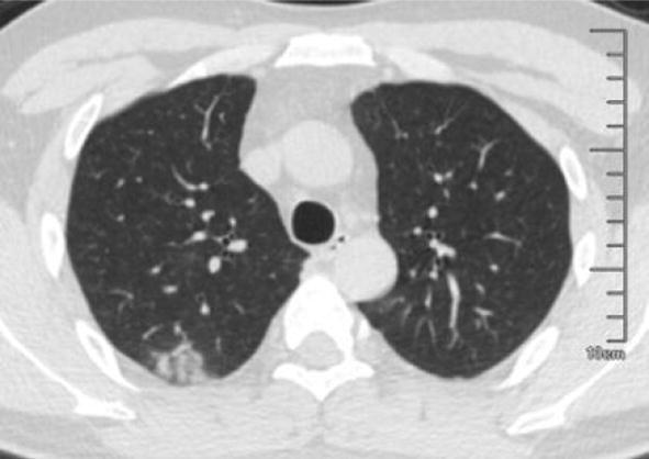
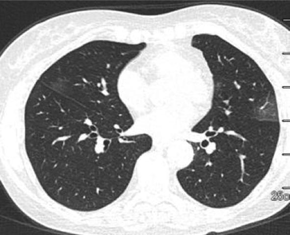
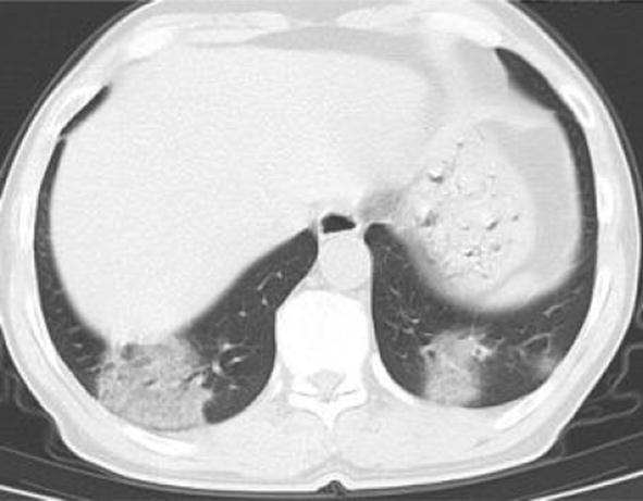
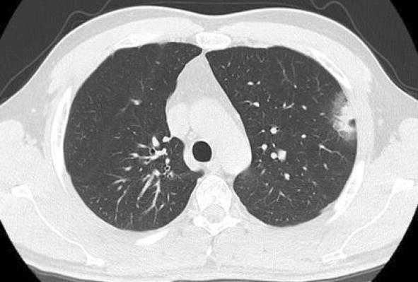
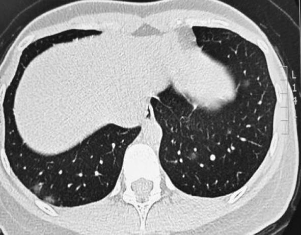
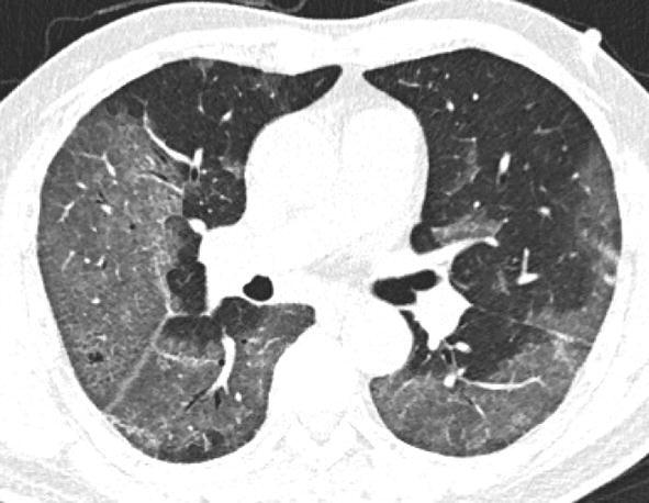
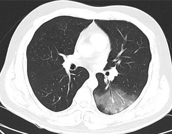
COVID-19 has become a public health emergency due to its rapid transmission. The appearance of pneumonia is one of the major clues for the diagnosis, progress and therapeutic evaluation. More and more literatures about imaging manifestations and related research have been reported. In order to know about the progress and prospective on imaging of COVID-19, this review focus on interpreting the CT findings, stating the potential pathological basis, proposing the challenge of patients with underlying diseases, differentiating with other diseases and suggesting the future research and clinical directions, which would be helpful for the radiologists in the clinical practice and research.

摘要

由于其快速传播,新型冠状病毒肺炎(COVID-19)已成为突发公共卫生事件。肺部炎症表现是诊断、病情进展及治疗评估的主要线索之一。越来越多关于其影像学表现及相关研究的文献被报道。为了了解COVID-19影像学方面的进展及前景,本综述着重解读CT表现、阐述潜在病理基础、提出合并基础疾病患者面临的挑战、与其他疾病进行鉴别并给出未来研究及临床方向,这将有助于放射科医生的临床实践与研究。

https://cdn.ncbi.nlm.nih.gov/pmc/blobs/f349/7149076/567daaf7113d/42058_2020_31_Fig20_HTML.jpg
https://cdn.ncbi.nlm.nih.gov/pmc/blobs/f349/7149076/9fc01a472e9e/42058_2020_31_Fig1_HTML.jpg
https://cdn.ncbi.nlm.nih.gov/pmc/blobs/f349/7149076/206311b4ae0c/42058_2020_31_Fig2_HTML.jpg
https://cdn.ncbi.nlm.nih.gov/pmc/blobs/f349/7149076/1a604ab03101/42058_2020_31_Fig3_HTML.jpg
https://cdn.ncbi.nlm.nih.gov/pmc/blobs/f349/7149076/e0e4dfd9d988/42058_2020_31_Fig4_HTML.jpg
https://cdn.ncbi.nlm.nih.gov/pmc/blobs/f349/7149076/bbad62b96a3e/42058_2020_31_Fig5_HTML.jpg
https://cdn.ncbi.nlm.nih.gov/pmc/blobs/f349/7149076/2e03b1570eb2/42058_2020_31_Fig6_HTML.jpg
https://cdn.ncbi.nlm.nih.gov/pmc/blobs/f349/7149076/835878befd48/42058_2020_31_Fig7_HTML.jpg
https://cdn.ncbi.nlm.nih.gov/pmc/blobs/f349/7149076/32d2492df30f/42058_2020_31_Fig8_HTML.jpg
https://cdn.ncbi.nlm.nih.gov/pmc/blobs/f349/7149076/2e4ef84f8560/42058_2020_31_Fig9_HTML.jpg
https://cdn.ncbi.nlm.nih.gov/pmc/blobs/f349/7149076/3c2277e0c12d/42058_2020_31_Fig10_HTML.jpg
https://cdn.ncbi.nlm.nih.gov/pmc/blobs/f349/7149076/c0ee33f1e237/42058_2020_31_Fig11_HTML.jpg
https://cdn.ncbi.nlm.nih.gov/pmc/blobs/f349/7149076/6b5fbb052092/42058_2020_31_Fig12_HTML.jpg
https://cdn.ncbi.nlm.nih.gov/pmc/blobs/f349/7149076/1fd47c0c97e8/42058_2020_31_Fig13_HTML.jpg
https://cdn.ncbi.nlm.nih.gov/pmc/blobs/f349/7149076/3a5015f4a044/42058_2020_31_Fig14_HTML.jpg
https://cdn.ncbi.nlm.nih.gov/pmc/blobs/f349/7149076/30990860cab8/42058_2020_31_Fig15_HTML.jpg
https://cdn.ncbi.nlm.nih.gov/pmc/blobs/f349/7149076/ad0fba83094f/42058_2020_31_Fig16_HTML.jpg
https://cdn.ncbi.nlm.nih.gov/pmc/blobs/f349/7149076/cac9f54c7744/42058_2020_31_Fig17_HTML.jpg
https://cdn.ncbi.nlm.nih.gov/pmc/blobs/f349/7149076/75f013f40b41/42058_2020_31_Fig18_HTML.jpg
https://cdn.ncbi.nlm.nih.gov/pmc/blobs/f349/7149076/82da75536951/42058_2020_31_Fig19_HTML.jpg
https://cdn.ncbi.nlm.nih.gov/pmc/blobs/f349/7149076/567daaf7113d/42058_2020_31_Fig20_HTML.jpg

相似文献

1
Progress and prospect on imaging diagnosis of COVID-19.新型冠状病毒肺炎影像学诊断的研究进展与展望
Chin J Acad Radiol. 2020;3(1):4-13. doi: 10.1007/s42058-020-00031-5. Epub 2020 Mar 18.
2
Artificial Intelligence Systems for Diagnosis and Clinical Classification of COVID-19.用于COVID-19诊断和临床分类的人工智能系统
Front Microbiol. 2021 Sep 27;12:729455. doi: 10.3389/fmicb.2021.729455. eCollection 2021.
3
CT Manifestations of Coronavirus Disease (COVID-19) Pneumonia and Influenza Virus Pneumonia: A Comparative Study.COVID-19 肺炎与流感病毒肺炎的 CT 表现:一项对比研究。
AJR Am J Roentgenol. 2021 Jan;216(1):71-79. doi: 10.2214/AJR.20.23304. Epub 2020 Jul 9.
4
Analysis of clinical features and imaging signs of COVID-19 with the assistance of artificial intelligence.人工智能辅助分析 COVID-19 的临床特征和影像征象。
Eur Rev Med Pharmacol Sci. 2020 Aug;24(15):8210-8218. doi: 10.26355/eurrev_202008_22510.
5
Differentiating pneumonia with and without COVID-19 using chest CT images: from qualitative to quantitative.使用胸部 CT 图像区分伴有和不伴有 COVID-19 的肺炎:从定性到定量。
J Xray Sci Technol. 2020;28(4):583-589. doi: 10.3233/XST-200689.
6
FaNet: fast assessment network for the novel coronavirus (COVID-19) pneumonia based on 3D CT imaging and clinical symptoms.FaNet:基于3D CT成像和临床症状的新型冠状病毒(COVID-19)肺炎快速评估网络
Appl Intell (Dordr). 2021;51(5):2838-2849. doi: 10.1007/s10489-020-01965-0. Epub 2020 Nov 14.
7
Two in one: Overlapping CT findings of COVID-19 and underlying lung diseases.两病重叠:COVID-19 与基础肺部疾病的 CT 表现重叠。
Clin Imaging. 2023 Jan;93:60-69. doi: 10.1016/j.clinimag.2022.11.005. Epub 2022 Nov 12.
8
Artificial intelligence for stepwise diagnosis and monitoring of COVID-19.人工智能在 COVID-19 的逐步诊断和监测中的应用。
Eur Radiol. 2022 Apr;32(4):2235-2245. doi: 10.1007/s00330-021-08334-6. Epub 2022 Jan 6.
9
Review of Artificial Intelligence Techniques in Imaging Data Acquisition, Segmentation, and Diagnosis for COVID-19.COVID-19 成像数据采集、分割和诊断中人工智能技术的综述。
IEEE Rev Biomed Eng. 2021;14:4-15. doi: 10.1109/RBME.2020.2987975. Epub 2021 Jan 22.
10
A Comparison of CT Manifestations Between Coronavirus Disease 2019 (COVID-19) and Other Types of Viral Pneumonia.新型冠状病毒病(COVID-19)与其他类型病毒性肺炎的 CT 表现比较。
Curr Med Imaging. 2021;17(11):1316-1323. doi: 10.2174/1573405617666210218092751.

引用本文的文献

1
Chest CT Scan Findings in Children with COVID-19: A Systematic Review.新型冠状病毒肺炎患儿胸部CT扫描结果:一项系统评价
Acta Med Philipp. 2024 Apr 30;58(7):110-128. doi: 10.47895/amp.v58i7.6385. eCollection 2024.
2
A dual-stage deep convolutional neural network for automatic diagnosis of COVID-19 and pneumonia from chest CT images.一种用于从胸部 CT 图像自动诊断 COVID-19 和肺炎的两阶段深度卷积神经网络。
Comput Biol Med. 2022 Oct;149:105806. doi: 10.1016/j.compbiomed.2022.105806. Epub 2022 Jul 19.
3
A Cross-Disciplinary View of Testing and Bioinformatic Analysis of SARS-CoV-2 and Other Human Respiratory Viruses in Pandemic Settings.

本文引用的文献

1
Severe Acute Respiratory Disease in a Huanan Seafood Market Worker: Images of an Early Casualty.一名华南海鲜市场工作人员的严重急性呼吸系统疾病:首例死亡病例影像
Radiol Cardiothorac Imaging. 2020 Feb 14;2(1):e200033. doi: 10.1148/ryct.2020200033. eCollection 2020 Feb.
2
Chest Imaging Appearance of COVID-19 Infection.新型冠状病毒肺炎感染的胸部影像学表现。
Radiol Cardiothorac Imaging. 2020 Feb 13;2(1):e200028. doi: 10.1148/ryct.2020200028. eCollection 2020 Feb.
3
COVID-19 Infection Presenting with CT Halo Sign.表现为CT晕征的新型冠状病毒肺炎感染
大流行背景下对严重急性呼吸综合征冠状病毒2(SARS-CoV-2)及其他人类呼吸道病毒进行检测和生物信息分析的跨学科视角
IEEE Access. 2021 Dec 6;9:163716-163734. doi: 10.1109/ACCESS.2021.3133417. eCollection 2021.
4
Neutralizing Antibodies Responses against SARS-CoV-2 in a Sardinian Cohort Group Up to 9 Months after BNT162b2 Vaccination.在撒丁岛队列组中,BNT162b2疫苗接种后长达9个月针对严重急性呼吸综合征冠状病毒2(SARS-CoV-2)的中和抗体反应
Vaccines (Basel). 2022 Mar 29;10(4):531. doi: 10.3390/vaccines10040531.
5
An Efficient Deep Learning Model to Detect COVID-19 Using Chest X-ray Images.利用胸部 X 光图像检测 COVID-19 的高效深度学习模型。
Int J Environ Res Public Health. 2022 Feb 11;19(4):2013. doi: 10.3390/ijerph19042013.
6
Automatic detection of COVID-19 from chest CT scan and chest X-Rays images using deep learning, transfer learning and stacking.利用深度学习、迁移学习和堆叠技术从胸部CT扫描和胸部X光图像中自动检测新冠肺炎。
Appl Intell (Dordr). 2022;52(2):2243-2259. doi: 10.1007/s10489-021-02393-4. Epub 2021 Jun 7.
7
Thymus Gland: A Double Edge Sword for Coronaviruses.胸腺:冠状病毒的双刃剑。
Vaccines (Basel). 2021 Oct 2;9(10):1119. doi: 10.3390/vaccines9101119.
8
COVID-19 imaging: Diagnostic approaches, challenges, and evolving advances.新型冠状病毒肺炎影像学:诊断方法、挑战及进展
World J Radiol. 2021 Jun 28;13(6):171-191. doi: 10.4329/wjr.v13.i6.171.
9
A stacked ensemble for the detection of COVID-19 with high recall and accuracy.一种具有高召回率和准确率的 COVID-19 检测堆叠集成方法。
Comput Biol Med. 2021 Aug;135:104608. doi: 10.1016/j.compbiomed.2021.104608. Epub 2021 Jun 30.
10
Can Clinical Symptoms and Laboratory Results Predict CT Abnormality? Initial Findings Using Novel Machine Learning Techniques in Children With COVID-19 Infections.临床症状和实验室检查结果能否预测CT异常?在新冠病毒感染儿童中使用新型机器学习技术的初步发现。
Front Med (Lausanne). 2021 Jun 14;8:699984. doi: 10.3389/fmed.2021.699984. eCollection 2021.
Radiol Cardiothorac Imaging. 2020 Feb 12;2(1):e200026. doi: 10.1148/ryct.2020200026. eCollection 2020 Feb.
4
Spectrum of Chest CT Findings in a Familial Cluster of COVID-19 Infection.新冠病毒感染家族聚集性病例的胸部CT表现谱
Radiol Cardiothorac Imaging. 2020 Feb 13;2(1):e200025. doi: 10.1148/ryct.2020200025. eCollection 2020 Feb.
5
Pathological findings of COVID-19 associated with acute respiratory distress syndrome.与急性呼吸窘迫综合征相关的新型冠状病毒肺炎的病理表现
Lancet Respir Med. 2020 Apr;8(4):420-422. doi: 10.1016/S2213-2600(20)30076-X. Epub 2020 Feb 18.
6
Time Course of Lung Changes at Chest CT during Recovery from Coronavirus Disease 2019 (COVID-19).新冠肺炎(COVID-19)康复过程中胸部 CT 肺部变化的时间进程。
Radiology. 2020 Jun;295(3):715-721. doi: 10.1148/radiol.2020200370. Epub 2020 Feb 13.
7
Pre- and Posttreatment Chest CT Findings: 2019 Novel Coronavirus (2019-nCoV) Pneumonia.治疗前和治疗后胸部CT表现:2019新型冠状病毒(2019-nCoV)肺炎
Radiology. 2020 Apr;295(1):21. doi: 10.1148/radiol.2020200323. Epub 2020 Feb 12.
8
Chest CT for Typical Coronavirus Disease 2019 (COVID-19) Pneumonia: Relationship to Negative RT-PCR Testing.胸部 CT 与典型 2019 冠状病毒病(COVID-19)肺炎:与阴性 RT-PCR 检测的关系。
Radiology. 2020 Aug;296(2):E41-E45. doi: 10.1148/radiol.2020200343. Epub 2020 Feb 12.
9
Use of Chest CT in Combination with Negative RT-PCR Assay for the 2019 Novel Coronavirus but High Clinical Suspicion.胸部CT联合2019新型冠状病毒逆转录聚合酶链反应检测结果为阴性但临床高度怀疑的应用
Radiology. 2020 Apr;295(1):22-23. doi: 10.1148/radiol.2020200330. Epub 2020 Feb 12.
10
Evolution of CT Manifestations in a Patient Recovered from 2019 Novel Coronavirus (2019-nCoV) Pneumonia in Wuhan, China.中国武汉一名从2019新型冠状病毒(2019-nCoV)肺炎康复患者的CT表现演变
Radiology. 2020 Apr;295(1):20. doi: 10.1148/radiol.2020200269. Epub 2020 Feb 7.