Taube Janis M, Akturk Guray, Angelo Michael, Engle Elizabeth L, Gnjatic Sacha, Greenbaum Shirley, Greenwald Noah F, Hedvat Cyrus V, Hollmann Travis J, Juco Jonathan, Parra Edwin R, Rebelatto Marlon C, Rimm David L, Rodriguez-Canales Jaime, Schalper Kurt A, Stack Edward C, Ferreira Cláudia S, Korski Konstanty, Lako Ana, Rodig Scott J, Schenck Emanuel, Steele Keith E, Surace Michael J, Tetzlaff Michael T, von Loga Katharina, Wistuba Ignacio I, Bifulco Carlo B
Department of Dermatology, Johns Hopkins School of Medicine, Bloomberg~Kimmel Institute for Cancer Immunotherapy, Baltimore, Maryland, USA
Tisch Cancer Institute, Icahn School of Medicine at Mount Sinai, New York, New York City, USA.
J Immunother Cancer. 2020 May;8(1). doi: 10.1136/jitc-2019-000155.
The interaction between the immune system and tumor cells is an important feature for the prognosis and treatment of cancer. Multiplex immunohistochemistry (mIHC) and multiplex immunofluorescence (mIF) analyses are emerging technologies that can be used to help quantify immune cell subsets, their functional state, and their spatial arrangement within the tumor microenvironment.
The Society for Immunotherapy of Cancer (SITC) convened a task force of pathologists and laboratory leaders from academic centers as well as experts from pharmaceutical and diagnostic companies to develop best practice guidelines for the optimization and validation of mIHC/mIF assays across platforms.
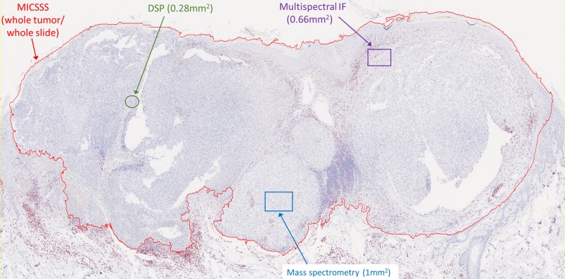
Representative outputs and the advantages and disadvantages of mIHC/mIF approaches, such as multiplexed chromogenic IHC, multiplexed immunohistochemical consecutive staining on single slide, mIF (including multispectral approaches), tissue-based mass spectrometry, and digital spatial profiling are discussed.
mIHC/mIF technologies are becoming standard tools for biomarker studies and are likely to enter routine clinical practice in the near future. Careful assay optimization and validation will help ensure outputs are robust and comparable across laboratories as well as potentially across mIHC/mIF platforms. Quantitative image analysis of mIHC/mIF output and data management considerations will be addressed in a complementary manuscript from this task force.
免疫系统与肿瘤细胞之间的相互作用是癌症预后和治疗的一个重要特征。多重免疫组化(mIHC)和多重免疫荧光(mIF)分析是新兴技术,可用于帮助量化肿瘤微环境中的免疫细胞亚群、它们的功能状态及其空间排列。
癌症免疫治疗协会(SITC)召集了一个由学术中心的病理学家和实验室负责人以及制药和诊断公司的专家组成的特别工作组,以制定跨平台优化和验证mIHC/mIF检测的最佳实践指南。
讨论了mIHC/mIF方法的代表性成果及其优缺点,如多重显色免疫组化、单张玻片上的多重免疫组化连续染色、mIF(包括多光谱方法)、基于组织的质谱分析和数字空间分析。
mIHC/mIF技术正成为生物标志物研究的标准工具,并可能在不久的将来进入常规临床实践。仔细的检测优化和验证将有助于确保各实验室之间以及潜在地跨mIHC/mIF平台的结果可靠且具有可比性。本特别工作组的一篇补充稿件将讨论mIHC/mIF结果的定量图像分析和数据管理方面的考虑因素。