Jiang Taoran, Yu Dewang, Wang Yuqi, Zan Tao, Wang Shuyi, Li Qingfeng
Department of Plastic and Reconstructive Surgery, Shanghai 9th People's Hospital, Shanghai Jiao Tong University School of Medicine, Shanghai, China.
School of Medical Instrument and Food Engineering, University of Shanghai for Science and Technology, Shanghai, China.
J Med Internet Res. 2020 Apr 17;22(4):e16852. doi: 10.2196/16852.
Vascular localization is crucial for perforator flap transfer. Augmented reality offers a novel method to seamlessly combine real information with virtual objects created by computed tomographic angiography to help the surgeon "see through" the skin and precisely localize the perforator. The head-mounted display augmented reality system HoloLens (Microsoft) could facilitate augmented reality-based perforator localization for a more convenient and safe procedure.
The aim of this study was to evaluate the precision of the HoloLens-based vascular localization system, as the most important performance indicator of a new localization system.
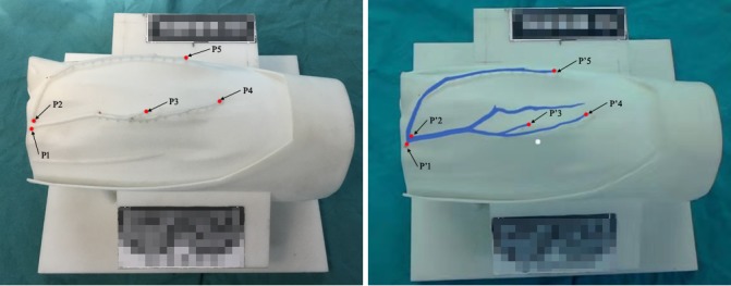
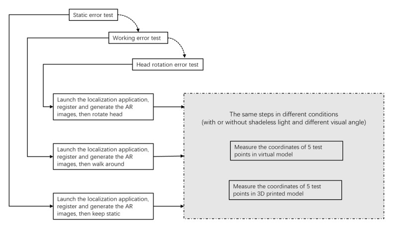
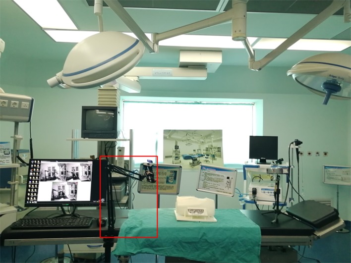
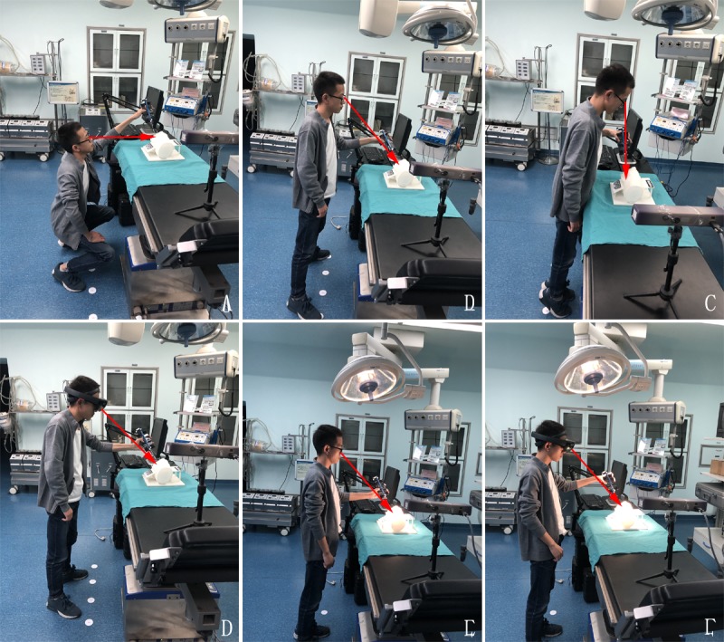
The precision of the HoloLens-based vascular localization system was tested in a simulated operating room under different conditions with a three-dimensional (3D) printed model. The coordinates of five pairs of points on the vascular map that could be easily identified on the 3D printed model and virtual model were detected by a probe, and the distance between the corresponding points was calculated as the navigation error.
The mean errors were determined under different conditions, with a minimum error of 1.35 mm (SD 0.43) and maximum error of 3.18 mm (SD 1.32), which were within the clinically acceptable range. There were no significant differences in the errors obtained under different visual angles, different light intensities, or different states (static or motion). However, the error was larger when tested with light compared with that tested without light.
This precision evaluation demonstrated that the HoloLens system can precisely localize the perforator and potentially help the surgeon accomplish the operation. The authors recommend using HoloLens-based surgical navigation without light.
血管定位对于穿支皮瓣移植至关重要。增强现实提供了一种新颖的方法,可将真实信息与计算机断层血管造影创建的虚拟对象无缝结合,帮助外科医生“看穿”皮肤并精确地定位穿支血管。头戴式显示器增强现实系统HoloLens(微软公司)可以促进基于增强现实的穿支血管定位,从而实现更便捷、安全的手术过程。
本研究旨在评估基于HoloLens的血管定位系统的精度,这是一种新定位系统最重要的性能指标。
使用三维(3D)打印模型,在不同条件下的模拟手术室中测试基于HoloLens的血管定位系统的精度。用探针检测3D打印模型和虚拟模型上易于识别的血管图谱上五对点的坐标,并计算对应点之间的距离作为导航误差。
测定了不同条件下的平均误差,最小误差为1.35毫米(标准差0.43),最大误差为3.18毫米(标准差1.32),均在临床可接受范围内。在不同视角、不同光照强度或不同状态(静态或动态)下获得的误差无显著差异。然而,与无光照测试相比,有光照测试时误差更大。
该精度评估表明,HoloLens系统能够精确地定位穿支血管,并可能有助于外科医生完成手术。作者建议在无光照条件下使用基于HoloLens的手术导航。