Widya Aji Resindra, Monno Yusuke, Okutomi Masatoshi, Suzuki Sho, Gotoda Takuji, Miki Kenji
1Department of Systems and Control EngineeringSchool of EngineeringTokyo Institute of TechnologyTokyo152-8550Japan.
2Division of Gastroenterology and HepatologyDepartment of MedicineNihon University School of MedicineTokyo101-8309Japan.
IEEE J Transl Eng Health Med. 2019 Oct 18;7:3300310. doi: 10.1109/JTEHM.2019.2946802. eCollection 2019.
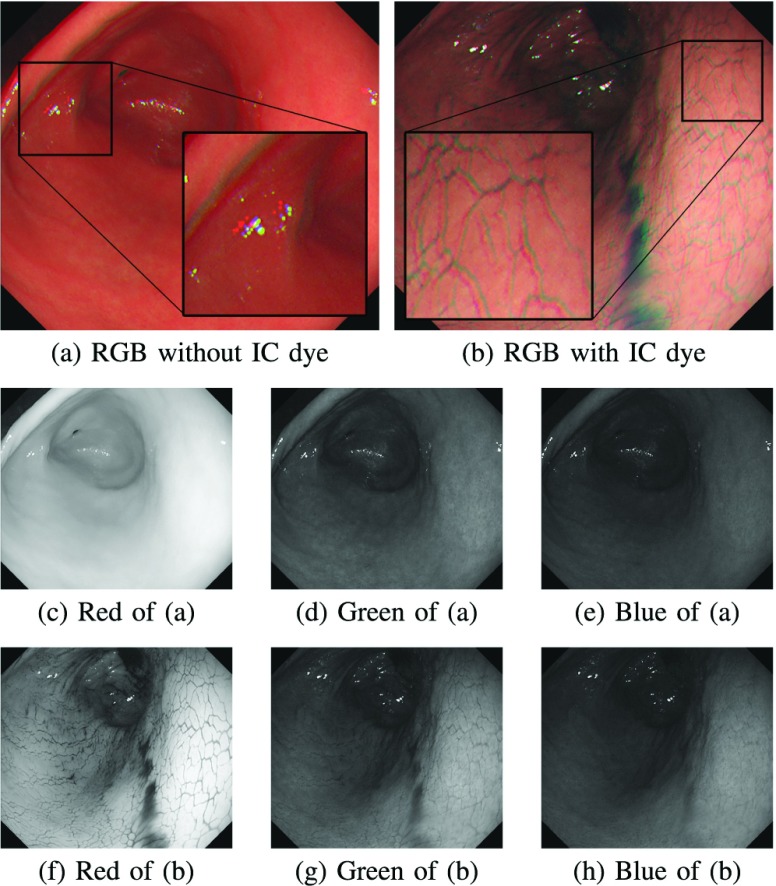
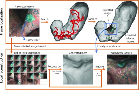
Gastric endoscopy is a common clinical practice that enables medical doctors to diagnose various lesions inside a stomach. In order to identify the location of a gastric lesion such as early cancer and a peptic ulcer within the stomach, this work addresses to reconstruct the color-textured 3D model of a whole stomach from a standard monocular endoscope video and localize any selected video frame to the 3D model. We examine how to enable structure-from-motion (SfM) to reconstruct the whole shape of a stomach from endoscope images, which is a challenging task due to the texture-less nature of the stomach surface. We specifically investigate the combined effect of chromo-endoscopy and color channel selection on SfM to increase the number of feature points. We also design a plane fitting-based algorithm for 3D point outliers removal to improve the 3D model quality. We show that whole stomach 3D reconstruction can be achieved (more than 90% of the frames can be reconstructed) by using red channel images captured under chromo-endoscopy by spreading indigo carmine (IC) dye on the stomach surface. In experimental results, we demonstrate the reconstructed 3D models for seven subjects and the application of lesion localization and reconstruction. The methodology and results presented in this paper could offer some valuable reference to other researchers and also could be an excellent tool for gastric surgeons in various computer-aided diagnosis applications.
胃镜检查是一种常见的临床操作,可使医生诊断胃内的各种病变。为了确定胃内病变(如早期癌症和消化性溃疡)的位置,这项工作致力于从标准单目内窥镜视频重建整个胃的彩色纹理三维模型,并将任何选定的视频帧定位到三维模型上。我们研究如何利用运动恢复结构(SfM)从内窥镜图像重建胃的整体形状,由于胃表面缺乏纹理,这是一项具有挑战性的任务。我们特别研究了染色内镜检查和颜色通道选择对SfM的综合影响,以增加特征点的数量。我们还设计了一种基于平面拟合的算法来去除三维点异常值,以提高三维模型质量。我们表明,通过在胃表面涂抹靛胭脂(IC)染料,利用染色内镜检查下拍摄的红色通道图像,可以实现整个胃的三维重建(超过90%的帧可以重建)。在实验结果中,我们展示了七名受试者的重建三维模型以及病变定位和重建的应用。本文提出的方法和结果可以为其他研究人员提供一些有价值的参考,也可以成为胃外科医生在各种计算机辅助诊断应用中的优秀工具。