• 文献检索
  • 文档翻译
  • 深度研究
  • 学术资讯
  • Suppr Zotero 插件Zotero 插件
  • 邀请有礼
  • 套餐&价格
  • 历史记录
应用&插件
Suppr Zotero 插件Zotero 插件浏览器插件Mac 客户端Windows 客户端微信小程序
定价
高级版会员购买积分包购买API积分包
服务
文献检索文档翻译深度研究API 文档MCP 服务
关于我们
关于 Suppr公司介绍联系我们用户协议隐私条款
关注我们

Suppr 超能文献

核心技术专利:CN118964589B侵权必究
粤ICP备2023148730 号-1Suppr @ 2026

文献检索

告别复杂PubMed语法,用中文像聊天一样搜索,搜遍4000万医学文献。AI智能推荐,让科研检索更轻松。

立即免费搜索

文件翻译

保留排版,准确专业,支持PDF/Word/PPT等文件格式,支持 12+语言互译。

免费翻译文档

深度研究

AI帮你快速写综述,25分钟生成高质量综述,智能提取关键信息,辅助科研写作。

立即免费体验

黑色素瘤肿瘤浸润 T 淋巴细胞的共享抗原表位及其同源 TCR 的景观图谱。

Landscape mapping of shared antigenic epitopes and their cognate TCRs of tumor-infiltrating T lymphocytes in melanoma.

机构信息

Tumor Immunotherapy Program, Campbell Family Institute for Breast Cancer Research, Princess Margaret Cancer Centre, University Health Network, Toronto, Canada.

Princess Margaret Cancer Centre, University Health Network, Toronto, Canada.

出版信息

Elife. 2020 Apr 21;9:e53244. doi: 10.7554/eLife.53244.

DOI:10.7554/eLife.53244
PMID:32314731
原文链接:https://pmc.ncbi.nlm.nih.gov/articles/PMC7234812/
Abstract

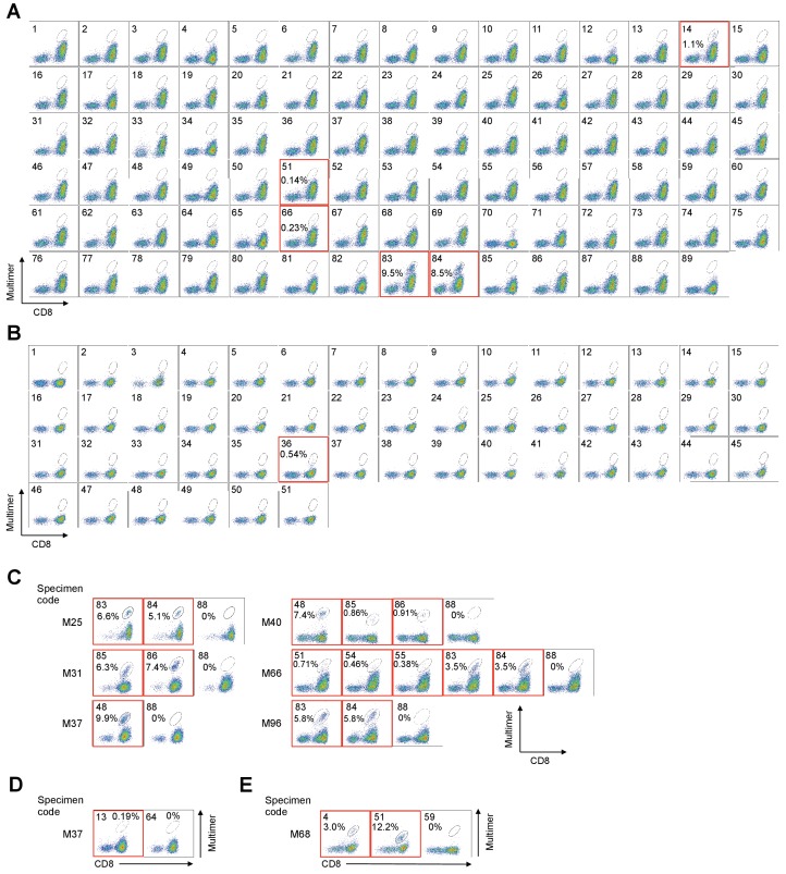
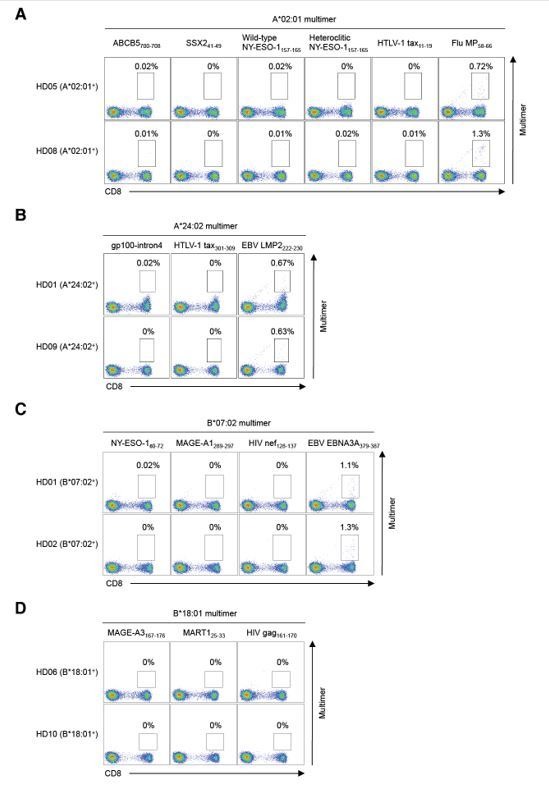
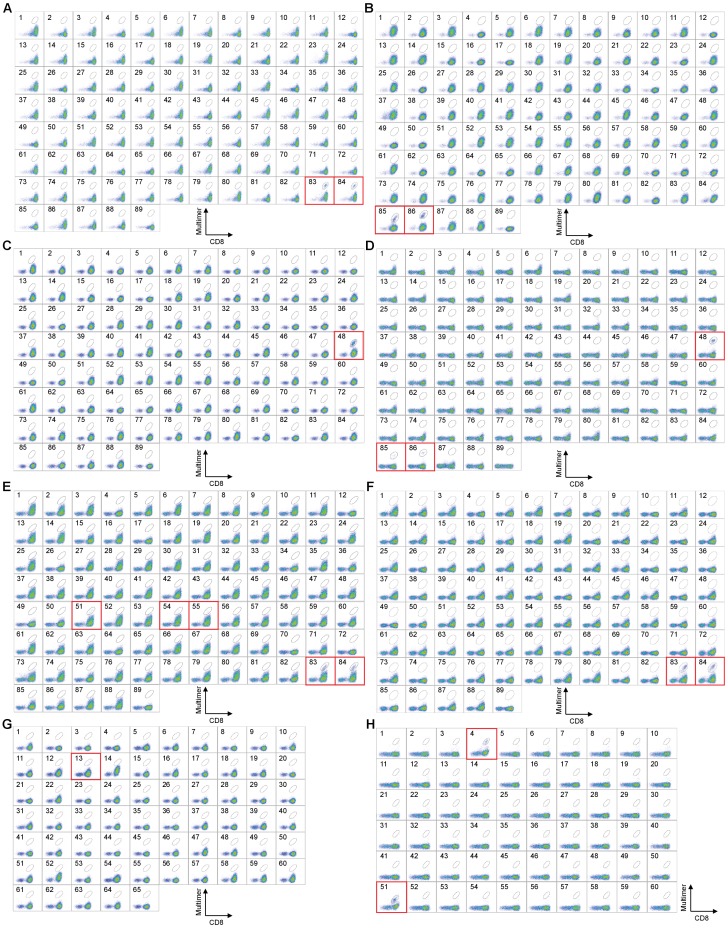
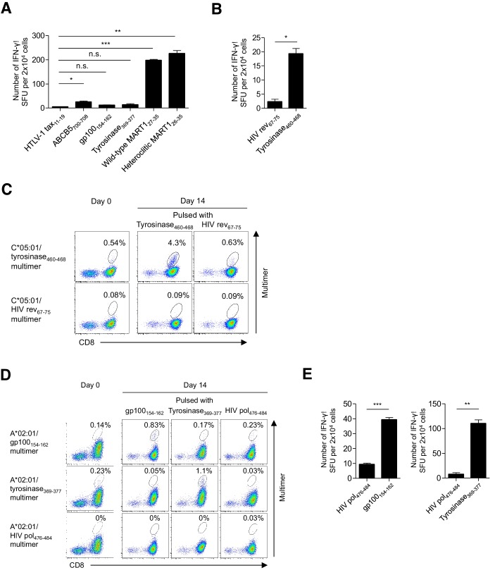
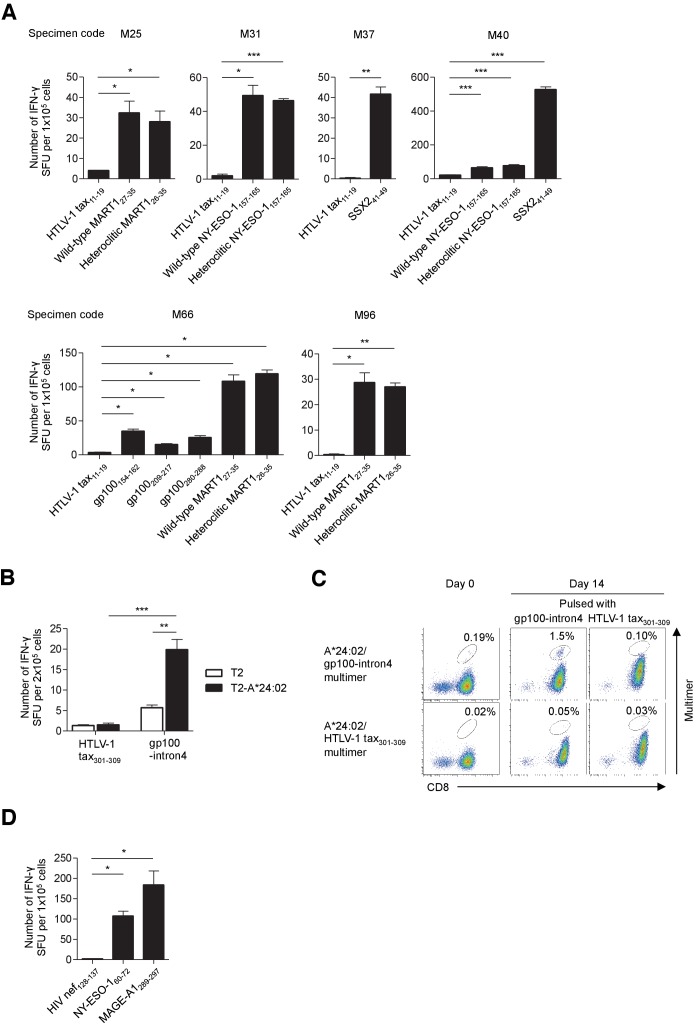
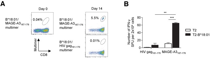
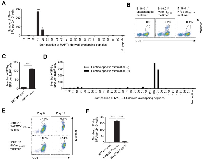
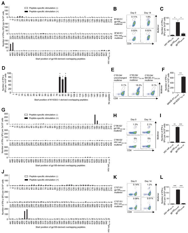
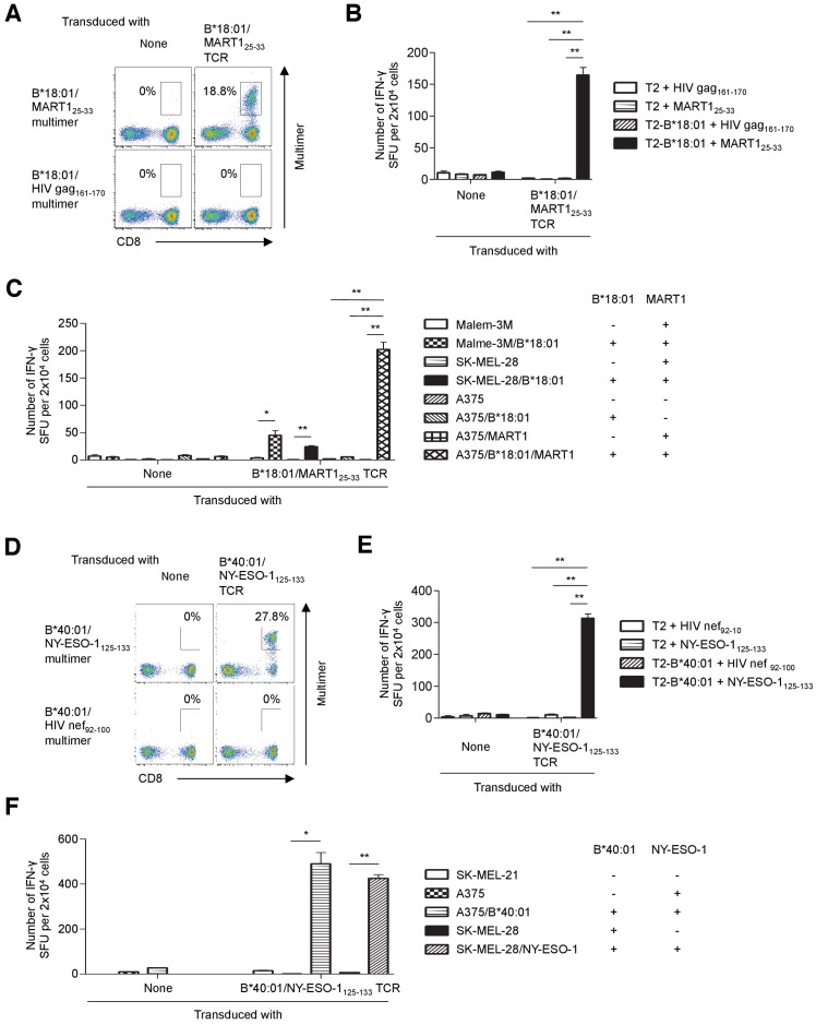
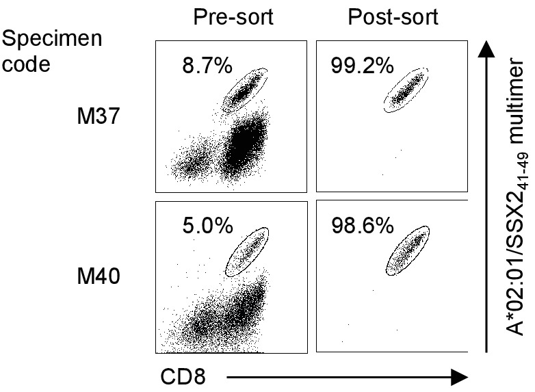
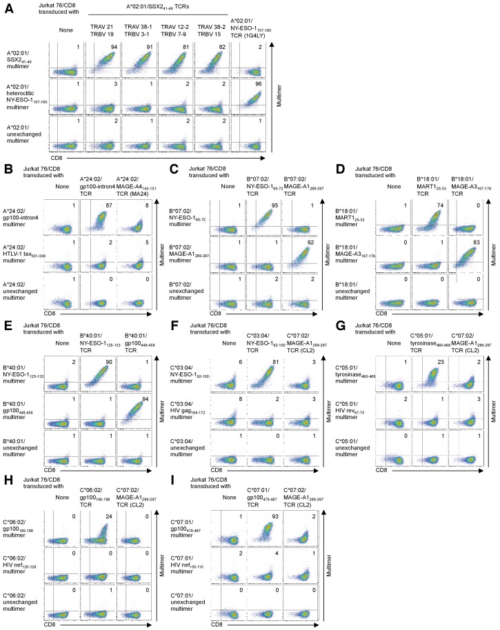
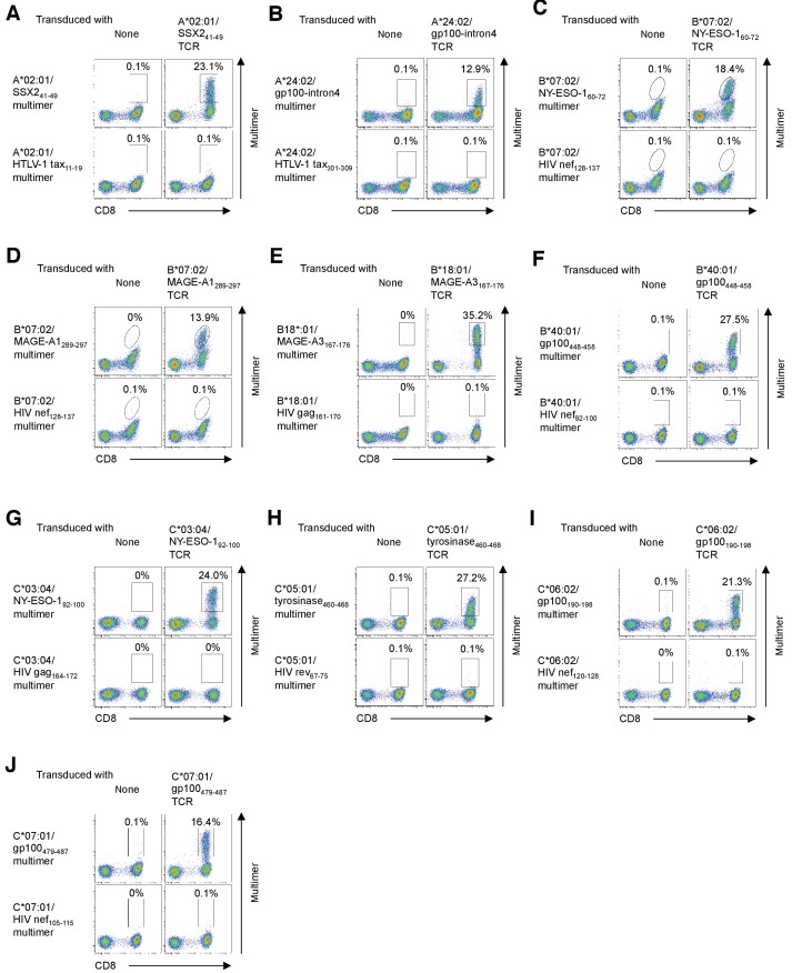
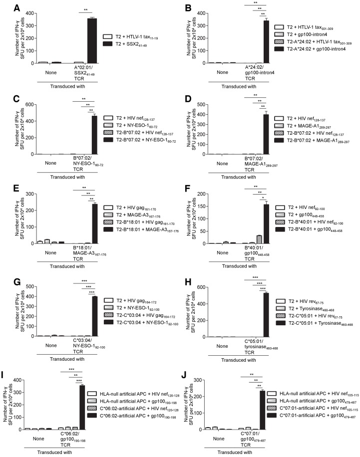
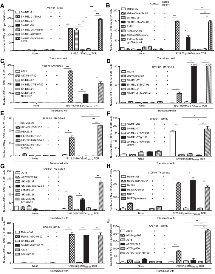
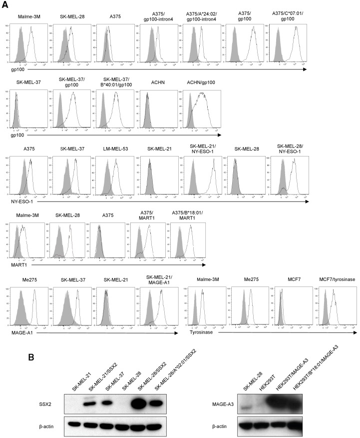
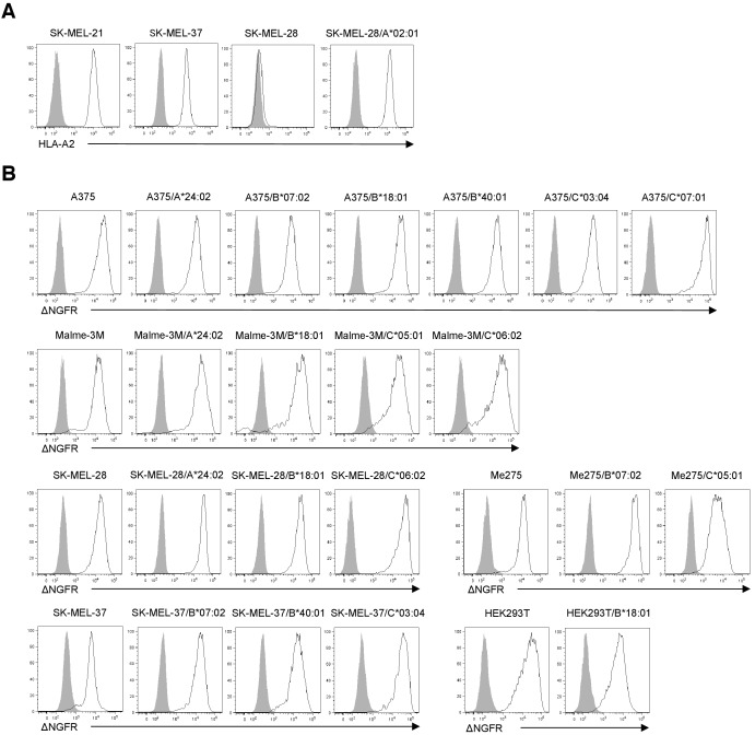
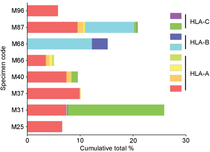
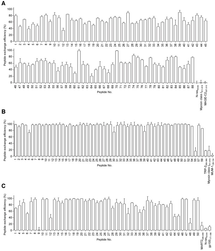
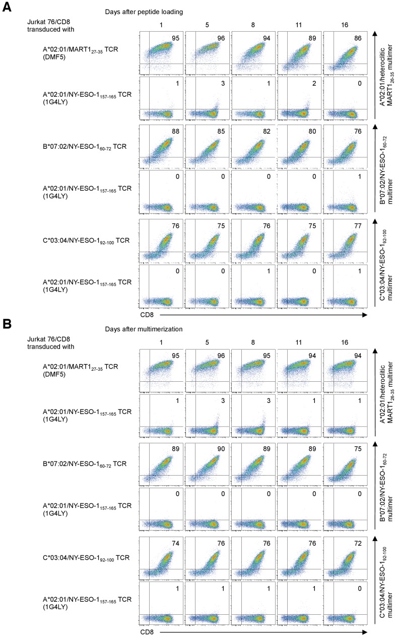
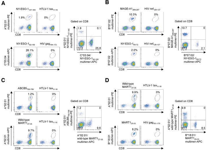
HLA-restricted T cell responses can induce antitumor effects in cancer patients. Previous human T cell research has largely focused on the few HLA alleles prevalent in a subset of ethnic groups. Here, using a panel of newly developed peptide-exchangeable peptide/HLA multimers and artificial antigen-presenting cells for 25 different class I alleles and greater than 800 peptides, we systematically and comprehensively mapped shared antigenic epitopes recognized by tumor-infiltrating T lymphocytes (TILs) from eight melanoma patients for all their class I alleles. We were able to determine the specificity, on average, of 12.2% of the TILs recognizing a mean of 3.1 shared antigen-derived epitopes across HLA-A, B, and C. Furthermore, we isolated a number of cognate T cell receptor genes with tumor reactivity. Our novel strategy allows for a more complete examination of the immune response and development of novel cancer immunotherapy not limited by HLA allele prevalence or tumor mutation burden.

摘要

HLA 限制性 T 细胞反应可在癌症患者中诱导抗肿瘤作用。以前的人类 T 细胞研究主要集中在少数在亚群中流行的 HLA 等位基因上。在这里,我们使用一组新开发的可交换肽/HLA 多聚体和人工抗原呈递细胞,针对 25 种不同的 I 类等位基因和超过 800 种肽,系统而全面地绘制了来自 8 名黑色素瘤患者的肿瘤浸润性 T 淋巴细胞 (TIL) 识别的共享抗原表位图谱。我们能够确定 12.2%的 TIL 的特异性,平均识别 HLA-A、B 和 C 上 3.1 个共享抗原衍生的表位。此外,我们还分离出了一些具有肿瘤反应性的同源 T 细胞受体基因。我们的新策略允许更全面地检查免疫反应,并开发不受 HLA 等位基因流行或肿瘤突变负担限制的新型癌症免疫疗法。

https://cdn.ncbi.nlm.nih.gov/pmc/blobs/0987/7234812/092341cb121f/elife-53244-resp-fig2.jpg
https://cdn.ncbi.nlm.nih.gov/pmc/blobs/0987/7234812/93738c323151/elife-53244-fig1.jpg
https://cdn.ncbi.nlm.nih.gov/pmc/blobs/0987/7234812/3d44e6545809/elife-53244-fig1-figsupp1.jpg
https://cdn.ncbi.nlm.nih.gov/pmc/blobs/0987/7234812/d02be1dfb848/elife-53244-fig2.jpg
https://cdn.ncbi.nlm.nih.gov/pmc/blobs/0987/7234812/deff8af85924/elife-53244-fig2-figsupp1.jpg
https://cdn.ncbi.nlm.nih.gov/pmc/blobs/0987/7234812/e4e9c788b8d1/elife-53244-fig3.jpg
https://cdn.ncbi.nlm.nih.gov/pmc/blobs/0987/7234812/a4da6eaf91a9/elife-53244-fig4.jpg
https://cdn.ncbi.nlm.nih.gov/pmc/blobs/0987/7234812/545cef19bede/elife-53244-fig4-figsupp1.jpg
https://cdn.ncbi.nlm.nih.gov/pmc/blobs/0987/7234812/63d1f345101d/elife-53244-fig5.jpg
https://cdn.ncbi.nlm.nih.gov/pmc/blobs/0987/7234812/eae09780dcc3/elife-53244-fig5-figsupp1.jpg
https://cdn.ncbi.nlm.nih.gov/pmc/blobs/0987/7234812/59221efd8376/elife-53244-fig5-figsupp2.jpg
https://cdn.ncbi.nlm.nih.gov/pmc/blobs/0987/7234812/fce9c58c11b5/elife-53244-fig5-figsupp3.jpg
https://cdn.ncbi.nlm.nih.gov/pmc/blobs/0987/7234812/252a4c317357/elife-53244-fig5-figsupp4.jpg
https://cdn.ncbi.nlm.nih.gov/pmc/blobs/0987/7234812/46769500d25e/elife-53244-fig5-figsupp5.jpg
https://cdn.ncbi.nlm.nih.gov/pmc/blobs/0987/7234812/6c00a10ac038/elife-53244-fig5-figsupp6.jpg
https://cdn.ncbi.nlm.nih.gov/pmc/blobs/0987/7234812/4cee6ab36eed/elife-53244-fig5-figsupp7.jpg
https://cdn.ncbi.nlm.nih.gov/pmc/blobs/0987/7234812/3e5d48bcbb4a/elife-53244-fig6.jpg
https://cdn.ncbi.nlm.nih.gov/pmc/blobs/0987/7234812/66f5f8b862be/elife-53244-fig6-figsupp1.jpg
https://cdn.ncbi.nlm.nih.gov/pmc/blobs/0987/7234812/b26cdd06c7a6/elife-53244-fig6-figsupp2.jpg
https://cdn.ncbi.nlm.nih.gov/pmc/blobs/0987/7234812/ae3b9ed53e1f/elife-53244-fig6-figsupp3.jpg
https://cdn.ncbi.nlm.nih.gov/pmc/blobs/0987/7234812/6abeec63a8cc/elife-53244-resp-fig1.jpg
https://cdn.ncbi.nlm.nih.gov/pmc/blobs/0987/7234812/092341cb121f/elife-53244-resp-fig2.jpg

相似文献

1
Landscape mapping of shared antigenic epitopes and their cognate TCRs of tumor-infiltrating T lymphocytes in melanoma.黑色素瘤肿瘤浸润 T 淋巴细胞的共享抗原表位及其同源 TCR 的景观图谱。
Elife. 2020 Apr 21;9:e53244. doi: 10.7554/eLife.53244.
2
Recognition of shared melanoma antigens in association with major HLA-A alleles by tumor infiltrating T lymphocytes from 123 patients with melanoma.123例黑色素瘤患者的肿瘤浸润性T淋巴细胞对与主要HLA - A等位基因相关的共享黑色素瘤抗原的识别。
J Immunother. 2000 Jan;23(1):17-27. doi: 10.1097/00002371-200001000-00004.
3
Identification of new melanoma epitopes on melanosomal proteins recognized by tumor infiltrating T lymphocytes restricted by HLA-A1, -A2, and -A3 alleles.在黑素小体蛋白上鉴定由HLA - A1、- A2和- A3等位基因限制的肿瘤浸润性T淋巴细胞所识别的新黑色素瘤表位。
J Immunol. 1998 Dec 15;161(12):6985-92.
4
Elucidating the immunological effects of 5-azacytidine treatment in patients with myelodysplastic syndrome and identifying new conditional ligands and T-cell epitopes of relevance in melanoma.阐明5-氮杂胞苷治疗骨髓增生异常综合征患者的免疫效应,并鉴定黑色素瘤中相关的新条件配体和T细胞表位。
Dan Med J. 2015 Aug;62(8):B5144.
5
T-cell receptor usage by melanoma-specific clonal and highly oligoclonal tumor-infiltrating lymphocyte lines.黑色素瘤特异性克隆性和高度寡克隆性肿瘤浸润淋巴细胞系对T细胞受体的使用情况。
Proc Natl Acad Sci U S A. 1994 Mar 29;91(7):2829-33. doi: 10.1073/pnas.91.7.2829.
6
Identification of five new HLA-B*3501-restricted epitopes derived from common melanoma-associated antigens, spontaneously recognized by tumor-infiltrating lymphocytes.鉴定出五个源自常见黑色素瘤相关抗原的新的HLA-B*3501限制性表位,这些表位可被肿瘤浸润淋巴细胞自发识别。
J Immunol. 2003 Dec 1;171(11):6283-9. doi: 10.4049/jimmunol.171.11.6283.
7
Broadening the repertoire of melanoma-associated T-cell epitopes.拓宽黑色素瘤相关T细胞表位的库。
Cancer Immunol Immunother. 2015 May;64(5):609-20. doi: 10.1007/s00262-015-1664-x. Epub 2015 Feb 18.
8
Melanocortin 1 Receptor-derived peptides are efficiently recognized by cytotoxic T lymphocytes from melanoma patients.黑素细胞刺激素 1 受体衍生肽可被黑素瘤患者的细胞毒性 T 淋巴细胞有效识别。
Immunobiology. 2014 Mar;219(3):189-97. doi: 10.1016/j.imbio.2013.10.002. Epub 2013 Oct 12.
9
Detection of naturally processed and HLA-A1-presented melanoma T-cell epitopes defined by CD8(+) T-cells' release of granulocyte-macrophage colony-stimulating factor but not by cytolysis.通过CD8(+) T细胞释放粒细胞巨噬细胞集落刺激因子而非细胞溶解作用来检测自然加工且由HLA - A1呈递的黑色素瘤T细胞表位。
Clin Cancer Res. 1996 Jan;2(1):87-95.
10
Recognition of neuroectodermal tumors by melanoma-specific cytotoxic T lymphocytes: evidence for antigen sharing by tumors derived from the neural crest.黑色素瘤特异性细胞毒性T淋巴细胞对神经外胚层肿瘤的识别:源自神经嵴的肿瘤存在抗原共享的证据。
Cancer Immunol Immunother. 1994 Aug;39(2):73-83. doi: 10.1007/BF01525312.

引用本文的文献

1
An SNP-dependent cancer-testis antigenic epitope serves as a promising immunotherapeutic target for cancer.一种单核苷酸多态性(SNP)依赖性癌胚抗原表位是一种很有前景的癌症免疫治疗靶点。
Oncoimmunology. 2025 Dec;14(1):2528110. doi: 10.1080/2162402X.2025.2528110. Epub 2025 Jul 9.
2
Interferon-α promotes HLA-B-restricted presentation of conventional and alternative antigens in human pancreatic β-cells.干扰素-α促进人胰腺β细胞中传统和替代抗原的HLA - B限制性呈递。
Nat Commun. 2025 Jan 17;16(1):765. doi: 10.1038/s41467-025-55908-9.
3
Establishment of potent TCR-T cells specific for cisplatin-resistance related tumor-associated antigen, CLSPN using codon-optimization.

本文引用的文献

1
Phase II clinical trial of adoptive cell therapy for patients with metastatic melanoma with autologous tumor-infiltrating lymphocytes and low-dose interleukin-2.自体肿瘤浸润淋巴细胞和低剂量白细胞介素-2过继细胞疗法治疗转移性黑色素瘤的 II 期临床试验。
Cancer Immunol Immunother. 2019 May;68(5):773-785. doi: 10.1007/s00262-019-02307-x. Epub 2019 Feb 11.
2
Low and variable tumor reactivity of the intratumoral TCR repertoire in human cancers.人类肿瘤中肿瘤内 TCR 库的低反应性和变异性。
Nat Med. 2019 Jan;25(1):89-94. doi: 10.1038/s41591-018-0266-5. Epub 2018 Dec 3.
3
Tracking antigen specific T-cells: Technological advancement and limitations.
采用密码子优化技术,建立针对顺铂耐药相关肿瘤相关抗原 CLSPN 的有效 TCR-T 细胞。
Hum Vaccin Immunother. 2024 Dec 31;20(1):2414542. doi: 10.1080/21645515.2024.2414542. Epub 2024 Nov 13.
4
B cell-based therapy produces antibodies that inhibit glioblastoma growth.基于 B 细胞的疗法产生抑制神经胶质瘤生长的抗体。
J Clin Invest. 2024 Aug 29;134(20):e177384. doi: 10.1172/JCI177384.
5
Characterisation of novel influenza-derived HLA-B*18:01-restricted epitopes.新型流感衍生的HLA - B*18:01限制性表位的特征分析
Clin Transl Immunology. 2024 May 10;13(5):e1509. doi: 10.1002/cti2.1509. eCollection 2024.
6
MediMer: a versatile do-it-yourself peptide-receptive MHC class I multimer platform for tumor neoantigen-specific T cell detection.MediMer:一种多功能 DIY 肽受体 MHC Ⅰ类多聚体平台,用于肿瘤新抗原特异性 T 细胞检测。
Front Immunol. 2024 Jan 4;14:1294565. doi: 10.3389/fimmu.2023.1294565. eCollection 2023.
7
Identification of antigenic epitopes recognized by tumor infiltrating lymphocytes in high grade serous ovarian cancer by multi-omics profiling of the auto-antigen repertoire.通过自身抗原库的多组学分析鉴定高级别浆液性卵巢癌肿瘤浸润淋巴细胞识别的抗原表位。
Cancer Immunol Immunother. 2023 Jul;72(7):2375-2392. doi: 10.1007/s00262-023-03413-7. Epub 2023 Mar 21.
8
The Cancer Epitope Database and Analysis Resource (CEDAR).癌症抗原数据库与分析资源(CEDAR)。
Nucleic Acids Res. 2023 Jan 6;51(D1):D845-D852. doi: 10.1093/nar/gkac902.
9
Targeting the antigen processing and presentation pathway to overcome resistance to immune checkpoint therapy.针对抗原加工和呈递途径克服免疫检查点治疗耐药性。
Front Immunol. 2022 Jul 22;13:948297. doi: 10.3389/fimmu.2022.948297. eCollection 2022.
10
T cells discriminate between groups C1 and C2 HLA-C.T 细胞区分 HLA-C 组 C1 和 C2。
Elife. 2022 May 19;11:e75670. doi: 10.7554/eLife.75670.
追踪抗原特异性 T 细胞:技术进展与局限性。
Biotechnol Adv. 2019 Jan-Feb;37(1):145-153. doi: 10.1016/j.biotechadv.2018.11.010. Epub 2018 Nov 30.
4
Co-expression of CD39 and CD103 identifies tumor-reactive CD8 T cells in human solid tumors.CD39 和 CD103 的共表达鉴定了人类实体瘤中肿瘤反应性 CD8 T 细胞。
Nat Commun. 2018 Jul 13;9(1):2724. doi: 10.1038/s41467-018-05072-0.
5
NY-ESO-1 Based Immunotherapy of Cancer: Current Perspectives.基于 NY-ESO-1 的癌症免疫治疗:当前观点。
Front Immunol. 2018 May 1;9:947. doi: 10.3389/fimmu.2018.00947. eCollection 2018.
6
DOT1L inhibition attenuates graft-versus-host disease by allogeneic T cells in adoptive immunotherapy models.DOT1L 抑制可通过过继免疫治疗模型中的同种异体 T 细胞减轻移植物抗宿主病。
Nat Commun. 2018 May 15;9(1):1915. doi: 10.1038/s41467-018-04262-0.
7
Personalized vaccines for cancer immunotherapy.用于癌症免疫治疗的个体化疫苗。
Science. 2018 Mar 23;359(6382):1355-1360. doi: 10.1126/science.aar7112.
8
A Believer's Overview of Cancer Immunosurveillance and Immunotherapy.癌症免疫监视与免疫治疗的信徒概览。
J Immunol. 2018 Jan 15;200(2):385-391. doi: 10.4049/jimmunol.1701302.
9
The dawn of vaccines for cancer prevention.癌症预防疫苗的曙光。
Nat Rev Immunol. 2018 Mar;18(3):183-194. doi: 10.1038/nri.2017.140. Epub 2017 Dec 27.
10
Is It Possible to Develop Cancer Vaccines to Neoantigens, What Are the Major Challenges, and How Can These Be Overcome? Neoantigens: Nothing New in Spite of the Name.是否有可能针对新抗原开发癌症疫苗,主要挑战是什么,以及如何克服这些挑战?新抗原:名副其实的“并非新生事物”。
Cold Spring Harb Perspect Biol. 2018 Nov 1;10(11):a028829. doi: 10.1101/cshperspect.a028829.