• 文献检索
  • 文档翻译
  • 深度研究
  • 学术资讯
  • Suppr Zotero 插件Zotero 插件
  • 邀请有礼
  • 套餐&价格
  • 历史记录
应用&插件
Suppr Zotero 插件Zotero 插件浏览器插件Mac 客户端Windows 客户端微信小程序
定价
高级版会员购买积分包购买API积分包
服务
文献检索文档翻译深度研究API 文档MCP 服务
关于我们
关于 Suppr公司介绍联系我们用户协议隐私条款
关注我们

Suppr 超能文献

核心技术专利:CN118964589B侵权必究
粤ICP备2023148730 号-1Suppr @ 2026

文献检索

告别复杂PubMed语法,用中文像聊天一样搜索,搜遍4000万医学文献。AI智能推荐,让科研检索更轻松。

立即免费搜索

文件翻译

保留排版,准确专业,支持PDF/Word/PPT等文件格式,支持 12+语言互译。

免费翻译文档

深度研究

AI帮你快速写综述,25分钟生成高质量综述,智能提取关键信息,辅助科研写作。

立即免费体验

一种用于立体定向放射外科的基于网络的脑转移瘤分割和标注平台。

A web-based brain metastases segmentation and labeling platform for stereotactic radiosurgery.

机构信息

Department of Radiation Oncology, The University of Texas Southwestern Medical Center, Dallas, TX, 75390, USA.

Biomedical Engineering Graduate Program, The University of Texas Southwestern Medical Center, Dallas, TX, 75390, USA.

出版信息

Med Phys. 2020 Aug;47(8):3263-3276. doi: 10.1002/mp.14201. Epub 2020 May 23.

DOI:10.1002/mp.14201
PMID:32333797
原文链接:https://pmc.ncbi.nlm.nih.gov/articles/PMC7567132/
Abstract

PURPOSE

Stereotactic radiosurgery (SRS) has become a standard of care for patients' with brain metastases (BMs). However, the manual multiple BMs delineation can be time-consuming and could create an efficiency bottleneck in SRS workflow. There is a clinical need for automatic delineation and quantitative evaluation tools. In this study, building on our previous developed deep learning-based segmentation algorithms, we developed a web-based automated BMs segmentation and labeling platform to assist the SRS clinical workflow.

METHOD

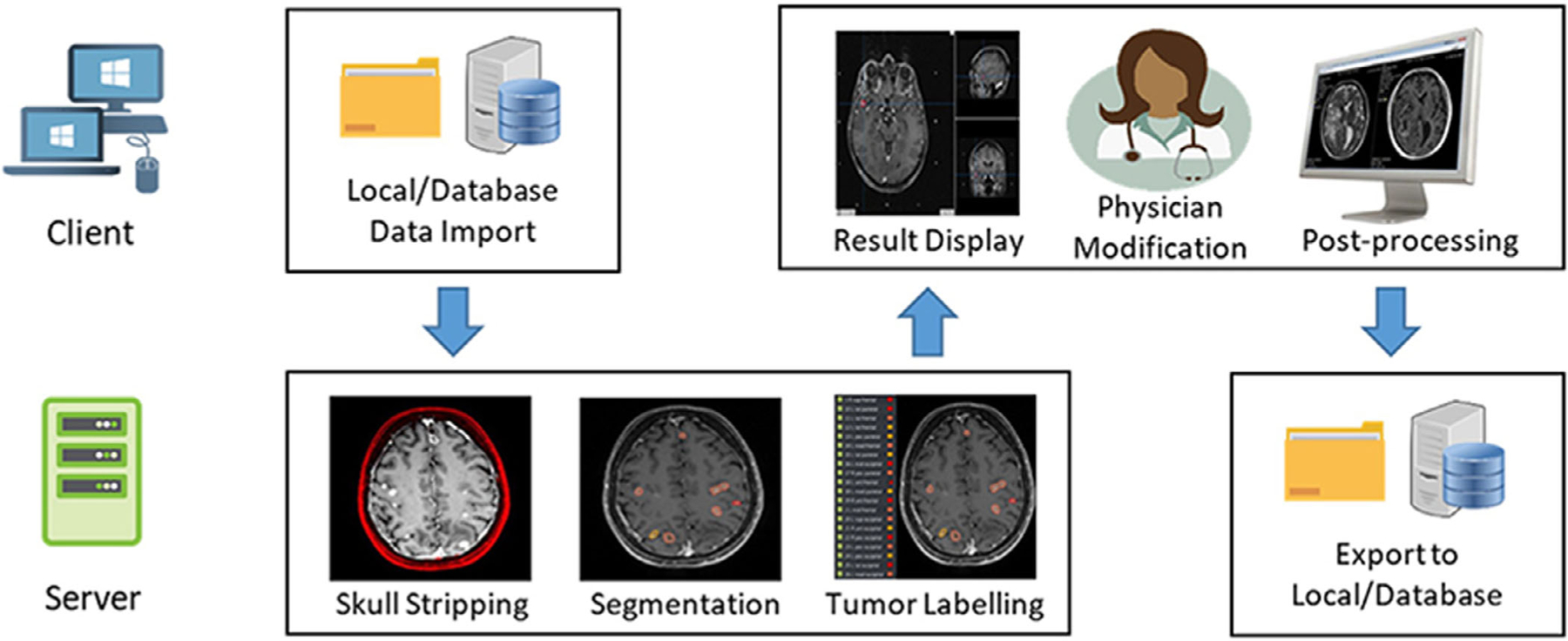
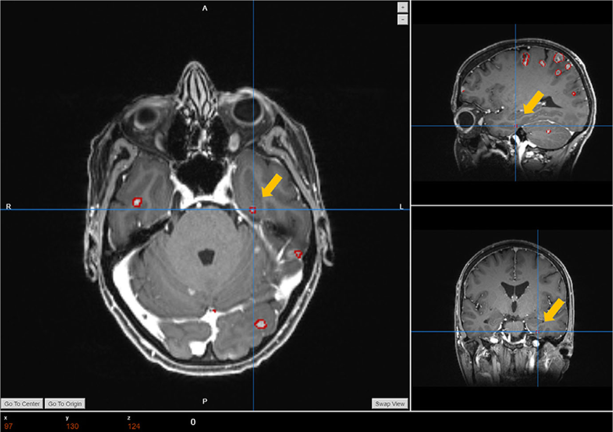
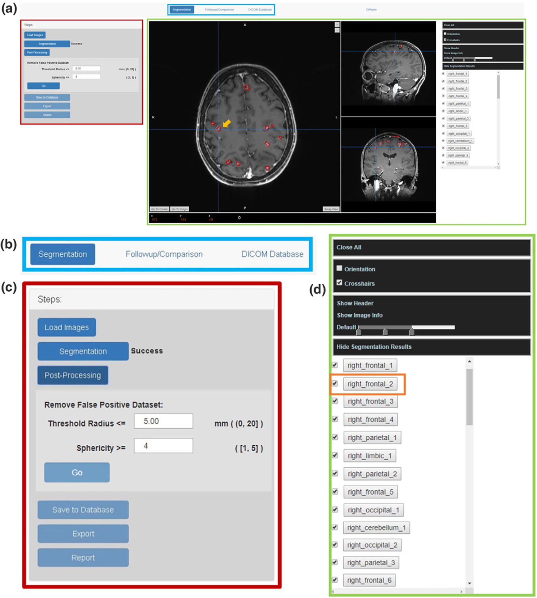
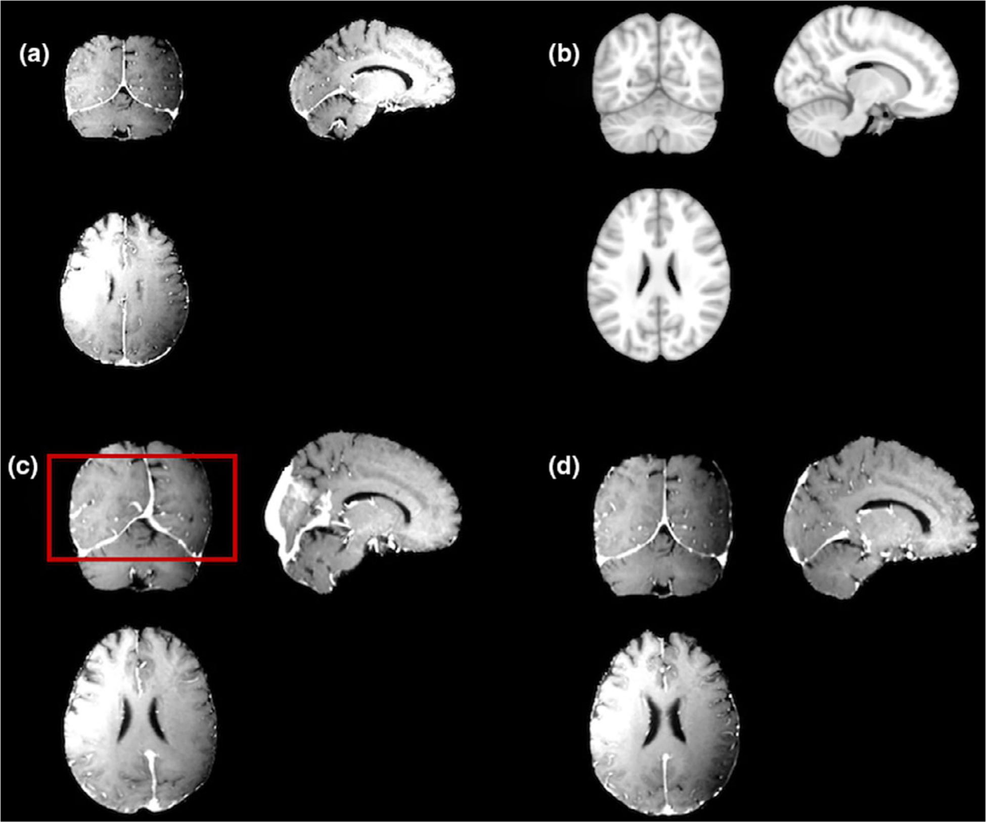
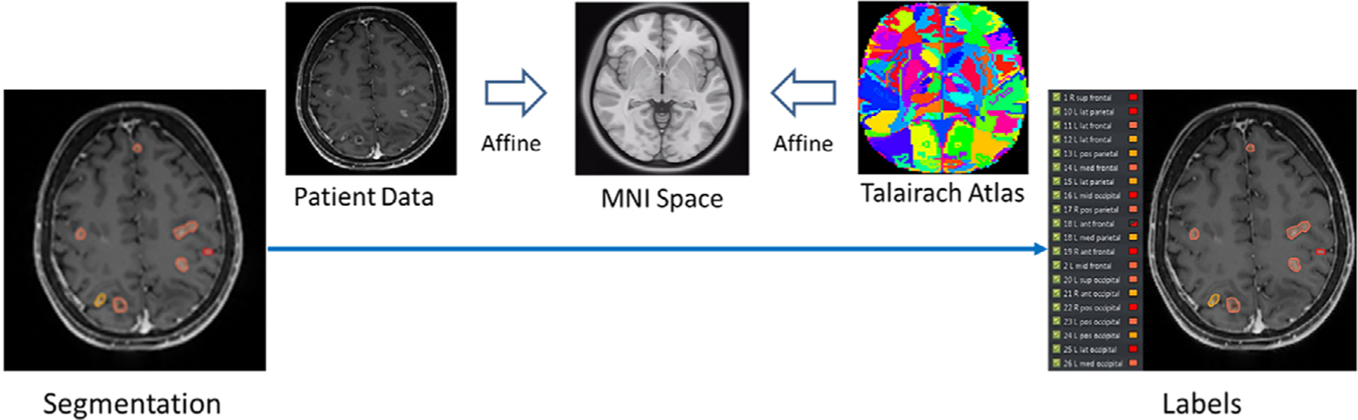
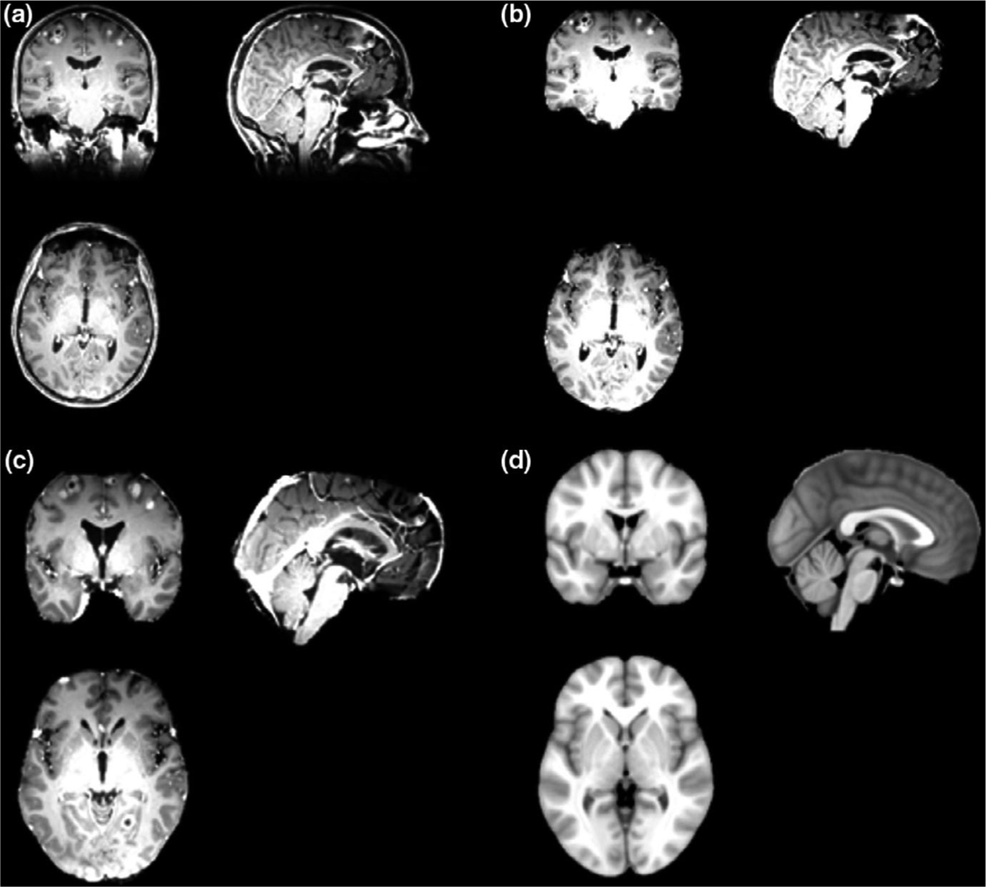
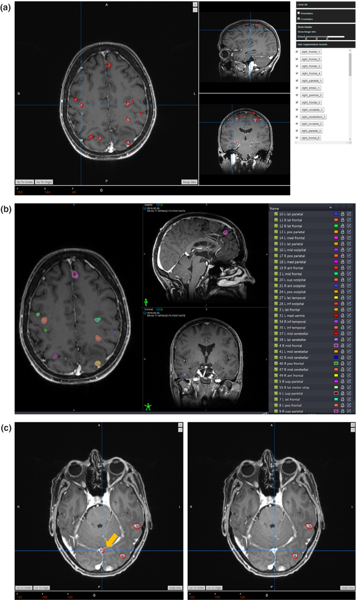
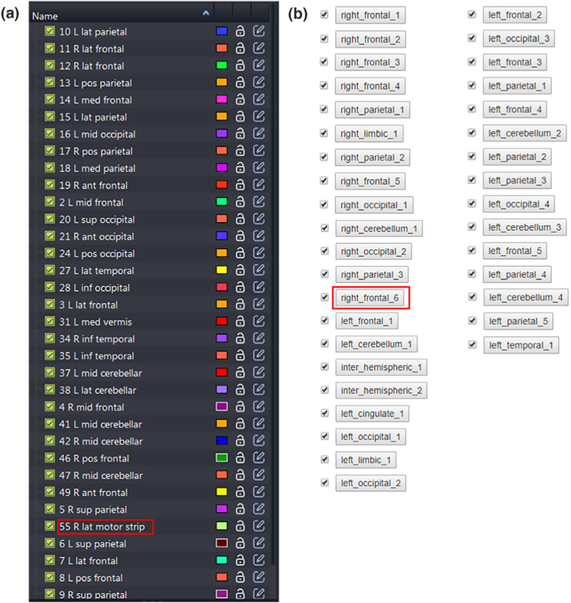
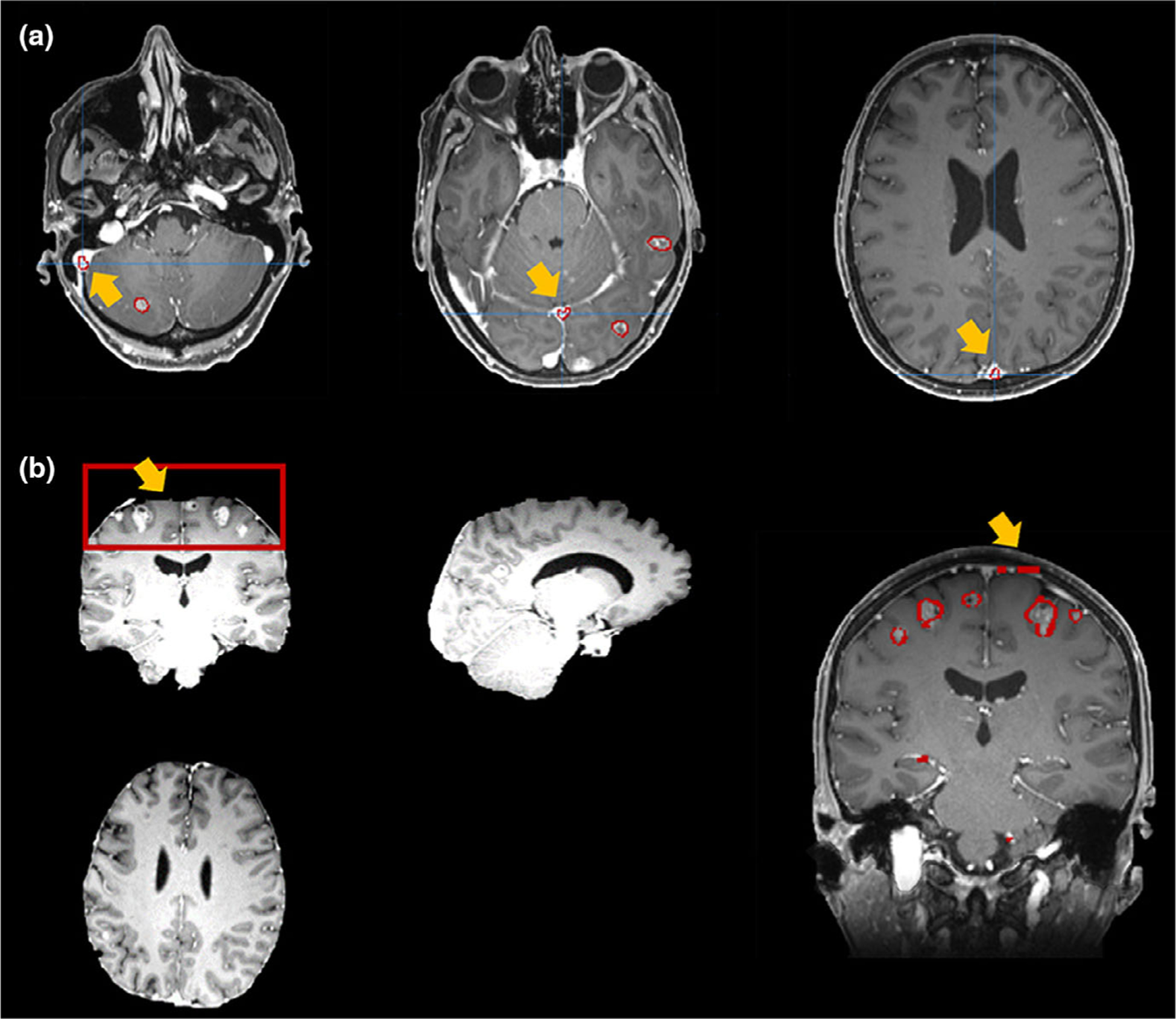
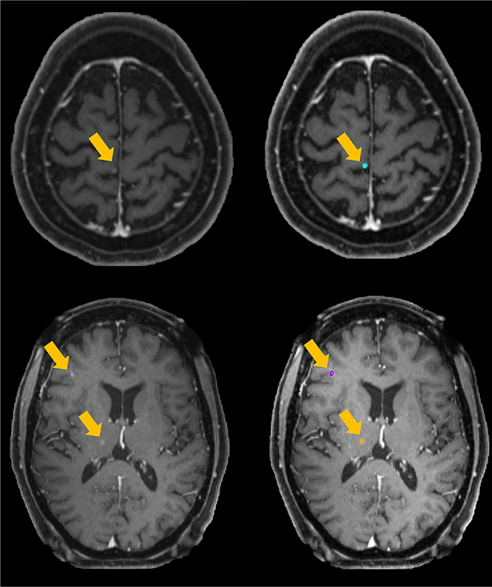
This platform was developed based on the Django framework, including a web client and a back-end server. The web client enables interactions as database access, data import, and image viewing. The server performs the segmentation and labeling tasks including: skull stripping; deep learning-based BMs segmentation; and affine registration-based BMs labeling. Additionally, the client can display BMs contours with corresponding atlas labels, and allows further postprocessing tasks including: (a) adjusting window levels; (b) displaying/hiding specific contours; (c) removing false-positive contours; (d) exporting contours as DICOM RTStruct files; etc. RESULTS: We evaluated this platform on 10 clinical cases with BMs number varied from 12-81 per case. The overall operation took about 4-5 min per patient. The segmentation accuracy was evaluated between the manual contour and automatic segmentation with several metrics. The averaged center of mass shift was 1.55 ± 0.36 mm, the Hausdorff distance was 2.98 ± 0.63 mm, the mean of surface-to-surface distance (SSD) was 1.06 ± 0.31 mm, and the standard deviation of SSD was 0.80 ± 0.16 mm. In addition, the initial averaged false-positive over union (FPoU) and false-negative rate (FNR) were 0.43 ± 0.19 and 0.15 ± 0.10 respectively. After case-specific postprocessing, the averaged FPoU and FNR were 0.19 ± 0.10 and 0.15 ± 0.10 respectively.

CONCLUSION

The evaluated web-based BMs segmentation and labeling platform can substantially improve the clinical efficiency compared to manual contouring. This platform can be a useful tool for assisting SRS treatment planning and treatment follow-up.

摘要

目的

立体定向放射外科(SRS)已成为脑转移瘤(BMs)患者的标准治疗方法。然而,手动勾画多个 BMs 较为耗时,可能会成为 SRS 工作流程中的效率瓶颈。因此,我们需要自动勾画和定量评估工具。在本研究中,我们在之前开发的基于深度学习的分割算法的基础上,开发了一个基于网络的自动 BMs 分割和标注平台,以辅助 SRS 临床工作流程。

方法

该平台基于 Django 框架开发,包括一个网络客户端和一个后端服务器。网络客户端实现了数据库访问、数据导入和图像查看等交互功能。服务器执行分割和标注任务,包括:颅骨剥离;基于深度学习的 BMs 分割;基于仿射配准的 BMs 标注。此外,客户端可以显示带有相应图谱标签的 BMs 轮廓,并允许进行进一步的后处理任务,包括:(a)调整窗宽;(b)显示/隐藏特定轮廓;(c)去除假阳性轮廓;(d)将轮廓导出为 DICOM RTStruct 文件等。

结果

我们在 10 例 BMs 数量从 12 到 81 不等的临床病例上评估了该平台。每位患者的整体操作时间约为 4-5 分钟。我们使用几种指标评估了手动轮廓和自动分割之间的分割准确性。平均质心偏移量为 1.55 ± 0.36mm,Hausdorff 距离为 2.98 ± 0.63mm,平均表面到表面距离(SSD)为 1.06 ± 0.31mm,SSD 标准差为 0.80 ± 0.16mm。此外,初始平均假阳性率(FPoU)和假阴性率(FNR)分别为 0.43 ± 0.19 和 0.15 ± 0.10。经过特定病例的后处理后,平均 FPoU 和 FNR 分别为 0.19 ± 0.10 和 0.15 ± 0.10。

结论

与手动勾画相比,评估的基于网络的 BMs 分割和标注平台可以显著提高临床效率。该平台可以成为辅助 SRS 治疗计划和治疗随访的有用工具。

https://cdn.ncbi.nlm.nih.gov/pmc/blobs/1d83/7567132/1198dfe579c1/nihms-1635861-f0010.jpg
https://cdn.ncbi.nlm.nih.gov/pmc/blobs/1d83/7567132/5c0f1f384279/nihms-1635861-f0001.jpg
https://cdn.ncbi.nlm.nih.gov/pmc/blobs/1d83/7567132/62ea2552cab8/nihms-1635861-f0002.jpg
https://cdn.ncbi.nlm.nih.gov/pmc/blobs/1d83/7567132/a69bed187802/nihms-1635861-f0003.jpg
https://cdn.ncbi.nlm.nih.gov/pmc/blobs/1d83/7567132/010970cbab0c/nihms-1635861-f0004.jpg
https://cdn.ncbi.nlm.nih.gov/pmc/blobs/1d83/7567132/baf8942a8481/nihms-1635861-f0005.jpg
https://cdn.ncbi.nlm.nih.gov/pmc/blobs/1d83/7567132/597d11b7f373/nihms-1635861-f0006.jpg
https://cdn.ncbi.nlm.nih.gov/pmc/blobs/1d83/7567132/0cf9ecfe25a8/nihms-1635861-f0007.jpg
https://cdn.ncbi.nlm.nih.gov/pmc/blobs/1d83/7567132/4d378e4b3840/nihms-1635861-f0008.jpg
https://cdn.ncbi.nlm.nih.gov/pmc/blobs/1d83/7567132/436a28677198/nihms-1635861-f0009.jpg
https://cdn.ncbi.nlm.nih.gov/pmc/blobs/1d83/7567132/1198dfe579c1/nihms-1635861-f0010.jpg

相似文献

1
A web-based brain metastases segmentation and labeling platform for stereotactic radiosurgery.一种用于立体定向放射外科的基于网络的脑转移瘤分割和标注平台。
Med Phys. 2020 Aug;47(8):3263-3276. doi: 10.1002/mp.14201. Epub 2020 May 23.
2
Deep-learning and radiomics ensemble classifier for false positive reduction in brain metastases segmentation.深度学习和放射组学集成分类器在脑转移瘤分割中降低假阳性率。
Phys Med Biol. 2022 Jan 19;67(2). doi: 10.1088/1361-6560/ac4667.
3
Stratified assessment of an FDA-cleared deep learning algorithm for automated detection and contouring of metastatic brain tumors in stereotactic radiosurgery.基于深度学习算法的自动检测和勾画转移瘤在立体定向放射外科中的应用:一项 FDA 认证的研究
Radiat Oncol. 2023 Apr 4;18(1):61. doi: 10.1186/s13014-023-02246-z.
4
Automatic metastatic brain tumor segmentation for stereotactic radiosurgery applications.用于立体定向放射外科应用的自动转移性脑肿瘤分割
Phys Med Biol. 2016 Dec 21;61(24):8440-8461. doi: 10.1088/0031-9155/61/24/8440. Epub 2016 Nov 15.
5
A general algorithm for distributed treatments of multiple brain metastases.一种用于多发性脑转移瘤分布式治疗的通用算法。
Med Phys. 2021 Apr;48(4):1832-1838. doi: 10.1002/mp.14722. Epub 2021 Feb 22.
6
Randomized multi-reader evaluation of automated detection and segmentation of brain tumors in stereotactic radiosurgery with deep neural networks.利用深度神经网络对立体定向放射外科中脑肿瘤的自动检测与分割进行随机多读者评估。
Neuro Oncol. 2021 Sep 1;23(9):1560-1568. doi: 10.1093/neuonc/noab071.
7
A deep convolutional neural network-based automatic delineation strategy for multiple brain metastases stereotactic radiosurgery.一种基于深度卷积神经网络的多脑转移瘤立体定向放射外科自动勾画策略。
PLoS One. 2017 Oct 6;12(10):e0185844. doi: 10.1371/journal.pone.0185844. eCollection 2017.
8
Automatic segmentation of brain metastases using T1 magnetic resonance and computed tomography images.基于 T1 磁共振和计算机断层扫描图像的脑转移瘤自动分割。
Phys Med Biol. 2021 Aug 26;66(17). doi: 10.1088/1361-6560/ac1835.
9
Computer-based radiological longitudinal evaluation of meningiomas following stereotactic radiosurgery.基于计算机的脑膜瘤立体定向放射外科治疗后放射影像学纵向评估。
Int J Comput Assist Radiol Surg. 2018 Feb;13(2):215-228. doi: 10.1007/s11548-017-1673-7. Epub 2017 Oct 14.
10
Stereotactic laser ablation as treatment for brain metastases that recur after stereotactic radiosurgery: a multiinstitutional experience.立体定向激光消融术治疗立体定向放射治疗后复发的脑转移瘤:一项多机构经验。
Neurosurg Focus. 2016 Oct;41(4):E11. doi: 10.3171/2016.7.FOCUS16227.

引用本文的文献

1
Deep learning-based overall survival prediction in patients with glioblastoma: An automatic end-to-end workflow using pre-resection basic structural multiparametric MRIs.基于深度学习的胶质母细胞瘤患者总生存期预测:使用术前基本结构多参数磁共振成像的自动端到端工作流程
Comput Biol Med. 2025 Feb;185:109436. doi: 10.1016/j.compbiomed.2024.109436. Epub 2024 Dec 4.
2
Where Does Auto-Segmentation for Brain Metastases Radiosurgery Stand Today?脑转移瘤放射外科手术的自动分割技术目前处于什么水平?
Bioengineering (Basel). 2024 May 3;11(5):454. doi: 10.3390/bioengineering11050454.
3
Automated, fast, robust brain extraction on contrast-enhanced T1-weighted MRI in presence of brain tumors: an optimized model based on multi-center datasets.

本文引用的文献

1
Tracking Metastatic Brain Tumors in Longitudinal Scans via Joint Image Registration and Labeling.通过联合图像配准和标记在纵向扫描中追踪转移性脑肿瘤
Spatiotemporal Image Anal Longitud Time Ser Image Data (2012). 2012 Oct;7570:124-136. doi: 10.1007/978-3-642-33555-6_11.
2
Automatic detection and segmentation of brain metastases on multimodal MR images with a deep convolutional neural network.基于深度卷积神经网络的多模态磁共振图像脑转移瘤的自动检测与分割。
Comput Biol Med. 2018 Apr 1;95:43-54. doi: 10.1016/j.compbiomed.2018.02.004. Epub 2018 Feb 9.
3
A deep convolutional neural network-based automatic delineation strategy for multiple brain metastases stereotactic radiosurgery.
在存在脑肿瘤的情况下,对对比增强 T1 加权 MRI 进行自动、快速、稳健的脑提取:基于多中心数据集的优化模型。
Eur Radiol. 2024 Feb;34(2):1190-1199. doi: 10.1007/s00330-023-10078-4. Epub 2023 Aug 24.
4
Adaptive Region-Specific Loss for Improved Medical Image Segmentation.自适应区域特定损失函数提高医学图像分割。
IEEE Trans Pattern Anal Mach Intell. 2023 Nov;45(11):13408-13421. doi: 10.1109/TPAMI.2023.3289667. Epub 2023 Oct 4.
5
RU-Net: skull stripping in rat brain MR images after ischemic stroke with rat U-Net.RU-Net:基于大鼠 U-Net 的大鼠脑磁共振成像缺血性卒中后颅骨剥离。
BMC Med Imaging. 2023 Mar 27;23(1):44. doi: 10.1186/s12880-023-00994-8.
6
Ensemble learning for glioma patients overall survival prediction using pre-operative MRIs.基于术前 MRI 的胶质母细胞瘤患者总体生存预测的集成学习。
Phys Med Biol. 2022 Dec 6;67(24). doi: 10.1088/1361-6560/aca375.
7
Automated detection and quantification of brain metastases on clinical MRI data using artificial neural networks.使用人工神经网络在临床MRI数据上自动检测和量化脑转移瘤。
Neurooncol Adv. 2022 Aug 23;4(1):vdac138. doi: 10.1093/noajnl/vdac138. eCollection 2022 Jan-Dec.
8
Deep-learning and radiomics ensemble classifier for false positive reduction in brain metastases segmentation.深度学习和放射组学集成分类器在脑转移瘤分割中降低假阳性率。
Phys Med Biol. 2022 Jan 19;67(2). doi: 10.1088/1361-6560/ac4667.
9
Evaluation of auto-segmentation accuracy of cloud-based artificial intelligence and atlas-based models.基于云的人工智能和图谱模型的自动分割准确性评估。
Radiat Oncol. 2021 Sep 9;16(1):175. doi: 10.1186/s13014-021-01896-1.
10
Saliency-guided deep learning network for automatic tumor bed volume delineation in post-operative breast irradiation.基于显著度引导的深度学习网络用于术后乳房放疗中肿瘤靶区自动勾画。
Phys Med Biol. 2021 Aug 27;66(17). doi: 10.1088/1361-6560/ac176d.
一种基于深度卷积神经网络的多脑转移瘤立体定向放射外科自动勾画策略。
PLoS One. 2017 Oct 6;12(10):e0185844. doi: 10.1371/journal.pone.0185844. eCollection 2017.
4
Efficient multi-scale 3D CNN with fully connected CRF for accurate brain lesion segmentation.高效多尺度 3D CNN 结合全连接条件随机场实现精准脑损伤分割。
Med Image Anal. 2017 Feb;36:61-78. doi: 10.1016/j.media.2016.10.004. Epub 2016 Oct 29.
5
Automatic metastatic brain tumor segmentation for stereotactic radiosurgery applications.用于立体定向放射外科应用的自动转移性脑肿瘤分割
Phys Med Biol. 2016 Dec 21;61(24):8440-8461. doi: 10.1088/0031-9155/61/24/8440. Epub 2016 Nov 15.
6
Optimization of Treatment Geometry to Reduce Normal Brain Dose in Radiosurgery of Multiple Brain Metastases with Single-Isocenter Volumetric Modulated Arc Therapy.采用单等中心容积调强弧形放疗优化治疗几何结构以降低多发脑转移瘤放射外科治疗中的正常脑剂量
Sci Rep. 2016 Sep 30;6:34511. doi: 10.1038/srep34511.
7
ISLES 2015 - A public evaluation benchmark for ischemic stroke lesion segmentation from multispectral MRI.ISLES 2015 - 多光谱 MRI 缺血性脑卒中病灶分割的公共评估基准。
Med Image Anal. 2017 Jan;35:250-269. doi: 10.1016/j.media.2016.07.009. Epub 2016 Jul 21.
8
A framework for automated contour quality assurance in radiation therapy including adaptive techniques.一种用于放射治疗中自动轮廓质量保证的框架,包括自适应技术。
Phys Med Biol. 2015 Jul 7;60(13):5199-209. doi: 10.1088/0031-9155/60/13/5199. Epub 2015 Jun 17.
9
Comparison of plan quality and delivery time between volumetric arc therapy (RapidArc) and Gamma Knife radiosurgery for multiple cranial metastases.容积弧形调强放疗(快速弧形放疗)与伽玛刀放射外科治疗多发性脑转移瘤的计划质量和治疗时间比较
Neurosurgery. 2014 Oct;75(4):409-17; discussion 417-8. doi: 10.1227/NEU.0000000000000448.
10
Accuracy quantification of a deformable image registration tool applied in a clinical setting.应用于临床环境的形变图像配准工具的准确性量化。
J Appl Clin Med Phys. 2014 Jan 6;15(1):4564. doi: 10.1120/jacmp.v15i1.4564.