• 文献检索
  • 文档翻译
  • 深度研究
  • 学术资讯
  • Suppr Zotero 插件Zotero 插件
  • 邀请有礼
  • 套餐&价格
  • 历史记录
应用&插件
Suppr Zotero 插件Zotero 插件浏览器插件Mac 客户端Windows 客户端微信小程序
定价
高级版会员购买积分包购买API积分包
服务
文献检索文档翻译深度研究API 文档MCP 服务
关于我们
关于 Suppr公司介绍联系我们用户协议隐私条款
关注我们

Suppr 超能文献

核心技术专利:CN118964589B侵权必究
粤ICP备2023148730 号-1Suppr @ 2026

文献检索

告别复杂PubMed语法,用中文像聊天一样搜索,搜遍4000万医学文献。AI智能推荐,让科研检索更轻松。

立即免费搜索

文件翻译

保留排版,准确专业,支持PDF/Word/PPT等文件格式,支持 12+语言互译。

免费翻译文档

深度研究

AI帮你快速写综述,25分钟生成高质量综述,智能提取关键信息,辅助科研写作。

立即免费体验

基于深度学习算法的自动检测和勾画转移瘤在立体定向放射外科中的应用:一项 FDA 认证的研究

Stratified assessment of an FDA-cleared deep learning algorithm for automated detection and contouring of metastatic brain tumors in stereotactic radiosurgery.

机构信息

Department of Radiation Oncology, Stanford University School of Medicine, 875 Blake Wilbur Drive, Stanford, CA, 94305, USA.

Vysioneer Inc, Cambridge, MA, USA.

出版信息

Radiat Oncol. 2023 Apr 4;18(1):61. doi: 10.1186/s13014-023-02246-z.

DOI:10.1186/s13014-023-02246-z
PMID:37016416
原文链接:https://pmc.ncbi.nlm.nih.gov/articles/PMC10074777/
Abstract

PURPOSE

Artificial intelligence-based tools can be leveraged to improve detection and segmentation of brain metastases for stereotactic radiosurgery (SRS). VBrain by Vysioneer Inc. is a deep learning algorithm with recent FDA clearance to assist in brain tumor contouring. We aimed to assess the performance of this tool by various demographic and clinical characteristics among patients with brain metastases treated with SRS.

MATERIALS AND METHODS

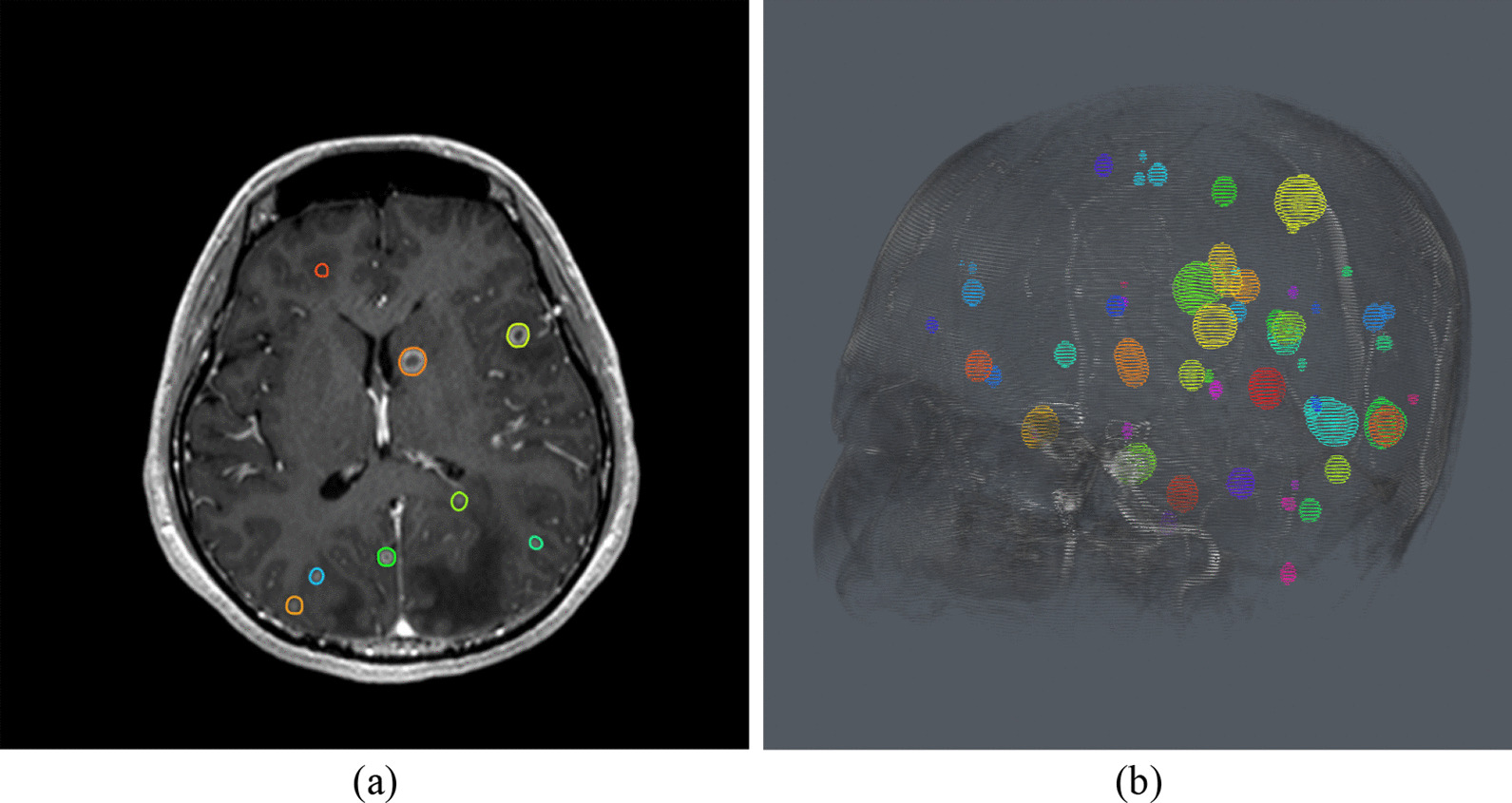
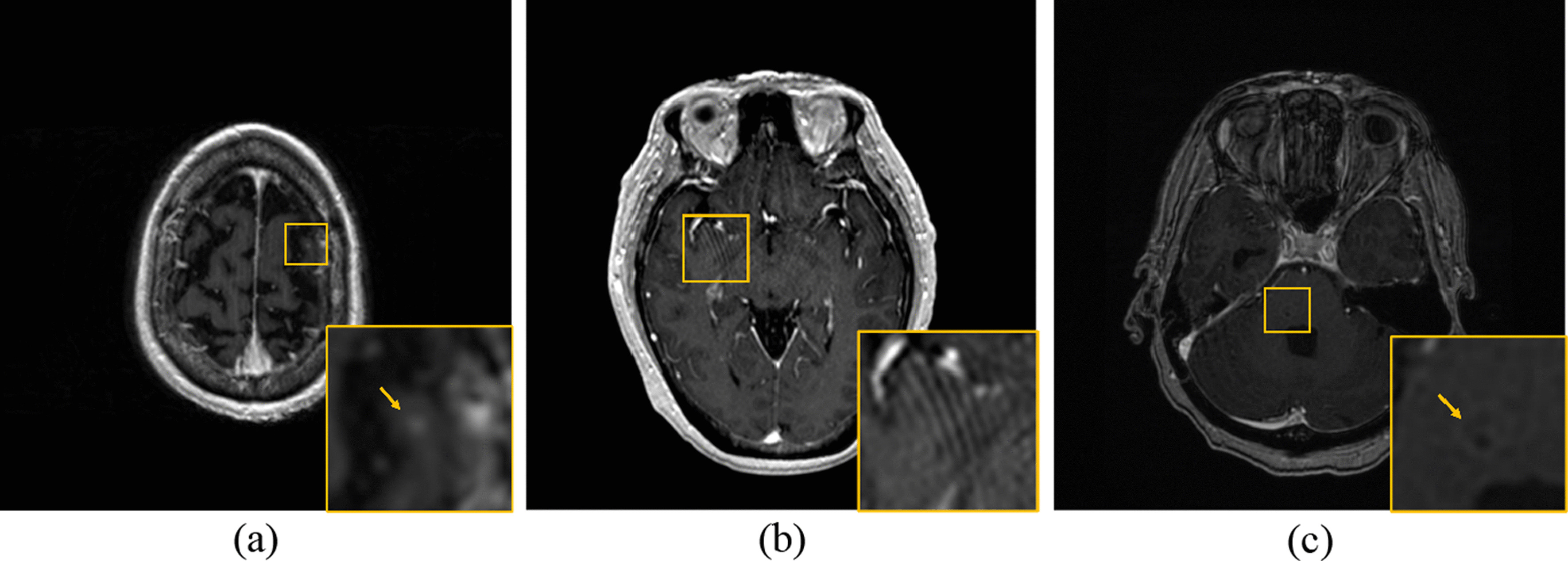
We randomly selected 100 patients with brain metastases who underwent initial SRS on the CyberKnife from 2017 to 2020 at a single institution. Cases with resection cavities were excluded from the analysis. Computed tomography (CT) and axial T1-weighted post-contrast magnetic resonance (MR) image data were extracted for each patient and uploaded to VBrain. A brain metastasis was considered "detected" when the VBrain- "predicted" contours overlapped with the corresponding physician contours ("ground-truth" contours). We evaluated performance of VBrain against ground-truth contours using the following metrics: lesion-wise Dice similarity coefficient (DSC), lesion-wise average Hausdorff distance (AVD), false positive count (FP), and lesion-wise sensitivity (%). Kruskal-Wallis tests were performed to assess the relationships between patient characteristics including sex, race, primary histology, age, and size and number of brain metastases, and performance metrics such as DSC, AVD, FP, and sensitivity.

RESULTS

We analyzed 100 patients with 435 intact brain metastases treated with SRS. Our cohort consisted of patients with a median number of 2 brain metastases (range: 1 to 52), median age of 69 (range: 19 to 91), and 50% male and 50% female patients. The primary site breakdown was 56% lung, 10% melanoma, 9% breast, 8% gynecological, 5% renal, 4% gastrointestinal, 2% sarcoma, and 6% other, while the race breakdown was 60% White, 18% Asian, 3% Black/African American, 2% Native Hawaiian or other Pacific Islander, and 17% other/unknown/not reported. The median tumor size was 0.112 c.c. (range: 0.010-26.475 c.c.). We found mean lesion-wise DSC to be 0.723, mean lesion-wise AVD to be 7.34% of lesion size (0.704 mm), mean FP count to be 0.72 tumors per case, and lesion-wise sensitivity to be 89.30% for all lesions. Moreover, mean sensitivity was found to be 99.07%, 97.59%, and 96.23% for lesions with diameter equal to and greater than 10 mm, 7.5 mm, and 5 mm, respectively. No other significant differences in performance metrics were observed across demographic or clinical characteristic groups.

CONCLUSION

In this study, a commercial deep learning algorithm showed promising results in segmenting brain metastases, with 96.23% sensitivity for metastases with diameters of 5 mm or higher. As the software is an assistive AI, future work of VBrain integration into the clinical workflow can provide further clinical and research insights.

摘要

目的

人工智能工具可用于提高立体定向放射外科(SRS)中脑转移瘤的检测和分割。Vysioneer 公司的 VBrain 是一种具有最近 FDA 批准的深度学习算法,可协助脑肿瘤勾画。我们旨在通过 SRS 治疗的脑转移患者的各种人口统计学和临床特征来评估该工具的性能。

材料和方法

我们随机选择了 2017 年至 2020 年在一家机构接受 CyberKnife 初始 SRS 的 100 例脑转移患者。排除了有切除术腔的病例。从每位患者中提取 CT 和轴向 T1 加权对比磁共振(MR)图像数据,并上传到 VBrain。当 VBrain-“预测”轮廓与相应医生的“地面真实”轮廓重叠时,我们认为脑转移瘤“被检测到”。我们使用以下指标评估 VBrain 对地面真实轮廓的性能:病变层面的 Dice 相似系数(DSC)、病变层面平均 Hausdorff 距离(AVD)、假阳性计数(FP)和病变层面灵敏度(%)。进行 Kruskal-Wallis 检验,以评估包括性别、种族、原发组织学、年龄以及脑转移瘤数量和大小等患者特征与 DSC、AVD、FP 和灵敏度等性能指标之间的关系。

结果

我们分析了 100 例接受 SRS 治疗的 435 个完整脑转移瘤患者。我们的队列包括中位数为 2 个脑转移瘤(范围:1-52)、中位数年龄为 69 岁(范围:19-91)、50%为男性和 50%为女性的患者。主要部位的分布为 56%为肺癌,10%为黑色素瘤,9%为乳腺癌,8%为妇科肿瘤,5%为肾癌,4%为胃肠道癌,2%为肉瘤,6%为其他,而种族分布为 60%为白人,18%为亚洲人,3%为黑人/非裔美国人,2%为夏威夷原住民或其他太平洋岛民,17%为其他/未知/未报告。肿瘤中位数大小为 0.112 c.c.(范围:0.010-26.475 c.c.)。我们发现平均病变层面 DSC 为 0.723,平均病变层面 AVD 为病变大小的 7.34%(0.704 mm),平均 FP 计数为每个病例 0.72 个肿瘤,病变层面灵敏度为所有病变的 89.30%。此外,对于直径等于或大于 10 mm、7.5 mm 和 5 mm 的病变,平均灵敏度分别为 99.07%、97.59%和 96.23%。在人口统计学或临床特征组中未观察到其他性能指标的显著差异。

结论

在这项研究中,一种商业深度学习算法在分割脑转移瘤方面表现出良好的效果,对于直径为 5 mm 或更大的转移瘤,灵敏度为 96.23%。由于该软件是一种辅助人工智能,因此未来 VBrain 集成到临床工作流程中可以提供进一步的临床和研究见解。

https://cdn.ncbi.nlm.nih.gov/pmc/blobs/8d58/10074777/e0bc9219a886/13014_2023_2246_Fig3_HTML.jpg
https://cdn.ncbi.nlm.nih.gov/pmc/blobs/8d58/10074777/1e6bf8e8ea24/13014_2023_2246_Fig1_HTML.jpg
https://cdn.ncbi.nlm.nih.gov/pmc/blobs/8d58/10074777/ba606ac656c8/13014_2023_2246_Fig2_HTML.jpg
https://cdn.ncbi.nlm.nih.gov/pmc/blobs/8d58/10074777/e0bc9219a886/13014_2023_2246_Fig3_HTML.jpg
https://cdn.ncbi.nlm.nih.gov/pmc/blobs/8d58/10074777/1e6bf8e8ea24/13014_2023_2246_Fig1_HTML.jpg
https://cdn.ncbi.nlm.nih.gov/pmc/blobs/8d58/10074777/ba606ac656c8/13014_2023_2246_Fig2_HTML.jpg
https://cdn.ncbi.nlm.nih.gov/pmc/blobs/8d58/10074777/e0bc9219a886/13014_2023_2246_Fig3_HTML.jpg

相似文献

1
Stratified assessment of an FDA-cleared deep learning algorithm for automated detection and contouring of metastatic brain tumors in stereotactic radiosurgery.基于深度学习算法的自动检测和勾画转移瘤在立体定向放射外科中的应用:一项 FDA 认证的研究
Radiat Oncol. 2023 Apr 4;18(1):61. doi: 10.1186/s13014-023-02246-z.
2
Randomized multi-reader evaluation of automated detection and segmentation of brain tumors in stereotactic radiosurgery with deep neural networks.利用深度神经网络对立体定向放射外科中脑肿瘤的自动检测与分割进行随机多读者评估。
Neuro Oncol. 2021 Sep 1;23(9):1560-1568. doi: 10.1093/neuonc/noab071.
3
Automatic segmentation of brain metastases using T1 magnetic resonance and computed tomography images.基于 T1 磁共振和计算机断层扫描图像的脑转移瘤自动分割。
Phys Med Biol. 2021 Aug 26;66(17). doi: 10.1088/1361-6560/ac1835.
4
A web-based brain metastases segmentation and labeling platform for stereotactic radiosurgery.一种用于立体定向放射外科的基于网络的脑转移瘤分割和标注平台。
Med Phys. 2020 Aug;47(8):3263-3276. doi: 10.1002/mp.14201. Epub 2020 May 23.
5
Computer-aided Detection of Brain Metastases in T1-weighted MRI for Stereotactic Radiosurgery Using Deep Learning Single-Shot Detectors.基于深度学习单发探测器的 T1 加权 MRI 立体定向放射手术中脑转移瘤的计算机辅助检测。
Radiology. 2020 May;295(2):407-415. doi: 10.1148/radiol.2020191479. Epub 2020 Mar 17.
6
MetNet: Computer-aided segmentation of brain metastases in post-contrast T1-weighted magnetic resonance imaging.MetNet:基于对比增强 T1 加权磁共振成像的脑转移瘤计算机辅助分割。
Radiother Oncol. 2020 Dec;153:189-196. doi: 10.1016/j.radonc.2020.09.016. Epub 2020 Sep 13.
7
Survival after surgery and stereotactic radiosurgery for patients with multiple intracranial metastases: results of a single-center retrospective study.多发颅内转移患者手术后和立体定向放射外科治疗后的生存:一项单中心回顾性研究的结果。
J Neurosurg. 2014 Oct;121(4):839-45. doi: 10.3171/2014.4.JNS13789. Epub 2014 May 23.
8
Impact of 2-staged stereotactic radiosurgery for treatment of brain metastases ≥ 2 cm.2 期立体定向放射外科治疗 ≥ 2 cm 的脑转移瘤的影响。
J Neurosurg. 2018 Aug;129(2):366-382. doi: 10.3171/2017.3.JNS162532. Epub 2017 Sep 22.
9
Federated Learning: A Cross-Institutional Feasibility Study of Deep Learning Based Intracranial Tumor Delineation Framework for Stereotactic Radiosurgery.联邦学习:基于深度学习的立体定向放射外科颅内肿瘤勾画框架的跨机构可行性研究。
J Magn Reson Imaging. 2024 Jun;59(6):1967-1975. doi: 10.1002/jmri.28950. Epub 2023 Aug 12.
10
Potential role for LINAC-based stereotactic radiosurgery for the treatment of 5 or more radioresistant melanoma brain metastases.基于直线加速器的立体定向放射外科治疗5个或更多放射性抵抗性黑色素瘤脑转移瘤的潜在作用。
J Neurosurg. 2015 Nov;123(5):1261-7. doi: 10.3171/2014.12.JNS141919. Epub 2015 Jul 3.

引用本文的文献

1
AI-Driven Innovations in Neuroradiology and Neurosurgery: Scoping Review of Current Evidence and Future Directions.人工智能驱动的神经放射学与神经外科创新:当前证据与未来方向的范围综述
Cancers (Basel). 2025 Aug 11;17(16):2625. doi: 10.3390/cancers17162625.
2
A comprehensive multifaceted technical evaluation framework for implementation of auto-segmentation models in radiotherapy.放疗中自动分割模型实施的综合多方面技术评估框架。
Commun Med (Lond). 2025 Jul 31;5(1):319. doi: 10.1038/s43856-025-01048-6.
3
A critical assessment of artificial intelligence in magnetic resonance imaging of cancer.

本文引用的文献

1
Deep-learning and radiomics ensemble classifier for false positive reduction in brain metastases segmentation.深度学习和放射组学集成分类器在脑转移瘤分割中降低假阳性率。
Phys Med Biol. 2022 Jan 19;67(2). doi: 10.1088/1361-6560/ac4667.
2
A new prognostic score for predicting survival in patients treated with robotic stereotactic radiotherapy for brain metastases.一种用于预测接受机器人立体定向放疗治疗脑转移瘤的患者生存情况的新预后评分。
Sci Rep. 2021 Oct 13;11(1):20347. doi: 10.1038/s41598-021-98847-3.
3
MRI pulse sequence integration for deep-learning-based brain metastases segmentation.
人工智能在癌症磁共振成像中的批判性评估。
Npj Imaging. 2025;3(1):15. doi: 10.1038/s44303-025-00076-0. Epub 2025 Apr 9.
4
Envisioning the Future of the Neurosurgical Operating Room with the Concept of the Medical Metaverse.用医学元宇宙概念展望神经外科手术室的未来。
J Korean Neurosurg Soc. 2025 Mar;68(2):137-149. doi: 10.3340/jkns.2024.0160. Epub 2024 Nov 4.
5
Novel radiotherapeutic strategies in the management of brain metastases: Challenging the dogma.新型放射治疗策略在脑转移瘤治疗中的应用:挑战传统观念。
Neuro Oncol. 2024 Mar 4;26(12 Suppl 2):S46-S55. doi: 10.1093/neuonc/noad260.
6
Artificial intelligence in neurology: opportunities, challenges, and policy implications.人工智能在神经科学中的应用:机遇、挑战与政策影响。
J Neurol. 2024 May;271(5):2258-2273. doi: 10.1007/s00415-024-12220-8. Epub 2024 Feb 17.
7
Advancing equitable and personalized cancer care: Novel applications and priorities of artificial intelligence for fairness and inclusivity in the patient care workflow.推进公平和个性化的癌症护理:人工智能在患者护理工作流程中的公平性和包容性方面的新应用和优先事项。
Eur J Cancer. 2024 Feb;198:113504. doi: 10.1016/j.ejca.2023.113504. Epub 2023 Dec 19.
8
Novel research and future prospects of artificial intelligence in cancer diagnosis and treatment.人工智能在癌症诊断和治疗中的新研究与未来展望。
J Hematol Oncol. 2023 Nov 27;16(1):114. doi: 10.1186/s13045-023-01514-5.
9
Segmentation of Brain Metastases Using Background Layer Statistics (BLAST).基于背景层统计信息的脑转移瘤分割(BLAST)。
AJNR Am J Neuroradiol. 2023 Oct;44(10):1135-1143. doi: 10.3174/ajnr.A7998. Epub 2023 Sep 21.
10
The Brain Tumor Segmentation - Metastases (BraTS-METS) Challenge 2023: Brain Metastasis Segmentation on Pre-treatment MRI.2023年脑肿瘤分割 - 转移瘤(BraTS-METS)挑战赛:治疗前MRI上的脑转移瘤分割
ArXiv. 2024 Dec 9:arXiv:2306.00838v3.
基于深度学习的脑转移瘤分割的 MRI 脉冲序列集成。
Med Phys. 2021 Oct;48(10):6020-6035. doi: 10.1002/mp.15136. Epub 2021 Aug 18.
4
The Stanford stereotactic radiosurgery experience on 7000 patients over 2 decades (1999-2018): looking far beyond the scalpel.斯坦福大学在20年(1999年至2018年)间对7000例患者进行立体定向放射外科手术的经验:超越手术刀的深远探索。
J Neurosurg. 2021 Apr 2;135(6):1725-1741. doi: 10.3171/2020.9.JNS201484. Print 2021 Dec 1.
5
Randomized multi-reader evaluation of automated detection and segmentation of brain tumors in stereotactic radiosurgery with deep neural networks.利用深度神经网络对立体定向放射外科中脑肿瘤的自动检测与分割进行随机多读者评估。
Neuro Oncol. 2021 Sep 1;23(9):1560-1568. doi: 10.1093/neuonc/noab071.
6
Automatic detection and segmentation of multiple brain metastases on magnetic resonance image using asymmetric UNet architecture.使用非对称 UNet 架构自动检测和分割磁共振图像上的多个脑转移瘤。
Phys Med Biol. 2021 Jan 13;66(1):015003. doi: 10.1088/1361-6560/abca53.
7
Tumor Primary Site and Histology Subtypes Role in Radiotherapeutic Management of Brain Metastases.肿瘤原发部位和组织学亚型在脑转移瘤放射治疗管理中的作用。
Front Oncol. 2020 Jul 7;10:781. doi: 10.3389/fonc.2020.00781. eCollection 2020.
8
Advanced Imaging of Brain Metastases: From Augmenting Visualization and Improving Diagnosis to Evaluating Treatment Response.脑转移瘤的高级成像:从增强可视化和改善诊断到评估治疗反应
Front Neurol. 2020 Apr 15;11:270. doi: 10.3389/fneur.2020.00270. eCollection 2020.
9
Deep convolutional neural networks for automated segmentation of brain metastases trained on clinical data.基于临床数据训练的用于脑转移瘤自动分割的深度卷积神经网络。
Radiat Oncol. 2020 Apr 20;15(1):87. doi: 10.1186/s13014-020-01514-6.
10
Consensus recommendations for a standardized brain tumor imaging protocol for clinical trials in brain metastases.脑转移瘤临床试验中标准化脑肿瘤成像方案的共识建议。
Neuro Oncol. 2020 Jun 9;22(6):757-772. doi: 10.1093/neuonc/noaa030.