• 文献检索
  • 文档翻译
  • 深度研究
  • 学术资讯
  • Suppr Zotero 插件Zotero 插件
  • 邀请有礼
  • 套餐&价格
  • 历史记录
应用&插件
Suppr Zotero 插件Zotero 插件浏览器插件Mac 客户端Windows 客户端微信小程序
定价
高级版会员购买积分包购买API积分包
服务
文献检索文档翻译深度研究API 文档MCP 服务
关于我们
关于 Suppr公司介绍联系我们用户协议隐私条款
关注我们

Suppr 超能文献

核心技术专利:CN118964589B侵权必究
粤ICP备2023148730 号-1Suppr @ 2026

文献检索

告别复杂PubMed语法,用中文像聊天一样搜索,搜遍4000万医学文献。AI智能推荐,让科研检索更轻松。

立即免费搜索

文件翻译

保留排版,准确专业,支持PDF/Word/PPT等文件格式,支持 12+语言互译。

免费翻译文档

深度研究

AI帮你快速写综述,25分钟生成高质量综述,智能提取关键信息,辅助科研写作。

立即免费体验

RU-Net:基于大鼠 U-Net 的大鼠脑磁共振成像缺血性卒中后颅骨剥离。

RU-Net: skull stripping in rat brain MR images after ischemic stroke with rat U-Net.

机构信息

Computational Biomedical Engineering Laboratory (CBEL), Department of Engineering Science and Ocean Engineering, National Taiwan University, No. 1 Sec. 4 Roosevelt Road, Daan, Taipei, 10617, Taiwan.

Department of Neurology and Stroke Center, National Taiwan University Hospital, Taipei, 10002, Taiwan.

出版信息

BMC Med Imaging. 2023 Mar 27;23(1):44. doi: 10.1186/s12880-023-00994-8.

DOI:10.1186/s12880-023-00994-8
PMID:36973775
原文链接:https://pmc.ncbi.nlm.nih.gov/articles/PMC10045128/
Abstract

BACKGROUND

Experimental ischemic stroke models play a fundamental role in interpreting the mechanism of cerebral ischemia and appraising the development of pathological extent. An accurate and automatic skull stripping tool for rat brain image volumes with magnetic resonance imaging (MRI) are crucial in experimental stroke analysis. Due to the deficiency of reliable rat brain segmentation methods and motivated by the demand for preclinical studies, this paper develops a new skull stripping algorithm to extract the rat brain region in MR images after stroke, which is named Rat U-Net (RU-Net).

METHODS

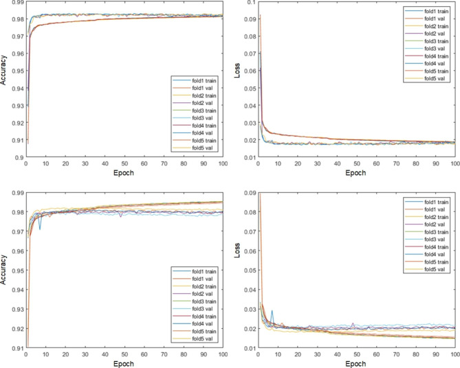
Based on a U-shape like deep learning architecture, the proposed framework integrates batch normalization with the residual network to achieve efficient end-to-end segmentation. A pooling index transmission mechanism between the encoder and decoder is exploited to reinforce the spatial correlation. Two different modalities of diffusion-weighted imaging (DWI) and T2-weighted MRI (T2WI) corresponding to two in-house datasets with each consisting of 55 subjects were employed to evaluate the performance of the proposed RU-Net.

RESULTS

Extensive experiments indicated great segmentation accuracy across diversified rat brain MR images. It was suggested that our rat skull stripping network outperformed several state-of-the-art methods and achieved the highest average Dice scores of 98.04% (p < 0.001) and 97.67% (p < 0.001) in the DWI and T2WI image datasets, respectively.

CONCLUSION

The proposed RU-Net is believed to be potential for advancing preclinical stroke investigation and providing an efficient tool for pathological rat brain image extraction, where accurate segmentation of the rat brain region is fundamental.

摘要

背景

实验性缺血性中风模型在解释脑缺血机制和评估病理程度发展方面起着重要作用。磁共振成像(MRI)大鼠脑图像具有准确、自动的颅骨剥离工具对于实验性中风分析至关重要。由于缺乏可靠的大鼠脑分割方法,并且受到临床前研究的需求的推动,本文开发了一种新的颅骨剥离算法,用于提取中风后大鼠脑磁共振图像的区域,命名为 Rat U-Net(RU-Net)。

方法

基于 U 形深度学习架构,该框架将批量归一化与残差网络集成在一起,以实现高效的端到端分割。利用编码器和解码器之间的池索引传输机制来增强空间相关性。两个不同的弥散加权成像(DWI)和 T2 加权磁共振成像(T2WI)模态对应于两个内部数据集,每个数据集由 55 个对象组成,用于评估所提出的 RU-Net 的性能。

结果

广泛的实验表明,在多样化的大鼠脑磁共振图像中具有出色的分割精度。建议我们的大鼠颅骨剥离网络优于几种最先进的方法,并在 DWI 和 T2WI 图像数据集上分别获得了 98.04%(p<0.001)和 97.67%(p<0.001)的平均 Dice 分数。

结论

所提出的 RU-Net 有望推进临床前中风研究,并为病理大鼠脑图像提取提供有效的工具,其中大鼠脑区域的准确分割是基础。

https://cdn.ncbi.nlm.nih.gov/pmc/blobs/9cbd/10045128/b57238ce0077/12880_2023_994_Fig11_HTML.jpg
https://cdn.ncbi.nlm.nih.gov/pmc/blobs/9cbd/10045128/86f8540920ac/12880_2023_994_Fig1_HTML.jpg
https://cdn.ncbi.nlm.nih.gov/pmc/blobs/9cbd/10045128/33d8c143436f/12880_2023_994_Fig2_HTML.jpg
https://cdn.ncbi.nlm.nih.gov/pmc/blobs/9cbd/10045128/fdb308f6f4ea/12880_2023_994_Fig3_HTML.jpg
https://cdn.ncbi.nlm.nih.gov/pmc/blobs/9cbd/10045128/33c46d613029/12880_2023_994_Fig4_HTML.jpg
https://cdn.ncbi.nlm.nih.gov/pmc/blobs/9cbd/10045128/0245df4028a4/12880_2023_994_Fig5_HTML.jpg
https://cdn.ncbi.nlm.nih.gov/pmc/blobs/9cbd/10045128/a343401fbafe/12880_2023_994_Fig6_HTML.jpg
https://cdn.ncbi.nlm.nih.gov/pmc/blobs/9cbd/10045128/b2bf21d1972f/12880_2023_994_Fig7_HTML.jpg
https://cdn.ncbi.nlm.nih.gov/pmc/blobs/9cbd/10045128/caffbf339fb7/12880_2023_994_Fig8_HTML.jpg
https://cdn.ncbi.nlm.nih.gov/pmc/blobs/9cbd/10045128/c0bf2e5cc73d/12880_2023_994_Fig9_HTML.jpg
https://cdn.ncbi.nlm.nih.gov/pmc/blobs/9cbd/10045128/c7691de015be/12880_2023_994_Fig10_HTML.jpg
https://cdn.ncbi.nlm.nih.gov/pmc/blobs/9cbd/10045128/b57238ce0077/12880_2023_994_Fig11_HTML.jpg

相似文献

1
RU-Net: skull stripping in rat brain MR images after ischemic stroke with rat U-Net.RU-Net:基于大鼠 U-Net 的大鼠脑磁共振成像缺血性卒中后颅骨剥离。
BMC Med Imaging. 2023 Mar 27;23(1):44. doi: 10.1186/s12880-023-00994-8.
2
Automatic brain extraction and hemisphere segmentation in rat brain MR images after stroke using deformable models.使用变形模型对脑卒中后大鼠脑磁共振图像进行自动脑提取和半球分割。
Med Phys. 2021 Oct;48(10):6036-6050. doi: 10.1002/mp.15157. Epub 2021 Sep 3.
3
Automated joint skull-stripping and segmentation with Multi-Task U-Net in large mouse brain MRI databases.基于多任务 U-Net 的大型鼠脑 MRI 数据库的自动关节颅骨剥离和分割。
Neuroimage. 2021 Apr 1;229:117734. doi: 10.1016/j.neuroimage.2021.117734. Epub 2021 Jan 14.
4
Automated Stroke Lesion Segmentation in Rat Brain MR Images Using an Encoder-Decoder Framework.使用编码器-解码器框架对大鼠脑磁共振图像进行自动中风病变分割
Annu Int Conf IEEE Eng Med Biol Soc. 2023 Jul;2023:1-4. doi: 10.1109/EMBC40787.2023.10340278.
5
Automatic Skull Stripping of Rat and Mouse Brain MRI Data Using U-Net.使用U-Net自动去除大鼠和小鼠脑MRI数据中的颅骨
Front Neurosci. 2020 Oct 7;14:568614. doi: 10.3389/fnins.2020.568614. eCollection 2020.
6
Robust skull stripping using multiple MR image contrasts insensitive to pathology.使用对病变不敏感的多个磁共振图像对比度进行稳健的颅骨剥离。
Neuroimage. 2017 Feb 1;146:132-147. doi: 10.1016/j.neuroimage.2016.11.017. Epub 2016 Nov 15.
7
Fully automated whole brain segmentation from rat MRI scans with a convolutional neural network.使用卷积神经网络从大鼠磁共振成像扫描中进行全自动全脑分割。
J Neurosci Methods. 2024 May;405:110078. doi: 10.1016/j.jneumeth.2024.110078. Epub 2024 Feb 8.
8
Study of multistep Dense U-Net-based automatic segmentation for head MRI scans.基于多步密集U-Net的头部磁共振成像扫描自动分割研究
Med Phys. 2024 Mar;51(3):2230-2238. doi: 10.1002/mp.16824. Epub 2023 Nov 13.
9
Semantic segmentation guided detector for segmentation, classification, and lesion mapping of acute ischemic stroke in MRI images.基于语义分割的引导检测器用于 MRI 图像中急性缺血性脑卒中的分割、分类和病灶定位。
Neuroimage Clin. 2022;35:103044. doi: 10.1016/j.nicl.2022.103044. Epub 2022 May 12.
10
Evaluation of Diffusion Lesion Volume Measurements in Acute Ischemic Stroke Using Encoder-Decoder Convolutional Network.基于编解码器卷积网络的急性缺血性脑卒中弥散病变体积测量评估。
Stroke. 2019 Jun;50(6):1444-1451. doi: 10.1161/STROKEAHA.118.024261. Epub 2019 May 16.

引用本文的文献

1
Flood change detection model based on an improved U-net network and multi-head attention mechanism.基于改进的U-net网络和多头注意力机制的洪水变化检测模型
Sci Rep. 2025 Jan 26;15(1):3295. doi: 10.1038/s41598-025-87851-6.
2
Deep learning segmentation model for quantification of infarct size in pigs with myocardial ischemia/reperfusion.用于定量评估猪心肌缺血/再灌注梗死面积的深度学习分割模型。
Basic Res Cardiol. 2024 Dec;119(6):923-936. doi: 10.1007/s00395-024-01081-x. Epub 2024 Sep 30.
3
Fully automated whole brain segmentation from rat MRI scans with a convolutional neural network.

本文引用的文献

1
Automatic Cerebral Hemisphere Segmentation in Rat MRI with Ischemic Lesions via Attention-based Convolutional Neural Networks.基于注意力卷积神经网络的大鼠 MRI 脑梗死病变自动半球分割。
Neuroinformatics. 2023 Jan;21(1):57-70. doi: 10.1007/s12021-022-09607-1. Epub 2022 Sep 30.
2
Differences in brain activity between normal and diabetic rats under isoflurane anesthesia: a resting-state functional MRI study.正常和糖尿病大鼠在异氟烷麻醉下脑活动的差异:一项静息态功能磁共振成像研究。
BMC Med Imaging. 2022 Aug 4;22(1):136. doi: 10.1186/s12880-022-00867-6.
3
Seed-based test-retest reliability of resting state functional magnetic resonance imaging at 3T and 7T.
使用卷积神经网络从大鼠磁共振成像扫描中进行全自动全脑分割。
J Neurosci Methods. 2024 May;405:110078. doi: 10.1016/j.jneumeth.2024.110078. Epub 2024 Feb 8.
4
Deep learning-based automated lesion segmentation on mouse stroke magnetic resonance images.基于深度学习的小鼠脑卒中磁共振图像自动病灶分割。
Sci Rep. 2023 Aug 16;13(1):13341. doi: 10.1038/s41598-023-39826-8.
基于种子的静息态功能磁共振成像在 3T 和 7T 下的测试-重测信度。
Med Phys. 2021 Oct;48(10):5756-5764. doi: 10.1002/mp.15210. Epub 2021 Sep 14.
4
Multi-level Kronecker Convolutional Neural Network (ML-KCNN) for Glioma Segmentation from Multi-modal MRI Volumetric Data.多模态 MRI 体数据集上基于多层 Kronecker 卷积神经网络(ML-KCNN)的脑胶质瘤分割
J Digit Imaging. 2021 Aug;34(4):905-921. doi: 10.1007/s10278-021-00486-7. Epub 2021 Jul 29.
5
Hemorrhagic stroke lesion segmentation using a 3D U-Net with squeeze-and-excitation blocks.使用带挤压激励块的 3D U-Net 进行出血性中风病变分割。
Comput Med Imaging Graph. 2021 Jun;90:101908. doi: 10.1016/j.compmedimag.2021.101908. Epub 2021 Apr 14.
6
Computed tomography image reconstruction using stacked U-Net.使用堆叠 U-Net 进行计算机断层扫描图像重建。
Comput Med Imaging Graph. 2021 Jun;90:101920. doi: 10.1016/j.compmedimag.2021.101920. Epub 2021 Apr 20.
7
Automated joint skull-stripping and segmentation with Multi-Task U-Net in large mouse brain MRI databases.基于多任务 U-Net 的大型鼠脑 MRI 数据库的自动关节颅骨剥离和分割。
Neuroimage. 2021 Apr 1;229:117734. doi: 10.1016/j.neuroimage.2021.117734. Epub 2021 Jan 14.
8
Automatic Skull Stripping of Rat and Mouse Brain MRI Data Using U-Net.使用U-Net自动去除大鼠和小鼠脑MRI数据中的颅骨
Front Neurosci. 2020 Oct 7;14:568614. doi: 10.3389/fnins.2020.568614. eCollection 2020.
9
CAB U-Net: An end-to-end category attention boosting algorithm for segmentation.CAB U-Net:一种用于分割的端到端类别注意力增强算法。
Comput Med Imaging Graph. 2020 Sep;84:101764. doi: 10.1016/j.compmedimag.2020.101764. Epub 2020 Jul 11.
10
A web-based brain metastases segmentation and labeling platform for stereotactic radiosurgery.一种用于立体定向放射外科的基于网络的脑转移瘤分割和标注平台。
Med Phys. 2020 Aug;47(8):3263-3276. doi: 10.1002/mp.14201. Epub 2020 May 23.