Boston Children's Hospital, Harvard Medical School, Boston, MA, United States of America.
Brigham and Women's Hospital, Harvard Medical School, Boston, MA, United States of America.
PLoS One. 2020 Apr 29;15(4):e0232376. doi: 10.1371/journal.pone.0232376. eCollection 2020.
To develop and test a deep learning algorithm to automatically detect cortical tubers in magnetic resonance imaging (MRI), to explore the utility of deep learning in rare disorders with limited data, and to generate an open-access deep learning standalone application.
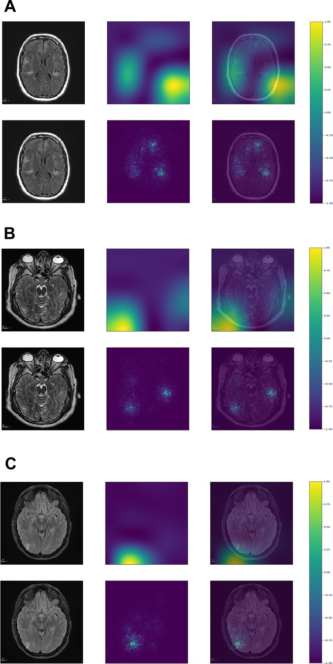
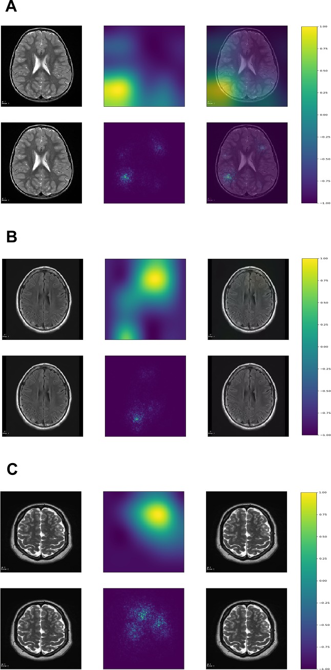
T2 and FLAIR axial images with and without tubers were extracted from MRIs of patients with tuberous sclerosis complex (TSC) and controls, respectively. We trained three different convolutional neural network (CNN) architectures on a training dataset and selected the one with the lowest binary cross-entropy loss in the validation dataset, which was evaluated on the testing dataset. We visualized image regions most relevant for classification with gradient-weighted class activation maps (Grad-CAM) and saliency maps.
114 patients with TSC and 114 controls were divided into a training set, a validation set, and a testing set. The InceptionV3 CNN architecture performed best in the validation set and was evaluated in the testing set with the following results: sensitivity: 0.95, specificity: 0.95, positive predictive value: 0.94, negative predictive value: 0.95, F1-score: 0.95, accuracy: 0.95, and area under the curve: 0.99. Grad-CAM and saliency maps showed that tubers resided in regions most relevant for image classification within each image. A stand-alone trained deep learning App was able to classify images using local computers with various operating systems.
This study shows that deep learning algorithms are able to detect tubers in selected MRI images, and deep learning can be prudently applied clinically to manually selected data in a rare neurological disorder.
开发和测试一种深度学习算法,以自动检测磁共振成像(MRI)中的皮质结节,探索深度学习在数据有限的罕见疾病中的应用,并生成一个开放获取的深度学习独立应用程序。
从结节性硬化症(TSC)患者和对照组的 MRI 中分别提取 T2 和 FLAIR 轴位图像,有和无结节。我们在训练数据集上训练了三种不同的卷积神经网络(CNN)架构,并在验证数据集上选择了二进制交叉熵损失最低的架构,然后在测试数据集上进行评估。我们使用梯度加权类激活图(Grad-CAM)和显著图可视化与分类最相关的图像区域。
114 例 TSC 患者和 114 例对照组患者被分为训练集、验证集和测试集。InceptionV3 CNN 架构在验证集上表现最好,并在测试集上进行了评估,结果如下:敏感性:0.95、特异性:0.95、阳性预测值:0.94、阴性预测值:0.95、F1 评分:0.95、准确性:0.95、曲线下面积:0.99。Grad-CAM 和显著图显示,结节位于每个图像中最相关的图像分类区域。一个独立训练的深度学习应用程序能够使用各种操作系统的本地计算机对图像进行分类。
本研究表明,深度学习算法能够检测选定 MRI 图像中的结节,并且可以谨慎地将深度学习应用于罕见神经疾病中手动选择的数据。