• 文献检索
  • 文档翻译
  • 深度研究
  • 学术资讯
  • Suppr Zotero 插件Zotero 插件
  • 邀请有礼
  • 套餐&价格
  • 历史记录
应用&插件
Suppr Zotero 插件Zotero 插件浏览器插件Mac 客户端Windows 客户端微信小程序
定价
高级版会员购买积分包购买API积分包
服务
文献检索文档翻译深度研究API 文档MCP 服务
关于我们
关于 Suppr公司介绍联系我们用户协议隐私条款
关注我们

Suppr 超能文献

核心技术专利:CN118964589B侵权必究
粤ICP备2023148730 号-1Suppr @ 2026

文献检索

告别复杂PubMed语法,用中文像聊天一样搜索,搜遍4000万医学文献。AI智能推荐,让科研检索更轻松。

立即免费搜索

文件翻译

保留排版,准确专业,支持PDF/Word/PPT等文件格式,支持 12+语言互译。

免费翻译文档

深度研究

AI帮你快速写综述,25分钟生成高质量综述,智能提取关键信息,辅助科研写作。

立即免费体验

使用可视化和文本解释来解释深度学习模型在脑胶质瘤生存分类中的应用。

Interpreting deep learning models for glioma survival classification using visualization and textual explanations.

机构信息

Department of Physics and Computational Radiology, Division of Radiology and Nuclear Medicine, Oslo University Hospital, Sognsvannsveien 20, 0372, Oslo, Norway.

Biomedical Data Science Laboratory,Instituto Universitario de Tecnologias de la Informacion Comunicaciones, Universitat Politècnica de València, 46022, Valencia, Spain.

出版信息

BMC Med Inform Decis Mak. 2023 Oct 18;23(1):225. doi: 10.1186/s12911-023-02320-2.

DOI:10.1186/s12911-023-02320-2
PMID:37853371
原文链接:https://pmc.ncbi.nlm.nih.gov/articles/PMC10583453/
Abstract

BACKGROUND

Saliency-based algorithms are able to explain the relationship between input image pixels and deep-learning model predictions. However, it may be difficult to assess the clinical value of the most important image features and the model predictions derived from the raw saliency map. This study proposes to enhance the interpretability of saliency-based deep learning model for survival classification of patients with gliomas, by extracting domain knowledge-based information from the raw saliency maps.

MATERIALS AND METHODS

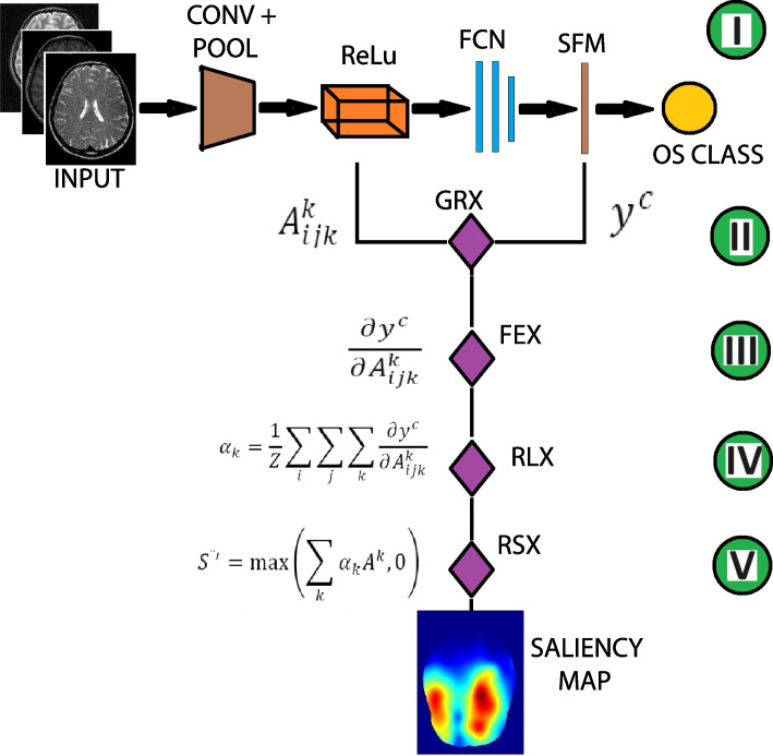
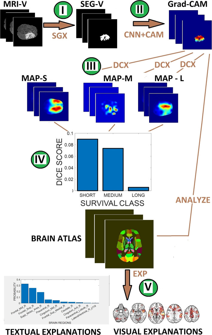
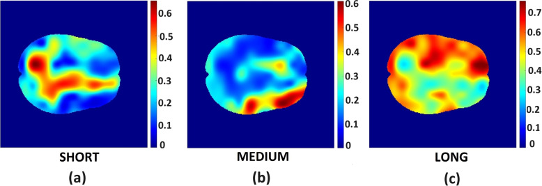
Our study includes presurgical T1-weighted (pre- and post-contrast), T2-weighted and T2-FLAIR MRIs of 147 glioma patients from the BraTs 2020 challenge dataset aligned to the SRI 24 anatomical atlas. Each image exam includes a segmentation mask and the information of overall survival (OS) from time of diagnosis (in days). This dataset was divided into training ([Formula: see text]) and validation ([Formula: see text]) datasets. The extent of surgical resection for all patients was gross total resection. We categorized the data into 42 short (mean [Formula: see text] days), 30 medium ([Formula: see text] days), and 46 long ([Formula: see text] days) survivors. A 3D convolutional neural network (CNN) trained on brain tumour MRI volumes classified all patients based on expected prognosis of either short-term, medium-term, or long-term survival. We extend the popular 2D Gradient-weighted Class Activation Mapping (Grad-CAM), for the generation of saliency map, to 3D and combined it with the anatomical atlas, to extract brain regions, brain volume and probability map that reveal domain knowledge-based information.

RESULTS

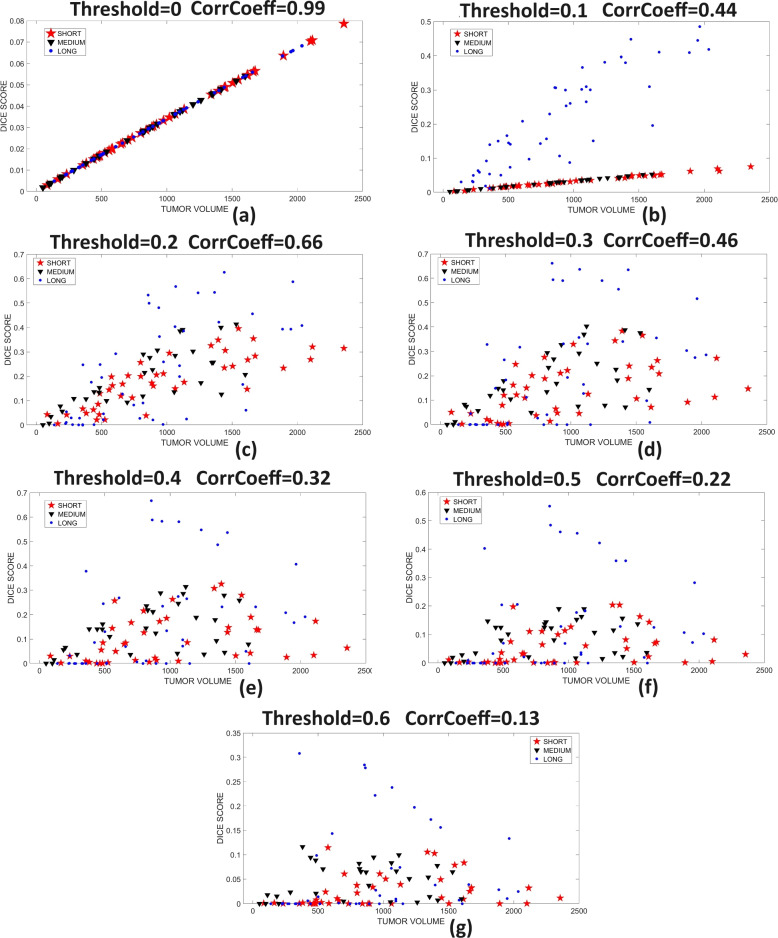
For each OS class, a larger tumor volume was associated with a shorter OS. There were 10, 7 and 27 tumor locations in brain regions that uniquely associate with the short-term, medium-term, and long-term survival, respectively. Tumors located in the transverse temporal gyrus, fusiform, and palladium are associated with short, medium and long-term survival, respectively. The visual and textual information displayed during OS prediction highlights tumor location and the contribution of different brain regions to the prediction of OS. This algorithm design feature assists the physician in analyzing and understanding different model prediction stages.

CONCLUSIONS

Domain knowledge-based information extracted from the saliency map can enhance the interpretability of deep learning models. Our findings show that tumors overlapping eloquent brain regions are associated with short patient survival.

摘要

背景

基于显著度的算法能够解释输入图像像素与深度学习模型预测之间的关系。然而,评估来自原始显著度图的最重要图像特征和模型预测的临床价值可能较为困难。本研究通过从原始显著度图中提取基于领域知识的信息,提出了一种增强基于显著度的深度学习模型在胶质瘤患者生存分类中的可解释性的方法。

材料和方法

我们的研究包括来自 BraTs 2020 挑战赛数据集的 147 名胶质瘤患者的术前 T1 加权(增强前后)、T2 加权和 T2-FLAIR MRI,这些患者与 SRI 24 解剖图谱对齐。每个图像检查包括一个分割掩模和从诊断时开始的总生存(OS)信息(以天为单位)。该数据集分为训练集([Formula: see text])和验证集([Formula: see text])。所有患者的手术切除范围均为大体全切除。我们将数据分为 42 名短期(平均 OS 时间为[Formula: see text]天)、30 名中期([Formula: see text]天)和 46 名长期([Formula: see text]天)生存者。一个基于脑肿瘤 MRI 体积的 3D 卷积神经网络(CNN)对所有患者进行分类,根据短期、中期或长期生存的预期预后进行分类。我们将流行的 2D 梯度加权类激活映射(Grad-CAM)扩展到 3D,并与解剖图谱相结合,以提取揭示基于领域知识的信息的脑区、脑体积和概率图。

结果

对于每个 OS 类别,较大的肿瘤体积与较短的 OS 相关。在与短期、中期和长期生存相关的脑区中,分别有 10、7 和 27 个肿瘤位置。位于横向颞叶、梭状回和钯的肿瘤分别与短期、中期和长期生存相关。在 OS 预测过程中显示的视觉和文本信息突出了肿瘤位置和不同脑区对 OS 预测的贡献。这种算法设计功能有助于医生分析和理解不同的模型预测阶段。

结论

从显著度图中提取的基于领域知识的信息可以增强深度学习模型的可解释性。我们的研究结果表明,与大脑功能区重叠的肿瘤与患者生存时间较短相关。

https://cdn.ncbi.nlm.nih.gov/pmc/blobs/6b15/10583453/cf368fc7a158/12911_2023_2320_Fig6_HTML.jpg
https://cdn.ncbi.nlm.nih.gov/pmc/blobs/6b15/10583453/51f533ebc08a/12911_2023_2320_Fig1_HTML.jpg
https://cdn.ncbi.nlm.nih.gov/pmc/blobs/6b15/10583453/1bd97c13f7b4/12911_2023_2320_Fig2_HTML.jpg
https://cdn.ncbi.nlm.nih.gov/pmc/blobs/6b15/10583453/d9a1a8a33cb9/12911_2023_2320_Fig3_HTML.jpg
https://cdn.ncbi.nlm.nih.gov/pmc/blobs/6b15/10583453/e4def27c95cd/12911_2023_2320_Fig4_HTML.jpg
https://cdn.ncbi.nlm.nih.gov/pmc/blobs/6b15/10583453/46704f6c76f1/12911_2023_2320_Fig5_HTML.jpg
https://cdn.ncbi.nlm.nih.gov/pmc/blobs/6b15/10583453/cf368fc7a158/12911_2023_2320_Fig6_HTML.jpg

相似文献

1
Interpreting deep learning models for glioma survival classification using visualization and textual explanations.使用可视化和文本解释来解释深度学习模型在脑胶质瘤生存分类中的应用。
BMC Med Inform Decis Mak. 2023 Oct 18;23(1):225. doi: 10.1186/s12911-023-02320-2.
2
Automated glioma grading on conventional MRI images using deep convolutional neural networks.使用深度卷积神经网络对传统MRI图像进行自动脑胶质瘤分级
Med Phys. 2020 Jul;47(7):3044-3053. doi: 10.1002/mp.14168. Epub 2020 May 11.
3
Deep learning in rare disease. Detection of tubers in tuberous sclerosis complex.深度学习在罕见病中的应用。结节性硬化症中结节的检测。
PLoS One. 2020 Apr 29;15(4):e0232376. doi: 10.1371/journal.pone.0232376. eCollection 2020.
4
DeepSeg: deep neural network framework for automatic brain tumor segmentation using magnetic resonance FLAIR images.DeepSeg:基于磁共振 FLAIR 图像的自动脑肿瘤分割的深度神经网络框架。
Int J Comput Assist Radiol Surg. 2020 Jun;15(6):909-920. doi: 10.1007/s11548-020-02186-z. Epub 2020 May 5.
5
What Does Deep Learning See? Insights From a Classifier Trained to Predict Contrast Enhancement Phase From CT Images.深度学习所见:从 CT 图像预测对比增强阶段的分类器训练所得到的见解。
AJR Am J Roentgenol. 2018 Dec;211(6):1184-1193. doi: 10.2214/AJR.18.20331. Epub 2018 Nov 7.
6
A deep dive into understanding tumor foci classification using multiparametric MRI based on convolutional neural network.基于卷积神经网络,深入探究利用多参数磁共振成像进行肿瘤病灶分类。
Med Phys. 2020 Sep;47(9):4077-4086. doi: 10.1002/mp.14255. Epub 2020 Jun 12.
7
Postoperative glioma segmentation in CT image using deep feature fusion model guided by multi-sequence MRIs.基于多序列 MRI 引导的深度特征融合模型的 CT 图像术后脑肿瘤分割。
Eur Radiol. 2020 Feb;30(2):823-832. doi: 10.1007/s00330-019-06441-z. Epub 2019 Oct 24.
8
fMRI volume classification using a 3D convolutional neural network robust to shifted and scaled neuronal activations.使用对移位和缩放神经元激活具有鲁棒性的 3D 卷积神经网络进行 fMRI 体积分类。
Neuroimage. 2020 Dec;223:117328. doi: 10.1016/j.neuroimage.2020.117328. Epub 2020 Sep 5.
9
An Intelligent Diagnosis Method of Brain MRI Tumor Segmentation Using Deep Convolutional Neural Network and SVM Algorithm.基于深度卷积神经网络和支持向量机算法的脑 MRI 肿瘤分割智能诊断方法。
Comput Math Methods Med. 2020 Jul 14;2020:6789306. doi: 10.1155/2020/6789306. eCollection 2020.
10
Saliency Map and Deep Learning in Binary Classification of Brain Tumours.显著图与深度学习在脑肿瘤二分类中的应用。
Sensors (Basel). 2023 May 7;23(9):4543. doi: 10.3390/s23094543.

引用本文的文献

1
Estimating overall survival of glioblastoma patients using clinical variables, tumor size, and location.利用临床变量、肿瘤大小和位置评估胶质母细胞瘤患者的总生存期。
Neurooncol Adv. 2025 Aug 3;7(1):vdaf154. doi: 10.1093/noajnl/vdaf154. eCollection 2025 Jan-Dec.
2
Predicting survival in malignant glioma using artificial intelligence.使用人工智能预测恶性胶质瘤的生存率。
Eur J Med Res. 2025 Jan 31;30(1):61. doi: 10.1186/s40001-025-02339-3.

本文引用的文献

1
Interpreting Deep Machine Learning Models: An Easy Guide for Oncologists.解读深度机器学习模型:肿瘤学家简易指南
IEEE Rev Biomed Eng. 2023;16:192-207. doi: 10.1109/RBME.2021.3131358. Epub 2023 Jan 5.
2
Explainable Deep Learning Models in Medical Image Analysis.医学图像分析中的可解释深度学习模型
J Imaging. 2020 Jun 20;6(6):52. doi: 10.3390/jimaging6060052.
3
The 2021 WHO Classification of Tumors of the Central Nervous System: a summary.2021 年世卫组织中枢神经系统肿瘤分类:概述。
Neuro Oncol. 2021 Aug 2;23(8):1231-1251. doi: 10.1093/neuonc/noab106.
4
A Multiparametric MRI-Based Radiomics Analysis to Efficiently Classify Tumor Subregions of Glioblastoma: A Pilot Study in Machine Learning.基于多参数磁共振成像的放射组学分析用于有效分类胶质母细胞瘤的肿瘤亚区域:机器学习的一项初步研究
J Clin Med. 2021 May 10;10(9):2030. doi: 10.3390/jcm10092030.
5
Survival of glioblastoma in relation to tumor location: a statistical tumor atlas of a population-based cohort.胶质母细胞瘤的生存与肿瘤位置的关系:基于人群队列的统计肿瘤图谱
Acta Neurochir (Wien). 2021 Jul;163(7):1895-1905. doi: 10.1007/s00701-021-04802-6. Epub 2021 Mar 20.
6
Predictive value of MGMT promoter methylation on the survival of TMZ treated -mutant glioblastoma.MGMT 启动子甲基化对替莫唑胺治疗的突变型胶质母细胞瘤患者生存的预测价值。
Cancer Biol Med. 2021 Feb 15;18(1):272-282. doi: 10.20892/j.issn.2095-3941.2020.0179.
7
Deep learning for glioblastoma segmentation using preoperative magnetic resonance imaging identifies volumetric features associated with survival.使用术前磁共振成像对胶质母细胞瘤进行分割的深度学习方法可以识别与生存相关的容积特征。
Acta Neurochir (Wien). 2020 Dec;162(12):3067-3080. doi: 10.1007/s00701-020-04483-7. Epub 2020 Jul 13.
8
On the Interpretability of Artificial Intelligence in Radiology: Challenges and Opportunities.论人工智能在放射学中的可解释性:挑战与机遇
Radiol Artif Intell. 2020 May 27;2(3):e190043. doi: 10.1148/ryai.2020190043.
9
MRI Atlas of IDH Wild-Type Supratentorial Glioblastoma: Probabilistic Maps of Phenotype, Management, and Outcomes.IDH 野生型幕上胶质母细胞瘤的 MRI 图谱:表型、治疗和预后的概率图。
Radiology. 2019 Dec;293(3):633-643. doi: 10.1148/radiol.2019190491. Epub 2019 Oct 8.
10
explAIner: A Visual Analytics Framework for Interactive and Explainable Machine Learning.Explainer:用于交互式和可解释机器学习的可视化分析框架。
IEEE Trans Vis Comput Graph. 2020 Jan;26(1):1064-1074. doi: 10.1109/TVCG.2019.2934629. Epub 2019 Aug 20.