Respiratory and Critical Care Center, Shanghai East Hospital Affiliated by Tongji University, China.
Respiratory Medical Center, First Hospital of Changsha, Hunan Province, China.
Respir Med. 2020 Jul;168:105989. doi: 10.1016/j.rmed.2020.105989. Epub 2020 Apr 22.
This retrospective study aims to illustrate the radiographic characteristics of Coronavirus Disease 2019 and the correlation with the clinical course.
195 hospitalized patients confirmed as Coronavirus Disease 2019 at First Hospital of Changsha, Hunan Province from December 31, 2019 to February 20, 2020 were enrolled. Chest computed tomography scan, clinical data and laboratory tests results were collected accordingly. Variable characteristics were recorded, radiographic evolution and outcome were analyzed along with the time course. Representative laboratory tests results were analyzed based on the image findings.
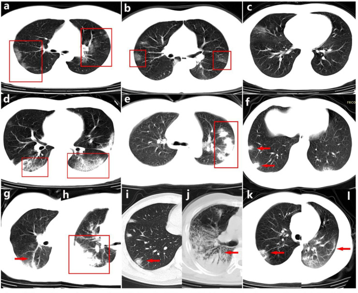
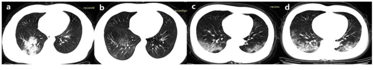
Majority of the patients showed bilateral (73.8%), multiple lobes involvements (75.9%), peripheral distribution (83.1%), ground-glass opacification (41.0%), increased vascular margins (63.1%), long axis parallelism (55.9%), patchy ground-glass opacities beneath the pleura (51.3%) and consolidation (45.6%). According to the repeated radiology analysis, patients of improving/stable group tended to have younger age compared with worsening group (45.3 ± 15.0 VS. 59.3 ± 13.5, P = 0.001). Based on the laboratory test results, patients with positive image findings shared elder age, 46.0 (35.0-60.0)VS.31.0 (12.0-37.0) P < 0.001, and higher chance developing fever(P < 0.05); higher level of lymphocytes, C-reactive protein, erythrocyte sedimentation rate and lactate dehydrogenase; lower level of white blood cells, neutrophil and albumin(P < 0.001).
There are several specific image changes along with the disease progression may be helpful in early recognition and differential diagnosis of Coronavirus Disease 2019. Comprehensive assessments of both imaging feature and laboratory test results may offer an intact knowledge of Coronavirus Disease 2019.
本回顾性研究旨在阐述 2019 年冠状病毒病的影像学特征及其与临床病程的相关性。
纳入 2019 年 12 月 31 日至 2020 年 2 月 20 日在湖南省长沙市第一医院确诊为 2019 年冠状病毒病的 195 例住院患者。收集胸部计算机断层扫描、临床资料和实验室检查结果。记录变量特征,分析放射学演变和结果随时间的变化。根据影像学表现分析有代表性的实验室检查结果。
大多数患者表现为双侧(73.8%)、多叶受累(75.9%)、外周分布(83.1%)、磨玻璃影(41.0%)、血管边缘增强(63.1%)、长轴平行(55.9%)、胸膜下斑片状磨玻璃影(51.3%)和实变(45.6%)。根据重复影像学分析,改善/稳定组患者的年龄较恶化组小(45.3±15.0 VS. 59.3±13.5,P=0.001)。根据实验室检查结果,有阳性影像学表现的患者年龄较大,46.0(35.0-60.0)VS.31.0(12.0-37.0),P<0.001,且更易发热(P<0.05);淋巴细胞、C 反应蛋白、红细胞沉降率和乳酸脱氢酶水平较高;白细胞、中性粒细胞和白蛋白水平较低(P<0.001)。
随着疾病的进展,有一些特定的影像学变化可能有助于早期识别和鉴别诊断 2019 年冠状病毒病。综合评估影像学特征和实验室检查结果可为 2019 年冠状病毒病提供全面的认识。