• 文献检索
  • 文档翻译
  • 深度研究
  • 学术资讯
  • Suppr Zotero 插件Zotero 插件
  • 邀请有礼
  • 套餐&价格
  • 历史记录
应用&插件
Suppr Zotero 插件Zotero 插件浏览器插件Mac 客户端Windows 客户端微信小程序
定价
高级版会员购买积分包购买API积分包
服务
文献检索文档翻译深度研究API 文档MCP 服务
关于我们
关于 Suppr公司介绍联系我们用户协议隐私条款
关注我们

Suppr 超能文献

核心技术专利:CN118964589B侵权必究
粤ICP备2023148730 号-1Suppr @ 2026

文献检索

告别复杂PubMed语法,用中文像聊天一样搜索,搜遍4000万医学文献。AI智能推荐,让科研检索更轻松。

立即免费搜索

文件翻译

保留排版,准确专业,支持PDF/Word/PPT等文件格式,支持 12+语言互译。

免费翻译文档

深度研究

AI帮你快速写综述,25分钟生成高质量综述,智能提取关键信息,辅助科研写作。

立即免费体验

具有六边形排列的视网膜下3D微电极的制造

Fabrication of Subretinal 3D Microelectrodes with Hexagonal Arrangement.

作者信息

Seo Hee Won, Kim Namju, Kim Sohee

机构信息

Department of Robotics Engineering, Daegu Gyeongbuk Institute of Science and Technology (DGIST), Daegu 42988, Korea.

出版信息

Micromachines (Basel). 2020 Apr 29;11(5):467. doi: 10.3390/mi11050467.

DOI:10.3390/mi11050467
PMID:32365472
原文链接:https://pmc.ncbi.nlm.nih.gov/articles/PMC7281732/
Abstract

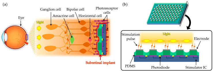
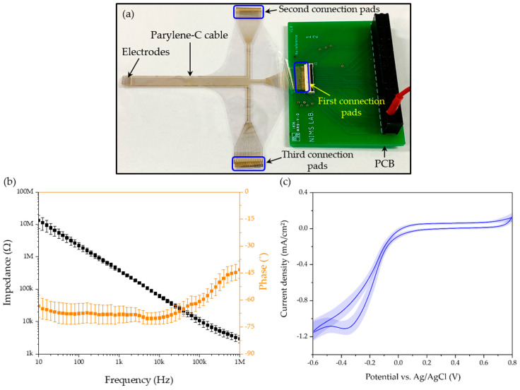
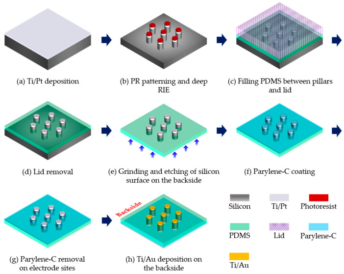
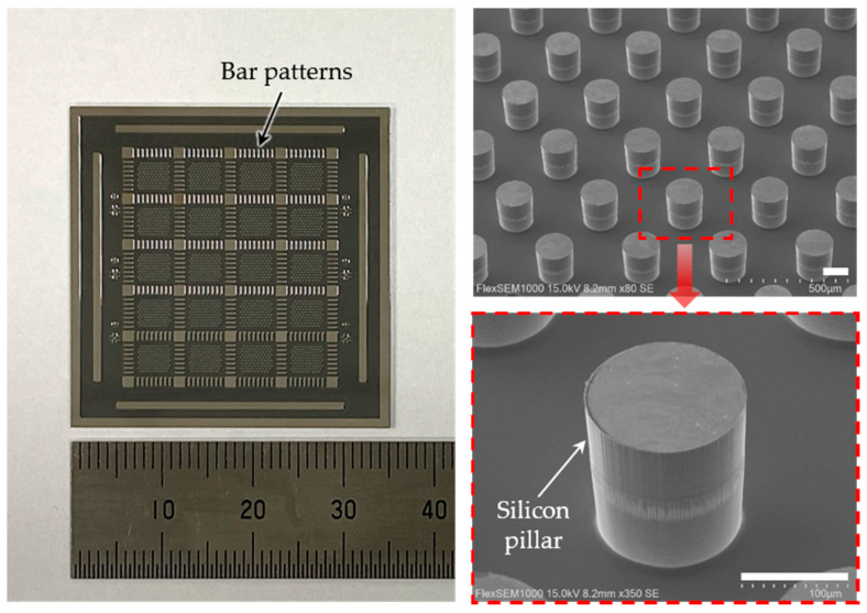
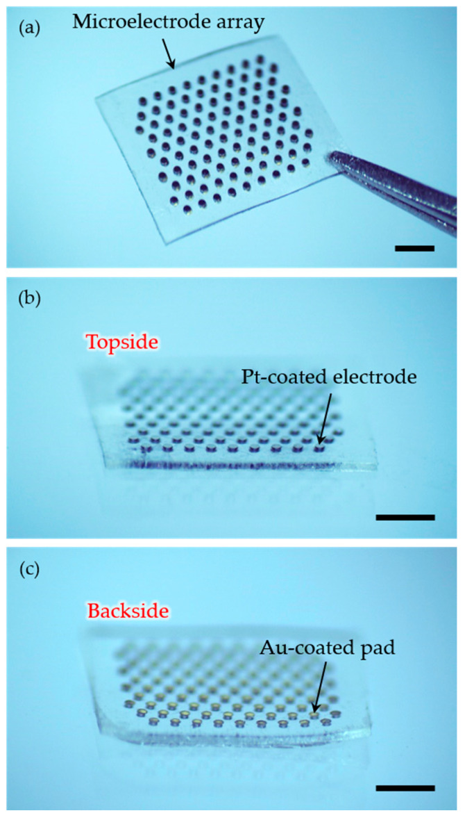
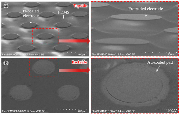
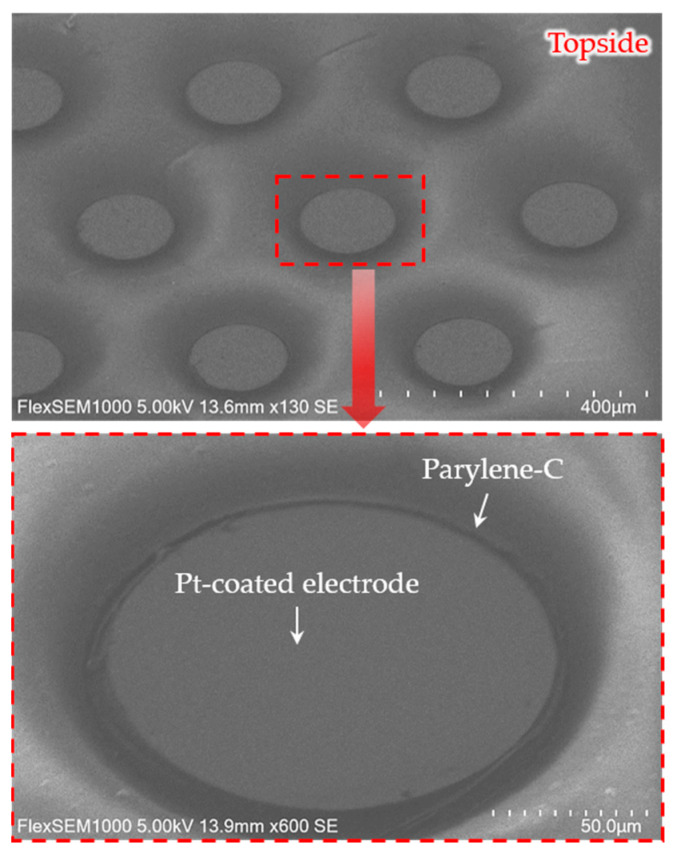
This study presents the fabrication of three-dimensional (3D) microelectrodes for subretinal stimulation, to accommodate adjacent return electrodes surrounding a stimulating electrode. For retinal prosthetic devices, the arrangement of return electrodes, the electrode size and spacing should be considered together, to reduce the undesired dissipation of electric currents. Here, we applied the hexagonal arrangement to the microelectrode array for the localized activation of retinal cells and better visual acuity. To provide stimuli more efficiently to non-spiking neurons, a 3D structure was created through a customized pressing process, utilizing the elastic property of the materials used in the fabrication processes. The diameter and pitch of the Pt-coated electrodes were 150 μm and 350 μm, respectively, and the height of the protruded electrodes was around 20 μm. The array consisted of 98 hexagonally arranged electrodes, supported by a flexible and transparent polydimethylsiloxane (PDMS) base, with a thickness of 140 μm. Also, the array was coated with 2 μm-thick parylene-C, except the active electrode sites, for more focused stimulation. Finally, the electrochemical properties of the fabricated microelectrodes were characterized, resulting in the mean impedance of 384.87 kΩ at 1 kHz and the charge storage capacity (CSC) of 2.83 mC·cm. The fabricated microelectrodes are to be combined with an integrated circuit (IC) for additional in vitro and in vivo experiments.

摘要

本研究展示了用于视网膜下刺激的三维(3D)微电极的制造方法,以容纳围绕刺激电极的相邻返回电极。对于视网膜假体装置,应综合考虑返回电极的排列、电极尺寸和间距,以减少电流的不必要耗散。在此,我们将六边形排列应用于微电极阵列,以实现视网膜细胞的局部激活并提高视力。为了更有效地向非尖峰神经元提供刺激,利用制造过程中所用材料的弹性特性,通过定制的压制工艺创建了三维结构。涂有铂的电极直径和间距分别为150μm和350μm,突出电极的高度约为20μm。该阵列由98个六边形排列的电极组成,由厚度为140μm的柔性透明聚二甲基硅氧烷(PDMS)基底支撑。此外,除了有源电极部位外,阵列还涂有2μm厚的聚对二甲苯-C,以实现更聚焦的刺激。最后,对制造的微电极的电化学特性进行了表征,在1kHz时平均阻抗为384.87kΩ,电荷存储容量(CSC)为2.83mC·cm 。制造的微电极将与集成电路(IC)结合,用于进一步的体外和体内实验。

https://cdn.ncbi.nlm.nih.gov/pmc/blobs/666d/7281732/e087a939d471/micromachines-11-00467-g007.jpg
https://cdn.ncbi.nlm.nih.gov/pmc/blobs/666d/7281732/b5b636d55b38/micromachines-11-00467-g001.jpg
https://cdn.ncbi.nlm.nih.gov/pmc/blobs/666d/7281732/629bf32916de/micromachines-11-00467-g002.jpg
https://cdn.ncbi.nlm.nih.gov/pmc/blobs/666d/7281732/823b61ac6b79/micromachines-11-00467-g003.jpg
https://cdn.ncbi.nlm.nih.gov/pmc/blobs/666d/7281732/13f544d2210c/micromachines-11-00467-g004.jpg
https://cdn.ncbi.nlm.nih.gov/pmc/blobs/666d/7281732/0e9e5d1fada1/micromachines-11-00467-g005.jpg
https://cdn.ncbi.nlm.nih.gov/pmc/blobs/666d/7281732/a6c7966df5d9/micromachines-11-00467-g006.jpg
https://cdn.ncbi.nlm.nih.gov/pmc/blobs/666d/7281732/e087a939d471/micromachines-11-00467-g007.jpg

相似文献

1
Fabrication of Subretinal 3D Microelectrodes with Hexagonal Arrangement.具有六边形排列的视网膜下3D微电极的制造
Micromachines (Basel). 2020 Apr 29;11(5):467. doi: 10.3390/mi11050467.
2
A 3D flexible microelectrode array for subretinal stimulation.用于视网膜下刺激的三维柔性微电极阵列。
J Neural Eng. 2019 Aug 21;16(5):056016. doi: 10.1088/1741-2552/ab36ab.
3
Focal stimulation of retinal ganglion cells using subretinal 3D microelectrodes with peripheral electrodes of opposite current.使用带有反向电流外周电极的视网膜下3D微电极对视网膜神经节细胞进行局部刺激。
Biomed Eng Lett. 2023 Dec 26;14(2):355-365. doi: 10.1007/s13534-023-00342-3. eCollection 2024 Mar.
4
Electrochemical characteristics of microelectrode designed for electrical stimulation.用于电刺激的微电极的电化学特性。
Biomed Eng Online. 2019 Aug 1;18(1):86. doi: 10.1186/s12938-019-0704-8.
5
Bottom-up SiO2 embedded carbon nanotube electrodes with superior performance for integration in implantable neural microsystems.用于植入式神经微系统集成的具有优异性能的自下而上 SiO2 嵌入碳纳米管电极。
ACS Nano. 2012 Jun 26;6(6):4615-28. doi: 10.1021/nn201609u. Epub 2012 May 21.
6
PDMS based multielectrode arrays for superior in-vitro retinal stimulation and recording.基于 PDMS 的多电极阵列,可实现卓越的体外视网膜刺激和记录。
Biomed Microdevices. 2017 Aug 25;19(4):75. doi: 10.1007/s10544-017-0221-0.
7
A double-sided fabrication process for intrafascicular parylene C based electrode arrays.一种用于束内聚对二甲苯C基电极阵列的双面制造工艺。
Annu Int Conf IEEE Eng Med Biol Soc. 2016 Aug;2016:2798-2801. doi: 10.1109/EMBC.2016.7591311.
8
Implantable nanostructured MEA with biphasic current stimulator for retinal prostheses.用于视网膜假体的具有双相电流刺激器的植入式纳米结构 MEAs。
Technol Health Care. 2023;31(5):1981-1995. doi: 10.3233/THC-235001.
9
Structural changes in the retina after implantation of subretinal three-dimensional implants in mini pigs.小型猪视网膜下植入三维植入物后视网膜的结构变化
Front Neurosci. 2022 Sep 30;16:1010445. doi: 10.3389/fnins.2022.1010445. eCollection 2022.
10
PEDOT-CNT coated electrodes stimulate retinal neurons at low voltage amplitudes and low charge densities.聚(3,4-乙撑二氧噻吩)-碳纳米管涂层电极在低电压幅度和低电荷密度下刺激视网膜神经元。
J Neural Eng. 2015 Feb;12(1):016014. doi: 10.1088/1741-2560/12/1/016014. Epub 2015 Jan 14.

引用本文的文献

1
Advances in Electrode Design and Physiological Considerations for Retinal Implants.视网膜植入物的电极设计进展及生理考量
Micromachines (Basel). 2025 May 21;16(5):598. doi: 10.3390/mi16050598.
2
3D electronic implants in subretinal space: Long-term follow-up in rodents.3D 电子植入物在视网膜下腔:啮齿动物的长期随访。
Biomaterials. 2024 Dec;311:122674. doi: 10.1016/j.biomaterials.2024.122674. Epub 2024 Jun 17.
3
Flexible high-density microelectrode arrays for closed-loop brain-machine interfaces: a review.用于闭环脑机接口的柔性高密度微电极阵列:综述

本文引用的文献

1
Improved visual acuity using a retinal implant and an optimized stimulation strategy.使用视网膜植入物和优化的刺激策略提高视力。
J Neural Eng. 2019 Dec 23;17(1):016018. doi: 10.1088/1741-2552/ab5299.
2
Protuberant Electrode Structures for Subretinal Electrical Stimulation: Modeling, Fabrication and Evaluation.用于视网膜下电刺激的突出电极结构:建模、制造与评估
Front Neurosci. 2019 Aug 27;13:885. doi: 10.3389/fnins.2019.00885. eCollection 2019.
3
The very large electrode array for retinal stimulation (VLARS)-A concept study.
Front Neurosci. 2024 Apr 15;18:1348434. doi: 10.3389/fnins.2024.1348434. eCollection 2024.
4
Focal stimulation of retinal ganglion cells using subretinal 3D microelectrodes with peripheral electrodes of opposite current.使用带有反向电流外周电极的视网膜下3D微电极对视网膜神经节细胞进行局部刺激。
Biomed Eng Lett. 2023 Dec 26;14(2):355-365. doi: 10.1007/s13534-023-00342-3. eCollection 2024 Mar.
5
Exploring retinal ganglion cells encoding to multi-modal stimulation using 3D microelectrodes arrays.使用3D微电极阵列探索视网膜神经节细胞对多模态刺激的编码。
Front Bioeng Biotechnol. 2023 Aug 1;11:1245082. doi: 10.3389/fbioe.2023.1245082. eCollection 2023.
6
Retinal Prostheses: Engineering and Clinical Perspectives for Vision Restoration.视网膜假体:视觉恢复的工程与临床视角。
Sensors (Basel). 2023 Jun 21;23(13):5782. doi: 10.3390/s23135782.
7
A flexible protruding microelectrode array for neural interfacing in bioelectronic medicine.一种用于生物电子医学中神经接口的柔性突出微电极阵列。
Microsyst Nanoeng. 2022 Dec 22;8:131. doi: 10.1038/s41378-022-00466-z. eCollection 2022.
8
Structural changes in the retina after implantation of subretinal three-dimensional implants in mini pigs.小型猪视网膜下植入三维植入物后视网膜的结构变化
Front Neurosci. 2022 Sep 30;16:1010445. doi: 10.3389/fnins.2022.1010445. eCollection 2022.
9
Artificial Visual Information Produced by Retinal Prostheses.视网膜假体产生的人工视觉信息。
Front Cell Neurosci. 2022 Jun 6;16:911754. doi: 10.3389/fncel.2022.911754. eCollection 2022.
10
Soft Devices for High-Resolution Neuro-Stimulation: The Interplay Between Low-Rigidity and Resolution.用于高分辨率神经刺激的柔性设备:低刚性与分辨率之间的相互作用
Front Med Technol. 2021 Jun 14;3:675744. doi: 10.3389/fmedt.2021.675744. eCollection 2021.
用于视网膜刺激的超大电极阵列(VLARS)-概念研究。
J Neural Eng. 2019 Nov 6;16(6):066031. doi: 10.1088/1741-2552/ab4113.
4
A 3D flexible microelectrode array for subretinal stimulation.用于视网膜下刺激的三维柔性微电极阵列。
J Neural Eng. 2019 Aug 21;16(5):056016. doi: 10.1088/1741-2552/ab36ab.
5
Characteristics of prosthetic vision in rats with subretinal flat and pillar electrode arrays.视网膜下平板和柱状电极阵列大鼠的假体视觉特征。
J Neural Eng. 2019 Oct 30;16(6):066027. doi: 10.1088/1741-2552/ab34b3.
6
Micro/Nano Technologies for High-Density Retinal Implant.用于高密度视网膜植入的微/纳米技术
Micromachines (Basel). 2019 Jun 22;10(6):419. doi: 10.3390/mi10060419.
7
An Intrafascicular Neural Interface With Enhanced Interconnection for Recording of Peripheral Nerve Signals.一种用于记录周围神经信号的增强连接的神经束内接口。
IEEE Trans Neural Syst Rehabil Eng. 2019 Jun;27(6):1312-1319. doi: 10.1109/TNSRE.2019.2917916. Epub 2019 May 21.
8
Electrode-size dependent thresholds in subretinal neuroprosthetic stimulation.电极大小依赖的视网膜下神经假体刺激阈值。
J Neural Eng. 2018 Aug;15(4):045003. doi: 10.1088/1741-2552/aac1c8. Epub 2018 May 2.
9
Design and validation of a foldable and photovoltaic wide-field epiretinal prosthesis.一种可折叠光伏宽视野视网膜外假体的设计与验证
Nat Commun. 2018 Mar 8;9(1):992. doi: 10.1038/s41467-018-03386-7.
10
Optimization of pillar electrodes in subretinal prosthesis for enhanced proximity to target neurons.优化视网膜下假体中的支柱电极,以增强与目标神经元的接近度。
J Neural Eng. 2018 Jun;15(3):036011. doi: 10.1088/1741-2552/aaac39. Epub 2018 Feb 1.