Department of Ophthalmology, Stanford University, Stanford, CA, USA; Hansen Experimental Physics Laboratory, Stanford University, Stanford, CA, USA.
Department of Physics, Stanford University, Stanford, CA, USA.
Biomaterials. 2024 Dec;311:122674. doi: 10.1016/j.biomaterials.2024.122674. Epub 2024 Jun 17.
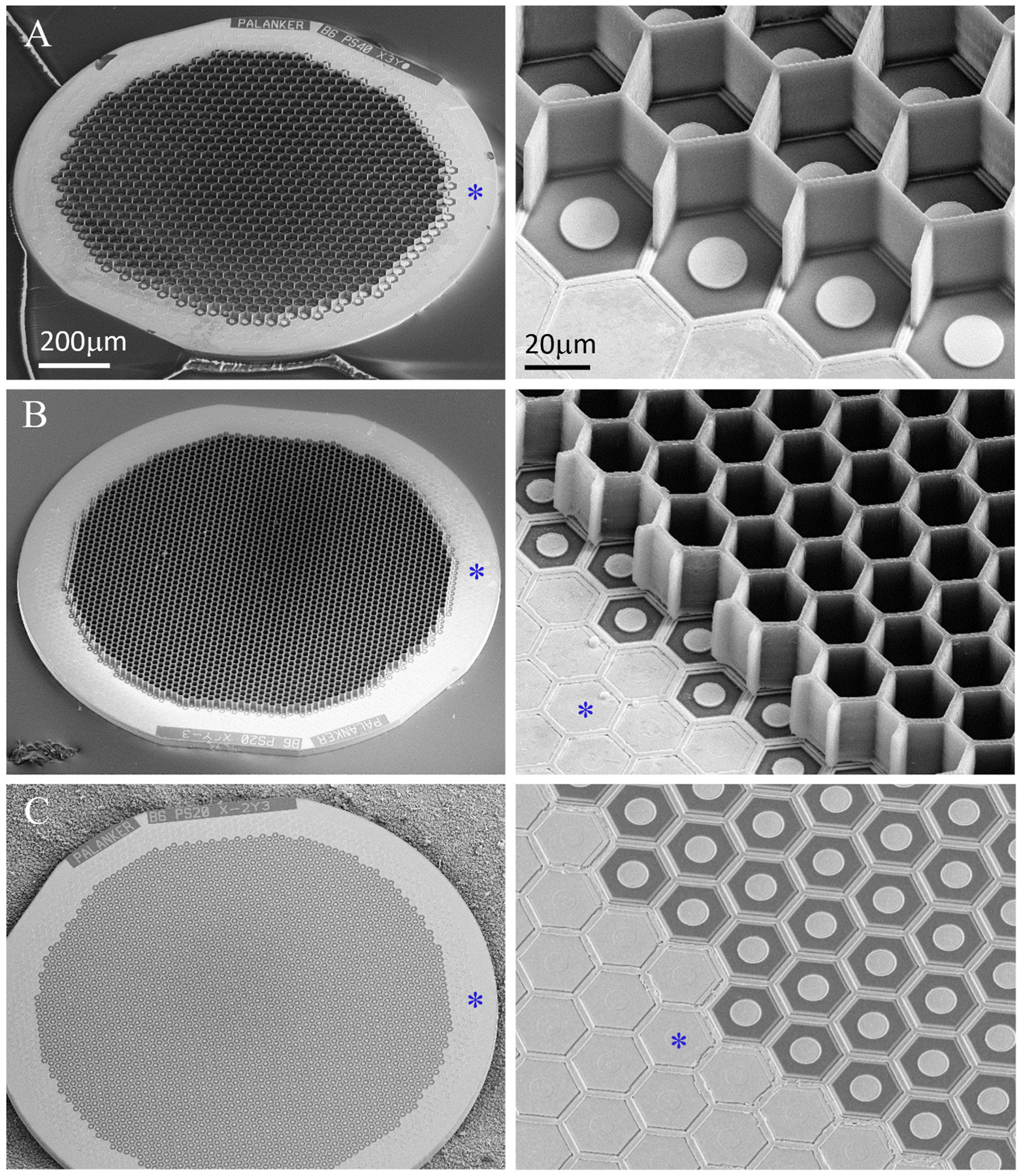
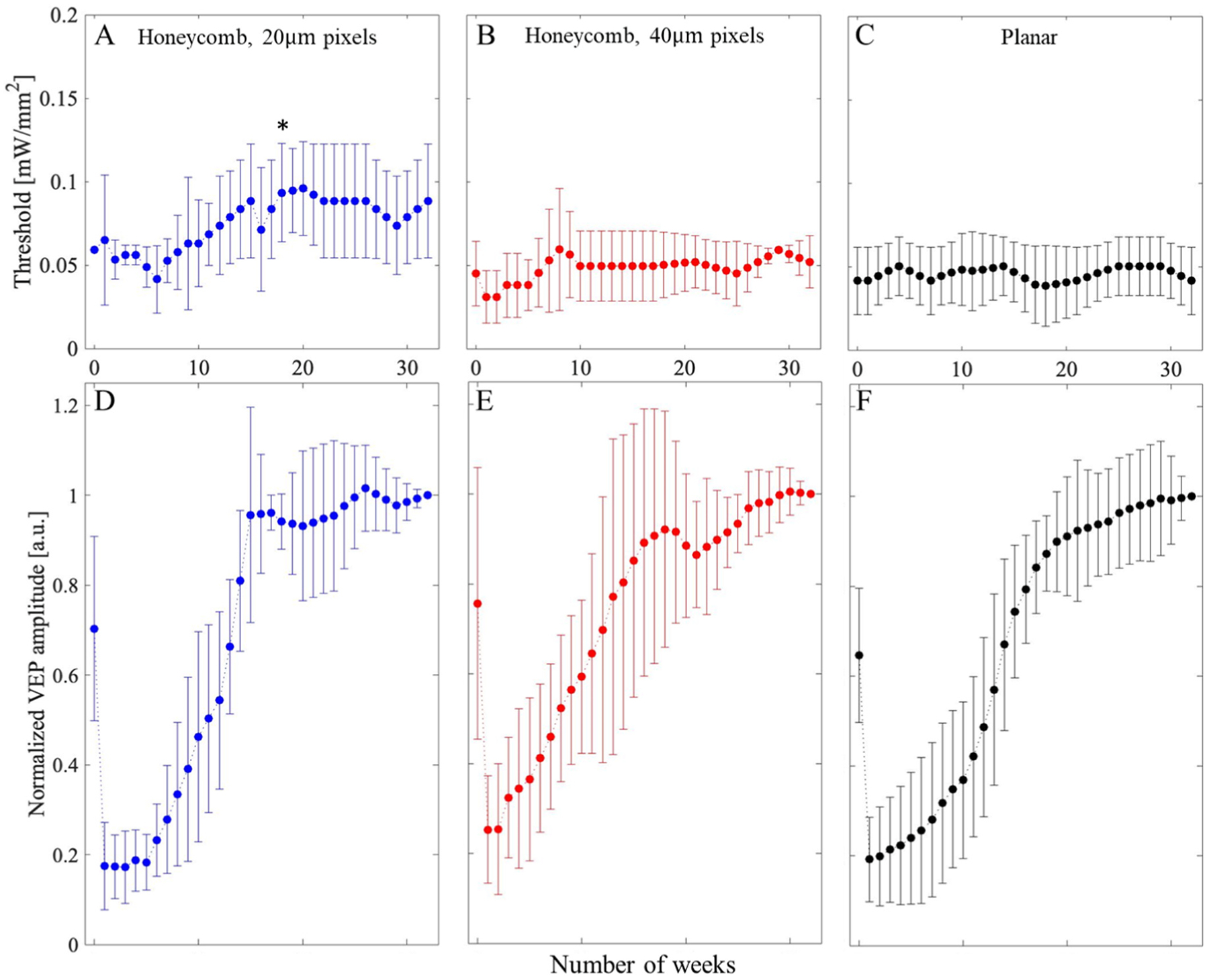
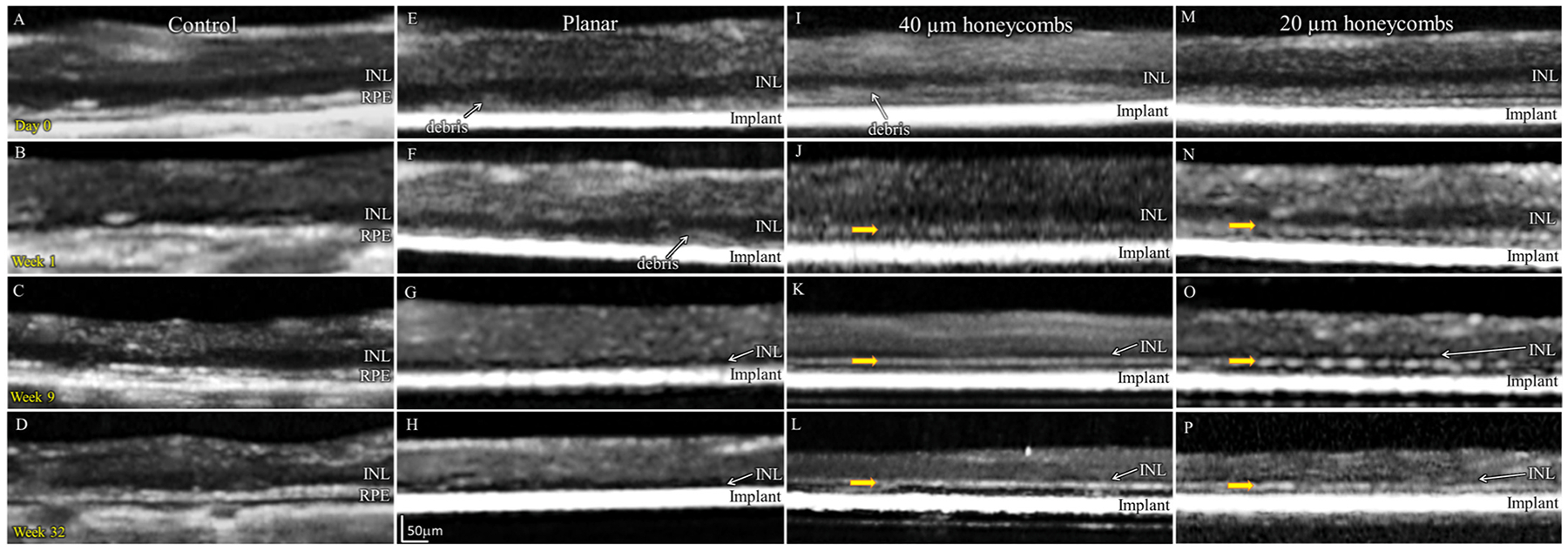
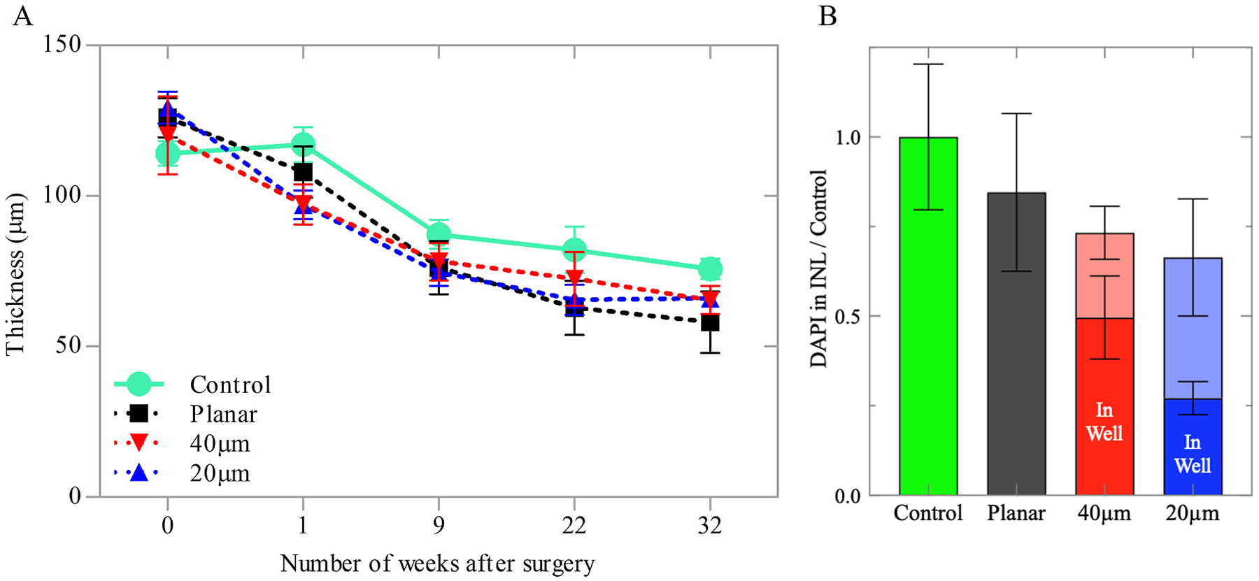
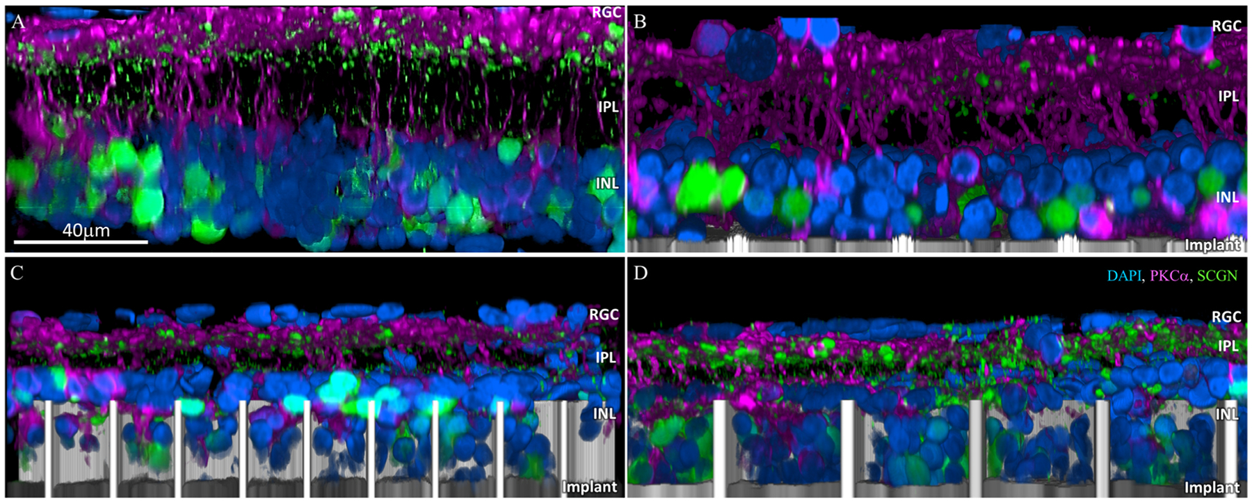
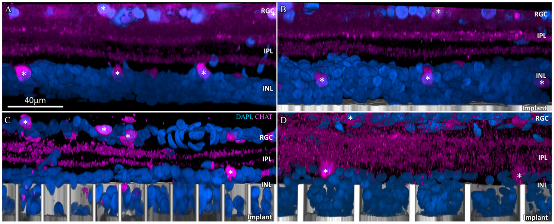
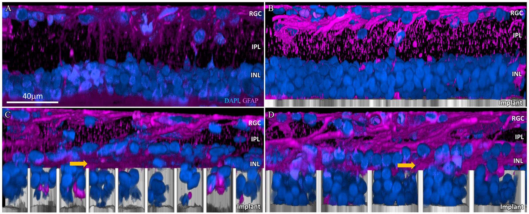
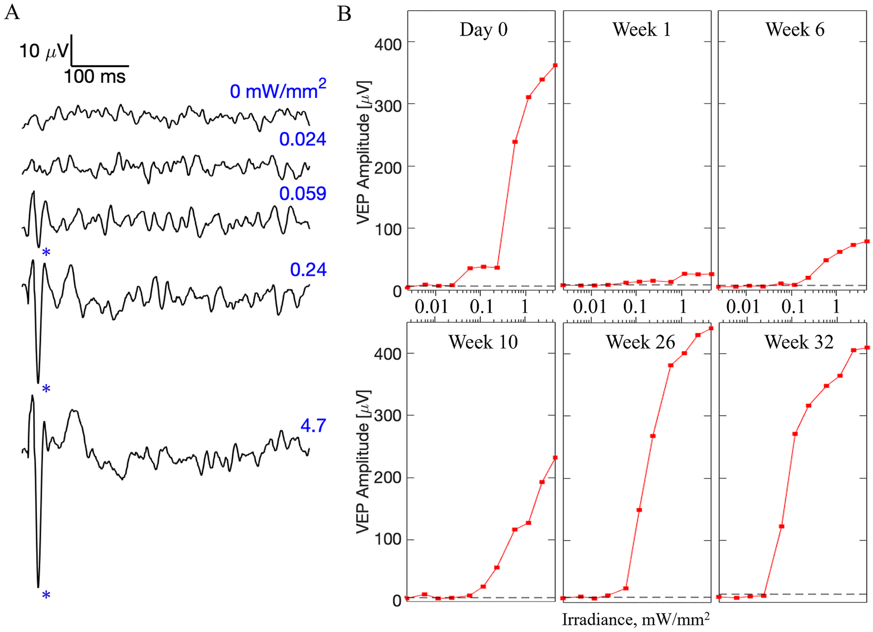
Clinical results with photovoltaic subretinal prosthesis (PRIMA) demonstrated restoration of sight via electrical stimulation of the interneurons in degenerated retina, with resolution matching the 100 μm pixel size. Since scaling the pixels below 75 μm in the current bipolar planar geometry will significantly limit the penetration depth of the electric field and increase stimulation threshold, we explore the possibility of using smaller pixels based on a novel 3-dimensional honeycomb-shaped design. We assessed the long-term biocompatibility and stability of these arrays in rats by investigating the anatomical integration of the retina with flat and 3D implants and response to electrical stimulation over lifetime - up to 32-36 weeks post-implantation in aged rats. With both flat and 3D implants, signals elicited in the visual cortex decreased after the day of implantation by more than 3-fold, and gradually recovered over the next 12-16 weeks. With 25 μm high honeycomb walls, the majority of bipolar cells migrate into the wells, while amacrine and ganglion cells remain above the cavities, which is essential for selective network-mediated stimulation of the retina. Retinal thickness and full-field stimulation threshold with 40 μm-wide honeycomb pixels were comparable to those with planar devices - 0.05 mW/mm with 10 ms pulses. However, fewer cells from the inner nuclear layer migrated into the 20 μm-wide wells, and stimulation threshold increased over 12-16 weeks, before stabilizing at about 0.08 mW/mm. Such threshold is still significantly lower than 1.8 mW/mm with a previous design of flat bipolar pixels, confirming the promise of the 3D honeycomb-based approach to high resolution subretinal prosthesis.
临床结果表明,通过对退化视网膜中的中间神经元进行电刺激,光伏型视网膜假体(PRIMA)可以恢复视力,其分辨率与 100μm 像素大小相匹配。由于在当前的双极平面几何结构中将像素缩小到 75μm 以下将显著限制电场的穿透深度并增加刺激阈值,因此我们探索了基于新型 3 维蜂窝状设计使用更小像素的可能性。我们通过研究视网膜与平面和 3D 植入物的解剖整合以及在寿命内(植入后长达 32-36 周)对电刺激的反应,评估了这些阵列在大鼠中的长期生物相容性和稳定性。对于平面和 3D 植入物,在植入后的第一天,视觉皮层中引出的信号减少了 3 倍以上,并且在接下来的 12-16 周内逐渐恢复。对于 25μm 高的蜂窝状壁,大多数双极细胞迁移到井中,而无长突细胞和节细胞则保留在腔上方,这对于选择性的网络介导的视网膜刺激至关重要。40μm 宽的蜂窝像素的视网膜厚度和全视野刺激阈值与平面器件相当-10ms 脉冲时为 0.05mW/mm。然而,与平面器件相比,来自内核层的更少细胞迁移到 20μm 宽的井中,并且刺激阈值在 12-16 周内增加,然后稳定在约 0.08mW/mm。这样的阈值仍然明显低于以前的平面双极像素设计的 1.8mW/mm,这证实了基于 3D 蜂窝的方法对于高分辨率视网膜假体的前景。