Xu Yuyun, Shu Zhenyu, Song Ge, Liu Yijun, Pang Peipei, Wen Xuehua, Gong Xiangyang
Department of Radiology, Zhejiang Provincial People's Hospital, Affiliated People's Hospital of Hangzhou Medical College, Hangzhou, China.
Department of Radiology, Zhejiang Cancer Hospital, Hangzhou, China.
Front Oncol. 2021 Mar 10;11:634452. doi: 10.3389/fonc.2021.634452. eCollection 2021.
This study aimed to develop and validate an integrated prediction model based on clinicoradiological data and computed tomography (CT)-radiomics for differentiating between benign and malignant parotid gland (PG) tumors multicentre cohorts.
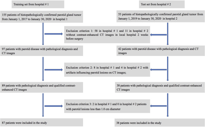
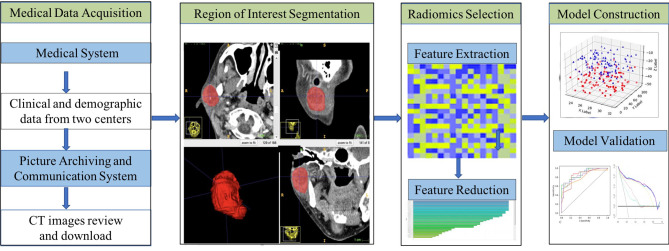
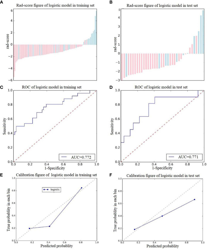
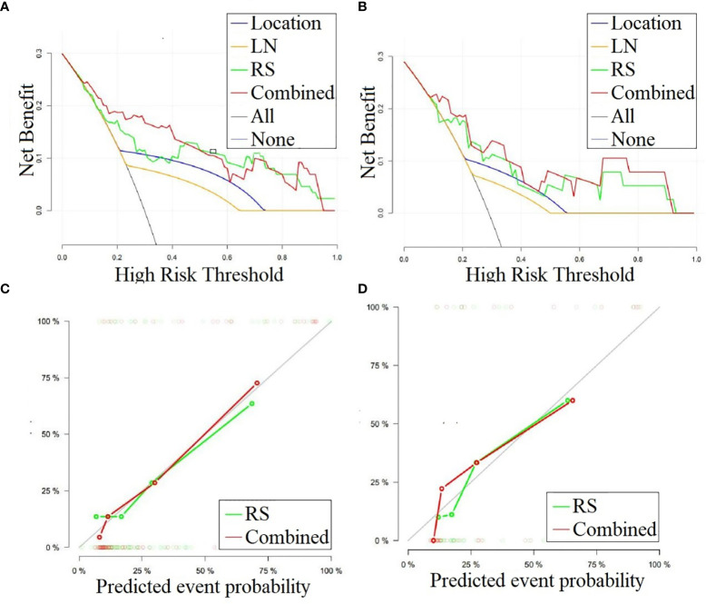
A cohort of 87 PG tumor patients from hospital #1 who were diagnosed between January 2017 and January 2020 were used for prediction model training. A total of 378 radiomic features were extracted from a single tumor region of interest (ROI) of each patient on each phase of CT images. Imaging features were extracted from plain CT and contrast-enhanced CT (CECT) images. After dimensionality reduction, a radiomics signature was constructed. A combination model was constructed by incorporating the rad-score and CT radiological features. An independent group of 38 patients from hospital #2 was used to validate the prediction models. The model performances were evaluated by receiver operating characteristic (ROC) curve analysis, and decision curve analysis (DCA) was used to evaluate the clinical effectiveness of the models. The radiomics signature model was constructed and the rad-score was calculated based on selected imaging features from plain CT and CECT images.
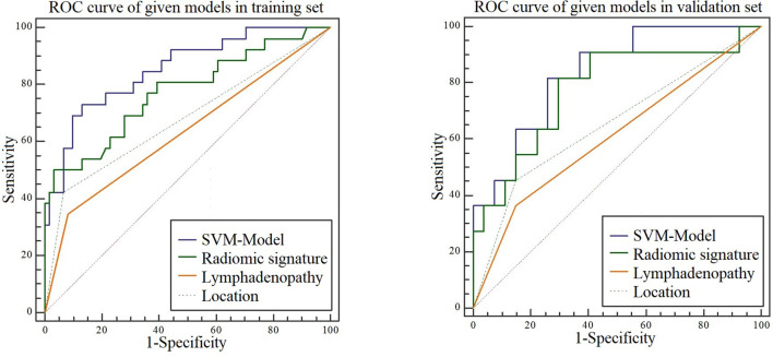
Analysis of variance and multivariable logistic regression analysis showed that location, lymph node metastases, and rad-score were independent predictors of tumor malignant status. The ROC curves showed that the accuracy of the support vector machine (SVM)-based prediction model, radiomics signature, location and lymph node status in the training set was 0.854, 0.772, 0.679, and 0.632, respectively; specificity was 0.869, 0.878, 0.734, and 0.773; and sensitivity was 0.731, 0.808, 0.723, and 0.742. In the test set, the accuracy was 0.835, 0.771, 0.653, and 0.608, respectively; the specificity was 0.741, 0.889, 0.852, and 0.812; and the sensitivity was 0.818, 0.790, 0.731, and 0.716.
The combination model based on the radiomics signature and CT radiological features is capable of evaluating the malignancy of PG tumors and can help clinicians guide clinical tumor management.
本研究旨在开发并验证一种基于临床放射学数据和计算机断层扫描(CT)影像组学的综合预测模型,用于在多中心队列中鉴别腮腺(PG)肿瘤的良恶性。
选取2017年1月至2020年1月期间在医院1诊断的87例PG肿瘤患者组成队列用于预测模型训练。在每位患者CT图像的每个阶段,从单个肿瘤感兴趣区域(ROI)提取总共378个影像组学特征。从平扫CT和增强CT(CECT)图像中提取影像特征。经过降维后,构建影像组学特征。通过纳入影像组学评分(rad-score)和CT放射学特征构建联合模型。使用来自医院2的38例患者独立组验证预测模型。通过受试者操作特征(ROC)曲线分析评估模型性能,并使用决策曲线分析(DCA)评估模型的临床有效性。基于从平扫CT和CECT图像中选择的影像特征构建影像组学特征模型并计算rad-score。
方差分析和多变量逻辑回归分析表明,位置、淋巴结转移和rad-score是肿瘤恶性状态的独立预测因素。ROC曲线显示,训练集中基于支持向量机(SVM)的预测模型、影像组学特征、位置和淋巴结状态的准确率分别为0.854、0.772、0.679和0.632;特异性分别为0.869、0.878、0.734和0.773;敏感性分别为0.73\1、0.808、0.723和0.742。在测试集中,准确率分别为0.835、0.771、0.653和0.608;特异性分别为0.741、0.889、0.852和0.812;敏感性分别为0.818、0.790、0.731和0.716。
基于影像组学特征和CT放射学特征的联合模型能够评估PG肿瘤的恶性程度,并可帮助临床医生指导临床肿瘤管理。