Department of Radiology, Chongqing General Hospital, University of Chinese Academy of Sciences, No.104 Pipashan Main St, Yuzhong District, Chongqing, 400014, China.
Molecular and Functional Imaging Laboratory, Chongqing General Hospital, University of Chinese Academy of Sciences, Chongqing, 400014, China.
BMC Med Imaging. 2020 Apr 15;20(1):38. doi: 10.1186/s12880-020-00442-x.
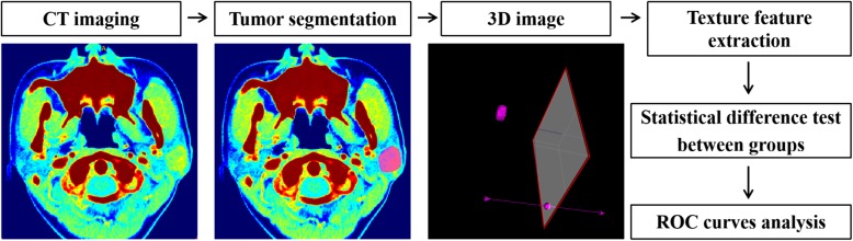
Our study aims to develop and validate diagnostic models of the common parotid tumors based on whole-volume CT textural image biomarkers (IBMs) in combination with clinical parameters at a single institution.
The study cohort was composed of 51 pleomorphic adenoma (PA) patients and 42 Warthin tumor (WT) patients. Clinical parameters and conventional image features were scored retrospectively and textural IBMs were extracted from CT images of arterial phase. Independent-samples t test or Chi-square test was used for evaluating the significance of the difference among clinical parameters, conventional CT image features, and textural IBMs. The diagnostic performance of univariate model and multivariate model was evaluated via receiver operating characteristic (ROC) curve and area under ROC curve (AUC).
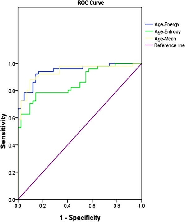
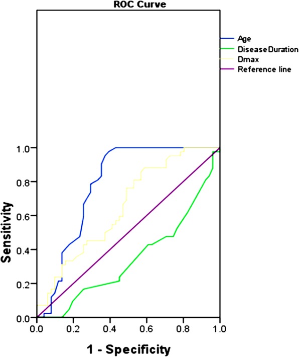
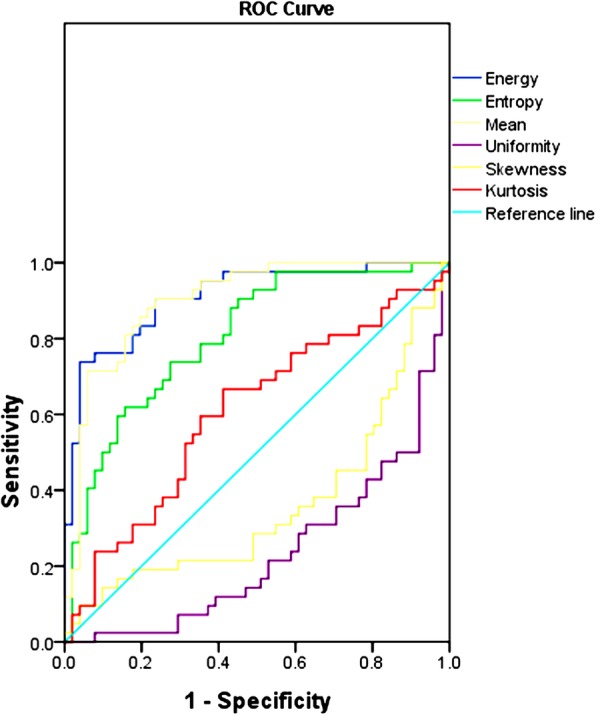
Significant differences were found in clinical parameters (age, gender, disease duration, smoking), conventional image features (site, maximum diameter, time-density curve, peripheral vessels sign) and textural IBMs (mean, uniformity, energy, entropy) between PA group and WT group (P<0.05). ROC analysis showed that clinical parameter (age) and quantitative textural IBMs (mean, energy, entropy) were able to categorize the patients into PA group and WT group, with the AUC of 0.784, 0.902, 0.910, 0.805, respectively. When IBMs were added in clinical model, the multivariate models including age-mean and age-energy performed significantly better than the univariate models with the improved AUC of 0.940, 0.944, respectively (P<0.001).
Both clinical parameter and CT textural IBMs can be used for the preoperative, noninvasive diagnosis of parotid PA and WT. The diagnostic performance of textural IBM model was obviously better than that of clinical model and conventional image model in this study. While the multivariate model consisted of clinical parameter and textural IBM had the optimal diagnostic performance, which would contribute to the better selection of individualized surgery program.
本研究旨在基于单机构的全容积 CT 纹理图像生物标志物(IBMs)和临床参数,开发和验证常见腮腺肿瘤的诊断模型。
研究队列由 51 例多形性腺瘤(PA)患者和 42 例沃辛瘤(WT)患者组成。回顾性评分临床参数和常规图像特征,并从动脉期 CT 图像中提取纹理 IBM。采用独立样本 t 检验或卡方检验评估临床参数、常规 CT 图像特征和纹理 IBM 之间的差异是否有统计学意义。通过受试者工作特征(ROC)曲线和 ROC 曲线下面积(AUC)评估单变量模型和多变量模型的诊断性能。
PA 组和 WT 组在临床参数(年龄、性别、病程、吸烟)、常规图像特征(部位、最大直径、时间密度曲线、周围血管征)和纹理 IBMs(均值、均匀度、能量、熵)方面存在显著差异(P<0.05)。ROC 分析表明,临床参数(年龄)和定量纹理 IBMs(均值、能量、熵)能够将患者分为 PA 组和 WT 组,AUC 分别为 0.784、0.902、0.910、0.805。当 IBMs 被添加到临床模型中时,包含年龄-均值和年龄-能量的多变量模型的 AUC 分别提高到 0.940 和 0.944,明显优于单变量模型(P<0.001)。
临床参数和 CT 纹理 IBMs 均可用于术前、无创性诊断腮腺 PA 和 WT。在本研究中,纹理 IBM 模型的诊断性能明显优于临床模型和常规图像模型。而包含临床参数和纹理 IBM 的多变量模型具有最佳的诊断性能,有助于更好地选择个体化手术方案。