Yee-de León Juan F, Soto-García Brenda, Aráiz-Hernández Diana, Delgado-Balderas Jesús Rolando, Esparza Miguel, Aguilar-Avelar Carlos, Wong-Campos J D, Chacón Franco, López-Hernández José Y, González-Treviño A Mauricio, Yee-de León José R, Zamora-Mendoza Jorge L, Alvarez Mario M, Trujillo-de Santiago Grissel, Gómez-Guerra Lauro S, Sánchez-Domínguez Celia N, Velarde-Calvillo Liza P, Abarca-Blanco Alejandro
Delee Corp., Mountain View, CA, 94041, USA.
Departamento de Bioquímica y Medicina Molecular, Facultad de Medicina, Universidad Autónoma de Nuevo León, Monterrey, 64460, Mexico.
Sci Rep. 2020 May 5;10(1):7543. doi: 10.1038/s41598-020-63672-7.
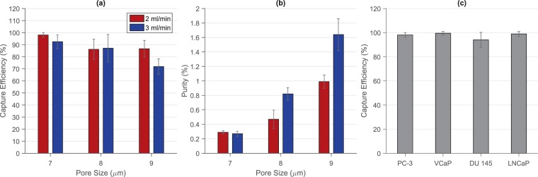
The detection and analysis of circulating tumor cells (CTCs) may enable a broad range of cancer-related applications, including the identification of acquired drug resistance during treatments. However, the non-scalable fabrication, prolonged sample processing times, and the lack of automation, associated with most of the technologies developed to isolate these rare cells, have impeded their transition into the clinical practice. This work describes a novel membrane-based microfiltration device comprised of a fully automated sample processing unit and a machine-vision-enabled imaging system that allows the efficient isolation and rapid analysis of CTCs from blood. The device performance was characterized using four prostate cancer cell lines, including PC-3, VCaP, DU-145, and LNCaP, obtaining high assay reproducibility and capture efficiencies greater than 93% after processing 7.5 mL blood samples spiked with 100 cancer cells. Cancer cells remained viable after filtration due to the minimal shear stress exerted over cells during the procedure, while the identification of cancer cells by immunostaining was not affected by the number of non-specific events captured on the membrane. We were also able to identify the androgen receptor (AR) point mutation T878A from 7.5 mL blood samples spiked with 50 LNCaP cells using RT-PCR and Sanger sequencing. Finally, CTCs were detected in 8 out of 8 samples from patients diagnosed with metastatic prostate cancer (mean ± SEM = 21 ± 2.957 CTCs/mL, median = 21 CTCs/mL), demonstrating the potential clinical utility of this device.
循环肿瘤细胞(CTCs)的检测与分析可实现广泛的癌症相关应用,包括在治疗过程中识别获得性耐药。然而,大多数用于分离这些稀有细胞的技术存在不可扩展的制造工艺、冗长的样本处理时间以及缺乏自动化等问题,阻碍了它们向临床实践的转化。这项工作描述了一种基于膜的新型微滤装置,该装置由一个全自动样本处理单元和一个具备机器视觉的成像系统组成,能够从血液中高效分离并快速分析CTCs。使用四种前列腺癌细胞系(包括PC-3、VCaP、DU-145和LNCaP)对该装置性能进行了表征,在处理添加了100个癌细胞的7.5 mL血样后,获得了高检测重现性,捕获效率大于93%。由于在过滤过程中施加在细胞上的剪切应力极小,癌细胞在过滤后仍保持活力,而通过免疫染色识别癌细胞不受膜上捕获的非特异性事件数量的影响。我们还能够使用逆转录聚合酶链反应(RT-PCR)和桑格测序从添加了50个LNCaP细胞的7.5 mL血样中鉴定出雄激素受体(AR)点突变T878A。最后,在8例被诊断为转移性前列腺癌患者的样本中,有8例检测到了CTCs(平均值±标准误=21±2.957个CTCs/mL,中位数=21个CTCs/mL),证明了该装置的潜在临床应用价值。