• 文献检索
  • 文档翻译
  • 深度研究
  • 学术资讯
  • Suppr Zotero 插件Zotero 插件
  • 邀请有礼
  • 套餐&价格
  • 历史记录
应用&插件
Suppr Zotero 插件Zotero 插件浏览器插件Mac 客户端Windows 客户端微信小程序
定价
高级版会员购买积分包购买API积分包
服务
文献检索文档翻译深度研究API 文档MCP 服务
关于我们
关于 Suppr公司介绍联系我们用户协议隐私条款
关注我们

Suppr 超能文献

核心技术专利:CN118964589B侵权必究
粤ICP备2023148730 号-1Suppr @ 2026

文献检索

告别复杂PubMed语法,用中文像聊天一样搜索,搜遍4000万医学文献。AI智能推荐,让科研检索更轻松。

立即免费搜索

文件翻译

保留排版,准确专业,支持PDF/Word/PPT等文件格式,支持 12+语言互译。

免费翻译文档

深度研究

AI帮你快速写综述,25分钟生成高质量综述,智能提取关键信息,辅助科研写作。

立即免费体验

一种严格无监督的 HEp-2 细胞图像分类深度学习方法。

A Strictly Unsupervised Deep Learning Method for HEp-2 Cell Image Classification.

机构信息

Department of IT Convergence and Application Engineering, Pukyong National University, Busan 48513, Korea.

Department of Computer Engineering, Dong-A University, Busan 49315, Korea.

出版信息

Sensors (Basel). 2020 May 9;20(9):2717. doi: 10.3390/s20092717.

DOI:10.3390/s20092717
PMID:32397567
原文链接:https://pmc.ncbi.nlm.nih.gov/articles/PMC7249201/
Abstract

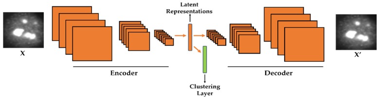
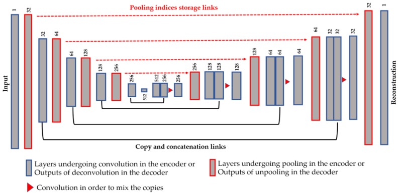
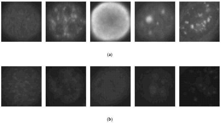
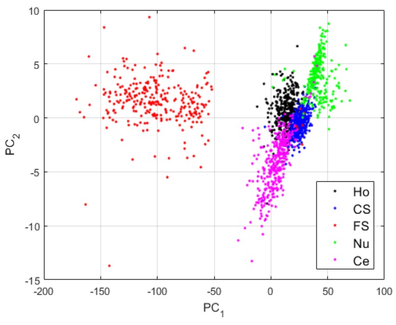
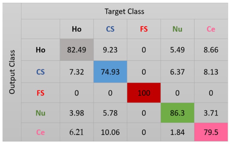
Classifying the images that portray the Human Epithelial cells of type 2 (HEp-2) represents one of the most important steps in the diagnosis procedure of autoimmune diseases. Performing this classification manually represents an extremely complicated task due to the heterogeneity of these cellular images. Hence, an automated classification scheme appears to be necessary. However, the majority of the available methods prefer to utilize the supervised learning approach for this problem. The need for thousands of images labelled manually can represent a difficulty with this approach. The first contribution of this work is to demonstrate that classifying HEp-2 cell images can also be done using the unsupervised learning paradigm. Unlike the majority of the existing methods, we propose here a deep learning scheme that performs both the feature extraction and the cells' discrimination through an end-to-end unsupervised paradigm. We propose the use of a deep convolutional autoencoder (DCAE) that performs feature extraction via an encoding-decoding scheme. At the same time, we embed in the network a clustering layer whose purpose is to automatically discriminate, during the feature learning process, the latent representations produced by the DCAE. Furthermore, we investigate how the quality of the network's reconstruction can affect the quality of the produced representations. We have investigated the effectiveness of our method on some benchmark datasets and we demonstrate here that the unsupervised learning, when done properly, performs at the same level as the actual supervised learning-based state-of-the-art methods in terms of accuracy.

摘要

对描绘 2 型人上皮细胞 (HEp-2) 的图像进行分类是自身免疫性疾病诊断过程中最重要的步骤之一。由于这些细胞图像的异质性,手动进行此类分类任务极其复杂。因此,需要一种自动化的分类方案。然而,大多数现有的方法都倾向于使用监督学习方法来解决这个问题。由于需要数千张手动标记的图像,因此这种方法可能存在困难。这项工作的第一个贡献是证明使用无监督学习范例也可以对 HEp-2 细胞图像进行分类。与大多数现有方法不同,我们在这里提出了一种深度学习方案,通过端到端的无监督范例同时执行特征提取和细胞识别。我们建议使用深度卷积自动编码器 (DCAE) 通过编码-解码方案进行特征提取。同时,我们在网络中嵌入一个聚类层,其目的是在特征学习过程中自动区分 DCAE 生成的潜在表示。此外,我们研究了网络重构的质量如何影响生成表示的质量。我们已经在一些基准数据集上调查了我们方法的有效性,并在此证明,在适当的情况下,无监督学习在准确性方面与实际基于监督学习的最先进方法的表现相当。

https://cdn.ncbi.nlm.nih.gov/pmc/blobs/03b3/7249201/b6c162816fe8/sensors-20-02717-g014.jpg
https://cdn.ncbi.nlm.nih.gov/pmc/blobs/03b3/7249201/2e6fc08a227c/sensors-20-02717-g001.jpg
https://cdn.ncbi.nlm.nih.gov/pmc/blobs/03b3/7249201/900814d0ba2f/sensors-20-02717-g002.jpg
https://cdn.ncbi.nlm.nih.gov/pmc/blobs/03b3/7249201/330308e43543/sensors-20-02717-g003.jpg
https://cdn.ncbi.nlm.nih.gov/pmc/blobs/03b3/7249201/4cdad4b592a1/sensors-20-02717-g004.jpg
https://cdn.ncbi.nlm.nih.gov/pmc/blobs/03b3/7249201/5d2c8a1e5b93/sensors-20-02717-g005.jpg
https://cdn.ncbi.nlm.nih.gov/pmc/blobs/03b3/7249201/12d960f8e247/sensors-20-02717-g006.jpg
https://cdn.ncbi.nlm.nih.gov/pmc/blobs/03b3/7249201/94a0c796f20e/sensors-20-02717-g007.jpg
https://cdn.ncbi.nlm.nih.gov/pmc/blobs/03b3/7249201/814221717e0f/sensors-20-02717-g008.jpg
https://cdn.ncbi.nlm.nih.gov/pmc/blobs/03b3/7249201/cee90f6b042e/sensors-20-02717-g009.jpg
https://cdn.ncbi.nlm.nih.gov/pmc/blobs/03b3/7249201/f0988559afc3/sensors-20-02717-g010.jpg
https://cdn.ncbi.nlm.nih.gov/pmc/blobs/03b3/7249201/659f15f36c80/sensors-20-02717-g011.jpg
https://cdn.ncbi.nlm.nih.gov/pmc/blobs/03b3/7249201/bc550360d11a/sensors-20-02717-g012.jpg
https://cdn.ncbi.nlm.nih.gov/pmc/blobs/03b3/7249201/7be6a55abf0e/sensors-20-02717-g013.jpg
https://cdn.ncbi.nlm.nih.gov/pmc/blobs/03b3/7249201/b6c162816fe8/sensors-20-02717-g014.jpg

相似文献

1
A Strictly Unsupervised Deep Learning Method for HEp-2 Cell Image Classification.一种严格无监督的 HEp-2 细胞图像分类深度学习方法。
Sensors (Basel). 2020 May 9;20(9):2717. doi: 10.3390/s20092717.
2
BIRADS features-oriented semi-supervised deep learning for breast ultrasound computer-aided diagnosis.基于 BI-RADS 特征的半监督深度学习在乳腺超声计算机辅助诊断中的应用。
Phys Med Biol. 2020 Jun 12;65(12):125005. doi: 10.1088/1361-6560/ab7e7d.
3
A Classification Method for the Cellular Images Based on Active Learning and Cross-Modal Transfer Learning.基于主动学习和跨模态迁移学习的细胞图像分类方法。
Sensors (Basel). 2021 Feb 20;21(4):1469. doi: 10.3390/s21041469.
4
Classification of lung adenocarcinoma transcriptome subtypes from pathological images using deep convolutional networks.利用深度卷积网络从病理图像中对肺腺癌转录组亚型进行分类。
Int J Comput Assist Radiol Surg. 2018 Dec;13(12):1905-1913. doi: 10.1007/s11548-018-1835-2. Epub 2018 Aug 29.
5
Convolutional sparse kernel network for unsupervised medical image analysis.卷积稀疏核网络在医学图像无监督分析中的应用。
Med Image Anal. 2019 Aug;56:140-151. doi: 10.1016/j.media.2019.06.005. Epub 2019 Jun 12.
6
Transfer learning of deep neural network representations for fMRI decoding.基于深度神经网络表示的 fMRI 解码的迁移学习。
J Neurosci Methods. 2019 Dec 1;328:108319. doi: 10.1016/j.jneumeth.2019.108319. Epub 2019 Oct 1.
7
Deep Learning Based Analysis of Histopathological Images of Breast Cancer.基于深度学习的乳腺癌组织病理学图像分析
Front Genet. 2019 Feb 19;10:80. doi: 10.3389/fgene.2019.00080. eCollection 2019.
8
Lung and Pancreatic Tumor Characterization in the Deep Learning Era: Novel Supervised and Unsupervised Learning Approaches.深度学习时代的肺部和胰腺肿瘤特征分析:新型监督式和非监督式学习方法。
IEEE Trans Med Imaging. 2019 Aug;38(8):1777-1787. doi: 10.1109/TMI.2019.2894349. Epub 2019 Jan 23.
9
HEp-2 Cell Image Classification With Deep Convolutional Neural Networks.基于深度卷积神经网络的人喉表皮样癌细胞(HEp-2)图像分类
IEEE J Biomed Health Inform. 2017 Mar;21(2):416-428. doi: 10.1109/JBHI.2016.2526603. Epub 2016 Feb 8.
10
A Tour of Unsupervised Deep Learning for Medical Image Analysis.医学图像分析的无监督深度学习之旅。
Curr Med Imaging. 2021;17(9):1059-1077. doi: 10.2174/1573405617666210127154257.

引用本文的文献

1
VOLTA: an enVironment-aware cOntrastive ceLl represenTation leArning for histopathology.VOLTA:一种环境感知的对比细胞表示学习方法,用于组织病理学。
Nat Commun. 2024 May 10;15(1):3942. doi: 10.1038/s41467-024-48062-1.
2
Comparison of the Capacity of Several Machine Learning Tools to Assist Immunofluorescence-Based Detection of Anti-Neutrophil Cytoplasmic Antibodies.几种机器学习工具辅助基于免疫荧光法检测抗中性粒细胞胞质抗体能力的比较。
Int J Mol Sci. 2024 Mar 13;25(6):3270. doi: 10.3390/ijms25063270.
3
Machine learning combined with radiomics and deep learning features extracted from CT images: a novel AI model to distinguish benign from malignant ovarian tumors.

本文引用的文献

1
SegNet: A Deep Convolutional Encoder-Decoder Architecture for Image Segmentation.SegNet:一种用于图像分割的深度卷积编解码器架构。
IEEE Trans Pattern Anal Mach Intell. 2017 Dec;39(12):2481-2495. doi: 10.1109/TPAMI.2016.2644615. Epub 2017 Jan 2.
2
HEp-2 Cell Image Classification With Deep Convolutional Neural Networks.基于深度卷积神经网络的人喉表皮样癌细胞(HEp-2)图像分类
IEEE J Biomed Health Inform. 2017 Mar;21(2):416-428. doi: 10.1109/JBHI.2016.2526603. Epub 2016 Feb 8.
3
Deep learning.深度学习。
机器学习结合从CT图像中提取的放射组学和深度学习特征:一种区分卵巢良性和恶性肿瘤的新型人工智能模型。
Insights Imaging. 2023 Apr 24;14(1):68. doi: 10.1186/s13244-023-01412-x.
4
Classification of HEp-2 Staining Pattern Images Using Adapted Multilayer Perceptron Neural Network-Based Intra-Class Variation of Cell Shape.基于细胞形态类内变异性的改进多层感知机神经网络对 HEp-2 染色模式图像的分类。
Sensors (Basel). 2023 Feb 15;23(4):2195. doi: 10.3390/s23042195.
5
Cell Phenotype Classification Based on Joint of Texture Information and Multilayer Feature Extraction in DenseNet.基于 DenseNet 中纹理信息和多层特征提取的细胞表型分类。
Comput Intell Neurosci. 2022 Nov 28;2022:6895833. doi: 10.1155/2022/6895833. eCollection 2022.
6
Artificial Intelligence and Deep Learning for Rheumatologists.人工智能与深度学习在风湿病学中的应用。
Arthritis Rheumatol. 2022 Dec;74(12):1893-1905. doi: 10.1002/art.42296. Epub 2022 Oct 26.
7
A Classification Method for the Cellular Images Based on Active Learning and Cross-Modal Transfer Learning.基于主动学习和跨模态迁移学习的细胞图像分类方法。
Sensors (Basel). 2021 Feb 20;21(4):1469. doi: 10.3390/s21041469.
8
Automatic Detection Method for Cancer Cell Nucleus Image Based on Deep-Learning Analysis and Color Layer Signature Analysis Algorithm.基于深度学习分析和颜色层特征分析算法的癌细胞核图像自动检测方法。
Sensors (Basel). 2020 Aug 7;20(16):4409. doi: 10.3390/s20164409.
Nature. 2015 May 28;521(7553):436-44. doi: 10.1038/nature14539.
4
Advanced statistical matrices for texture characterization: application to cell classification.高级纹理特征描述统计矩阵:在细胞分类中的应用。
IEEE Trans Biomed Eng. 2014 Mar;61(3):630-7. doi: 10.1109/TBME.2013.2284600. Epub 2013 Oct 4.
5
Benchmarking HEp-2 cells classification methods.HEp-2 细胞分类方法的基准测试。
IEEE Trans Med Imaging. 2013 Oct;32(10):1878-89. doi: 10.1109/TMI.2013.2268163. Epub 2013 Jun 18.
6
A completed modeling of local binary pattern operator for texture classification.完成了用于纹理分类的局部二值模式算子的建模。
IEEE Trans Image Process. 2010 Jun;19(6):1657-63. doi: 10.1109/TIP.2010.2044957. Epub 2010 Mar 8.
7
Indirect immunofluorescence in autoimmune diseases: assessment of digital images for diagnostic purpose.自身免疫性疾病中的间接免疫荧光:用于诊断目的的数字图像评估
Cytometry B Clin Cytom. 2007 Nov;72(6):472-7. doi: 10.1002/cyto.b.20356.
8
Reducing the dimensionality of data with neural networks.使用神经网络降低数据维度。
Science. 2006 Jul 28;313(5786):504-7. doi: 10.1126/science.1127647.
9
Principal component analysis for clustering gene expression data.用于聚类基因表达数据的主成分分析。
Bioinformatics. 2001 Sep;17(9):763-74. doi: 10.1093/bioinformatics/17.9.763.