• 文献检索
  • 文档翻译
  • 深度研究
  • 学术资讯
  • Suppr Zotero 插件Zotero 插件
  • 邀请有礼
  • 套餐&价格
  • 历史记录
应用&插件
Suppr Zotero 插件Zotero 插件浏览器插件Mac 客户端Windows 客户端微信小程序
定价
高级版会员购买积分包购买API积分包
服务
文献检索文档翻译深度研究API 文档MCP 服务
关于我们
关于 Suppr公司介绍联系我们用户协议隐私条款
关注我们

Suppr 超能文献

核心技术专利:CN118964589B侵权必究
粤ICP备2023148730 号-1Suppr @ 2026

文献检索

告别复杂PubMed语法,用中文像聊天一样搜索,搜遍4000万医学文献。AI智能推荐,让科研检索更轻松。

立即免费搜索

文件翻译

保留排版,准确专业,支持PDF/Word/PPT等文件格式,支持 12+语言互译。

免费翻译文档

深度研究

AI帮你快速写综述,25分钟生成高质量综述,智能提取关键信息,辅助科研写作。

立即免费体验

c-Jun过表达通过人脐带间充质干细胞加速糖尿病大鼠伤口愈合

c-Jun Overexpression Accelerates Wound Healing in Diabetic Rats by Human Umbilical Cord-Derived Mesenchymal Stem Cells.

作者信息

Yue Chun, Guo Zi, Luo Yufang, Yuan Jingjing, Wan Xinxing, Mo Zhaohui

机构信息

Department of Endocrinology and Metabolism, Third Xiangya Hospital of Central South University, Changsha, China.

Diabetic Foot Research Center, Third Xiangya Hospital of Central South University, Changsha, China.

出版信息

Stem Cells Int. 2020 Jan 14;2020:7430968. doi: 10.1155/2020/7430968. eCollection 2020.

DOI:10.1155/2020/7430968
PMID:32399050
原文链接:https://pmc.ncbi.nlm.nih.gov/articles/PMC7201444/
Abstract

OBJECTIVE

Mesenchymal stem cells (MSCs) are considered a promising therapy for wound healing. Here, we explored the role of c-Jun in diabetic wound healing using human umbilical cord-derived MSCs (hUC-MSCs).

METHODS

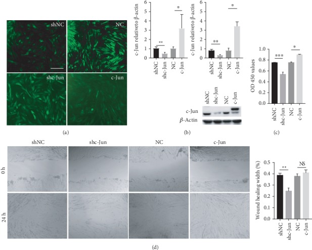
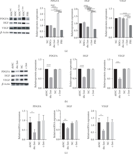
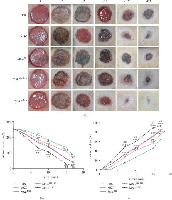
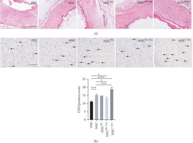
Freshly isolated hUC-MSCs were subjected to extensive subcultivation. The cell proliferative and migratory capacities were assessed by the Cell Counting Kit-8 and scratch assays, respectively. c-Jun expression was evaluated by RT-PCR and western blot analysis. The function of c-Jun was investigated with lentivirus transduction-based gene silencing and overexpression. Diabetes mellitus was induced in SD rats on a high-glucose/fat diet by streptozocin administration. Wounds were created on the dorsal skin. The effects of c-Jun silencing and overexpression on wound closure by hUC-MSCs were examined. Reepithelialization and angiogenesis were assessed by histological and immunohistochemical analysis, respectively. Platelet-derived growth factor A (PDGFA), hepatocyte growth factor (HGF), and vascular endothelial growth factor (VEGF) levels were determined by western blot analysis.

RESULTS

hUC-MSCs showed gradually decreased cell proliferation, migration, and c-Jun expression during subcultivation. c-Jun silencing inhibited cell proliferation and migration, while c-Jun overexpression enhanced proliferation but not migration. Compared with untransduced hUC-MSCs, local subcutaneous injection of c-Jun-overexpressing hUC-MSCs accelerated wound closure, enhanced angiogenesis and reepithelialization at the wound bed, and increased PDGFA and HGF levels in wound tissues.

CONCLUSION

c-Jun overexpression promoted hUC-MSC proliferation and migration and accelerated diabetic wound closure, reepithelization, and angiogenesis by hUC-MSCs . These beneficial effects of c-Jun overexpression in diabetic wound healing by hUC-MSCs were at least partially mediated by increased PDGFA and HGF levels in wound tissues.

摘要

目的

间充质干细胞(MSCs)被认为是一种有前景的伤口愈合治疗方法。在此,我们利用人脐带间充质干细胞(hUC-MSCs)探讨了c-Jun在糖尿病伤口愈合中的作用。

方法

对新鲜分离的hUC-MSCs进行广泛传代培养。分别通过细胞计数试剂盒-8和划痕试验评估细胞增殖和迁移能力。通过RT-PCR和蛋白质印迹分析评估c-Jun表达。利用基于慢病毒转导的基因沉默和过表达研究c-Jun的功能。通过给高脂高糖饮食的SD大鼠注射链脲佐菌素诱导糖尿病。在背部皮肤制造伤口。检测c-Jun沉默和过表达对hUC-MSCs伤口闭合的影响。分别通过组织学和免疫组织化学分析评估再上皮化和血管生成。通过蛋白质印迹分析测定血小板衍生生长因子A(PDGFA)、肝细胞生长因子(HGF)和血管内皮生长因子(VEGF)水平。

结果

hUC-MSCs在传代培养过程中细胞增殖、迁移和c-Jun表达逐渐下降。c-Jun沉默抑制细胞增殖和迁移,而c-Jun过表达增强增殖但不增强迁移。与未转导的hUC-MSCs相比,局部皮下注射过表达c-Jun的hUC-MSCs加速了伤口闭合,增强了伤口床的血管生成和再上皮化,并增加了伤口组织中PDGFA和HGF水平。

结论

c-Jun过表达促进hUC-MSC增殖和迁移,并加速hUC-MSCs介导的糖尿病伤口闭合、再上皮化和血管生成。c-Jun过表达在hUC-MSCs糖尿病伤口愈合中的这些有益作用至少部分是由伤口组织中PDGFA和HGF水平增加介导的。

https://cdn.ncbi.nlm.nih.gov/pmc/blobs/33b0/7201444/f6b27bef198e/SCI2020-7430968.005.jpg
https://cdn.ncbi.nlm.nih.gov/pmc/blobs/33b0/7201444/c563b480a79f/SCI2020-7430968.002.jpg
https://cdn.ncbi.nlm.nih.gov/pmc/blobs/33b0/7201444/cbdf70d168f2/SCI2020-7430968.003.jpg
https://cdn.ncbi.nlm.nih.gov/pmc/blobs/33b0/7201444/d8f6f4152498/SCI2020-7430968.004.jpg
https://cdn.ncbi.nlm.nih.gov/pmc/blobs/33b0/7201444/f6b27bef198e/SCI2020-7430968.005.jpg

相似文献

1
c-Jun Overexpression Accelerates Wound Healing in Diabetic Rats by Human Umbilical Cord-Derived Mesenchymal Stem Cells.c-Jun过表达通过人脐带间充质干细胞加速糖尿病大鼠伤口愈合
Stem Cells Int. 2020 Jan 14;2020:7430968. doi: 10.1155/2020/7430968. eCollection 2020.
2
JAM-A Overexpression in Human Umbilical Cord-Derived Mesenchymal Stem Cells Accelerated the Angiogenesis of Diabetic Wound By Enhancing Both Paracrine Function and Survival of Mesenchymal Stem Cells.JAM-A 在人脐带间充质干细胞中的过表达通过增强间充质干细胞的旁分泌功能和存活来加速糖尿病创面的血管生成。
Stem Cell Rev Rep. 2023 Jul;19(5):1554-1575. doi: 10.1007/s12015-023-10518-0. Epub 2023 Apr 15.
3
Role of quercetin and rutin in enhancing the therapeutic potential of mesenchymal stem cells for cold induced burn wound.槲皮素和芦丁在增强间充质干细胞对冷致烧伤创面治疗潜力中的作用
Regen Ther. 2022 Aug 23;21:225-238. doi: 10.1016/j.reth.2022.07.011. eCollection 2022 Dec.
4
-Formed Fibrin Hydrogel Scaffold Loaded With Human Umbilical Cord Mesenchymal Stem Cells Promotes Skin Wound Healing.载有人脐带间充质干细胞的纤维蛋白水凝胶支架促进皮肤伤口愈合。
Cell Transplant. 2023 Jan-Dec;32:9636897231156215. doi: 10.1177/09636897231156215.
5
Self-Assembled Nano-Peptide Hydrogels with Human Umbilical Cord Mesenchymal Stem Cell Spheroids Accelerate Diabetic Skin Wound Healing by Inhibiting Inflammation and Promoting Angiogenesis.自组装纳米肽水凝胶与人脐带间充质干细胞球协同作用,通过抑制炎症和促进血管生成加速糖尿病皮肤伤口愈合。
Int J Nanomedicine. 2022 May 30;17:2459-2474. doi: 10.2147/IJN.S363777. eCollection 2022.
6
Human umbilical cord mesenchymal stem cells transplantation promotes cutaneous wound healing of severe burned rats.人脐带间充质干细胞移植促进严重烧伤大鼠皮肤伤口愈合。
PLoS One. 2014 Feb 20;9(2):e88348. doi: 10.1371/journal.pone.0088348. eCollection 2014.
7
Efficacy of the therapy of 5-aminolevulinic acid photodynamic therapy combined with human umbilical cord mesenchymal stem cells on methicillin-resistant Staphylococcus aureus-infected wound in a diabetic mouse model.5-氨基酮戊酸光动力疗法联合人脐带间充质干细胞治疗糖尿病小鼠耐甲氧西林金黄色葡萄球菌感染创面的疗效。
Photodiagnosis Photodyn Ther. 2021 Dec;36:102480. doi: 10.1016/j.pdpdt.2021.102480. Epub 2021 Aug 8.
8
Zinc enhances the cell adhesion, migration, and self-renewal potential of human umbilical cord derived mesenchymal stem cells.锌可增强人脐带间充质干细胞的细胞黏附、迁移及自我更新能力。
World J Stem Cells. 2023 Jul 26;15(7):751-767. doi: 10.4252/wjsc.v15.i7.751.
9
Effect of hTIMP-1 overexpression in human umbilical cord mesenchymal stem cells on the repair of pancreatic islets in type-1 diabetic mice.人脐带间充质干细胞过表达 hTIMP-1 对 1 型糖尿病小鼠胰岛修复的影响。
Cell Biol Int. 2021 May;45(5):1038-1049. doi: 10.1002/cbin.11548. Epub 2021 Feb 4.
10
Role and effect of vein-transplanted human umbilical cord mesenchymal stem cells in the repair of diabetic foot ulcers in rats.静脉移植人脐带间充质干细胞在大鼠糖尿病足溃疡修复中的作用和影响。
Acta Biochim Biophys Sin (Shanghai). 2020 Jun 20;52(6):620-630. doi: 10.1093/abbs/gmaa039.

引用本文的文献

1
Engineering strategies to enhance the research progress of mesenchymal stem cells in wound healing.促进间充质干细胞在伤口愈合中研究进展的工程策略。
Stem Cell Res Ther. 2025 Jul 1;16(1):342. doi: 10.1186/s13287-025-04471-7.
2
Freeze-Derived Anisotropic Porous Microparticles for Engineered Mesenchymal Stem Cell Loading and Wound Healing.用于工程化间充质干细胞负载与伤口愈合的冷冻衍生各向异性多孔微颗粒
Research (Wash D C). 2025 Apr 22;8:0668. doi: 10.34133/research.0668. eCollection 2025.
3
The Role of Mesenchymal Stem Cells in Treating Diabetic Kidney Disease: Immunomodulatory Effects and Kidney Regeneration.

本文引用的文献

1
Characterization and Therapeutic Application of Mesenchymal Stem Cells with Neuromesodermal Origin from Human Pluripotent Stem Cells.人多能干细胞来源的具有神经中胚层特性的间充质干细胞的鉴定和治疗应用。
Theranostics. 2019 Feb 28;9(6):1683-1697. doi: 10.7150/thno.30487. eCollection 2019.
2
Mesenchymal Stem Cells Coated by the Extracellular Matrix Promote Wound Healing in Diabetic Rats.细胞外基质包被的间充质干细胞促进糖尿病大鼠伤口愈合
Stem Cells Int. 2019 Jan 28;2019:9564869. doi: 10.1155/2019/9564869. eCollection 2019.
3
In vitro comparative study of human mesenchymal stromal cells from dermis and adipose tissue for application in skin wound healing.
间充质干细胞在治疗糖尿病肾病中的作用:免疫调节作用与肾脏再生
Int J Med Sci. 2025 Mar 3;22(7):1720-1735. doi: 10.7150/ijms.103806. eCollection 2025.
4
Inhibition of microRNA-139-5p Improves Fibroblasts Viability and Enhances Wound Repair in Diabetic Rats Through AP-1 (c-Fos/c-Jun).抑制微小RNA-139-5p可通过AP-1(c-Fos/c-Jun)改善糖尿病大鼠成纤维细胞活力并增强伤口修复能力。
Diabetes Metab Syndr Obes. 2025 Jan 30;18:237-248. doi: 10.2147/DMSO.S496556. eCollection 2025.
5
Human Umbilical Cord-Mesenchymal Stem Cells Combined With Low Dosage Nintedanib Rather Than Using Alone Mitigates Pulmonary Fibrosis in Mice.人脐带间充质干细胞联合低剂量尼达尼布而非单独使用可减轻小鼠肺纤维化。
Stem Cells Int. 2025 Jan 7;2025:9445735. doi: 10.1155/sci/9445735. eCollection 2025.
6
Refrigerated amniotic membrane maintains its therapeutic qualities for 48 hours.冷藏羊膜的治疗特性可维持48小时。
Front Bioeng Biotechnol. 2024 Nov 6;12:1455397. doi: 10.3389/fbioe.2024.1455397. eCollection 2024.
7
Cellular and molecular roles of reactive oxygen species in wound healing.活性氧在伤口愈合中的细胞和分子作用。
Commun Biol. 2024 Nov 19;7(1):1534. doi: 10.1038/s42003-024-07219-w.
8
Inflammation-Modulating Biomedical Interventions for Diabetic Wound Healing: An Overview of Preclinical and Clinical Studies.用于糖尿病伤口愈合的炎症调节生物医学干预措施:临床前和临床研究综述
ACS Omega. 2024 Nov 1;9(45):44860-44875. doi: 10.1021/acsomega.4c02251. eCollection 2024 Nov 12.
9
Advances in stem cell therapy for diabetic foot.糖尿病足干细胞治疗的进展
Front Genet. 2024 Sep 3;15:1427205. doi: 10.3389/fgene.2024.1427205. eCollection 2024.
10
Pretreatment can alleviate programmed cell death in mesenchymal stem cells.预处理可减轻间充质干细胞中的程序性细胞死亡。
World J Stem Cells. 2024 Aug 26;16(8):773-779. doi: 10.4252/wjsc.v16.i8.773.
真皮和脂肪来源的人间质基质细胞在皮肤创伤愈合中的体外比较研究。
J Tissue Eng Regen Med. 2019 May;13(5):729-741. doi: 10.1002/term.2820. Epub 2019 Mar 21.
4
Mesenchymal stem/stromal cells genetically engineered to produce vascular endothelial growth factor for revascularization in wound healing and ischemic conditions.经过基因工程改造以产生血管内皮生长因子的间充质干/基质细胞,用于伤口愈合和缺血性疾病中的血管再生。
Transfusion. 2019 Feb;59(S1):893-897. doi: 10.1111/trf.14914. Epub 2018 Nov 1.
5
In vivo migration of mesenchymal stem cells to burn injury sites and their therapeutic effects in a living mouse model.体内间充质干细胞向烧伤损伤部位的迁移及其在活体小鼠模型中的治疗效果。
J Control Release. 2018 Jun 10;279:79-88. doi: 10.1016/j.jconrel.2018.04.020. Epub 2018 Apr 12.
6
Tenascin-C induces migration and invasion through JNK/c-Jun signalling in pancreatic cancer.腱生蛋白-C通过JNK/c-Jun信号通路诱导胰腺癌的迁移和侵袭。
Oncotarget. 2017 Aug 10;8(43):74406-74422. doi: 10.18632/oncotarget.20160. eCollection 2017 Sep 26.
7
Platelet-derived growth factor receptor beta identifies mesenchymal stem cells with enhanced engraftment to tissue injury and pro-angiogenic property.血小板衍生生长因子受体 β 鉴定出具有增强组织损伤和促血管生成特性的间充质干细胞。
Cell Mol Life Sci. 2018 Feb;75(3):547-561. doi: 10.1007/s00018-017-2641-7. Epub 2017 Sep 19.
8
Aging of bone marrow- and umbilical cord-derived mesenchymal stromal cells during expansion.骨髓和脐带间充质基质细胞在扩增过程中的老化
Cytotherapy. 2017 Jul;19(7):798-807. doi: 10.1016/j.jcyt.2017.03.071. Epub 2017 Apr 24.
9
Native and solubilized decellularized extracellular matrix: A critical assessment of their potential for improving the expansion of mesenchymal stem cells.天然脱细胞细胞外基质和可溶脱细胞细胞外基质:对其提高间充质干细胞扩增潜力的评估。
Acta Biomater. 2017 Jun;55:1-12. doi: 10.1016/j.actbio.2017.04.014. Epub 2017 Apr 12.
10
SRT1720 promotes survival of aged human mesenchymal stem cells via FAIM: a pharmacological strategy to improve stem cell-based therapy for rat myocardial infarction.SRT1720通过FAIM促进衰老的人间充质干细胞存活:一种改善大鼠心肌梗死干细胞治疗的药理学策略。
Cell Death Dis. 2017 Apr 6;8(4):e2731. doi: 10.1038/cddis.2017.107.