Glimmerveen Astrid B, Keijzer Hanneke M, Ruijter Barry J, Tjepkema-Cloostermans Marleen C, van Putten Michel J A M, Hofmeijer Jeannette
Department of Neurology, Rijnstate Hospital, Arnhem, Netherlands.
Department of Intensive Care Medicine and Neurology, Donders Institute for Brain Cognition, and Behaviour, Radboud University Medical Center, Nijmegen, Netherlands.
Front Neurol. 2020 Apr 28;11:335. doi: 10.3389/fneur.2020.00335. eCollection 2020.
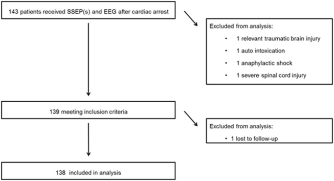
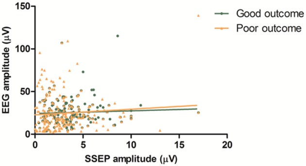
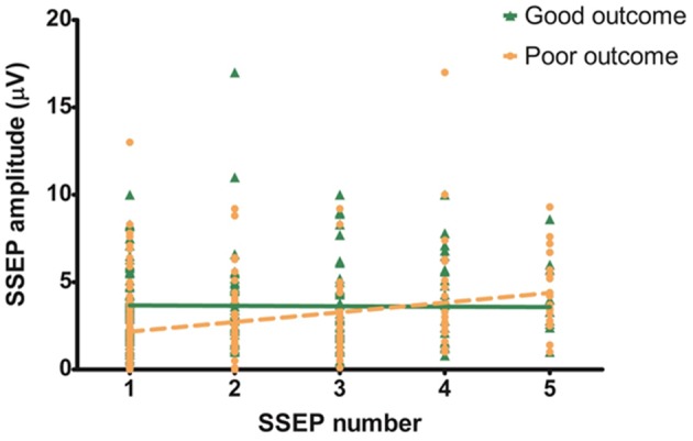
We present relations of SSEP amplitude with neurological outcome and of SSEP amplitude with EEG amplitude in comatose patients after cardiac arrest. This is a analysis of a prospective cohort study in comatose patients after cardiac arrest. Amplitude of SSEP recordings obtained within 48-72 h, and EEG patterns obtained at 12 and 24h after cardiac arrest were related to good (CPC 1-2) or poor (CPC 3-5) outcome at 6 months. In 39% of the study population multiple SSEP measurements were performed. Additionally, SSEP amplitude was related to mean EEG amplitude. We included 138 patients (77% poor outcome). Absent SSEP responses, a N20 amplitude <0.4 μV within 48-72 h, and suppressed or synchronous EEG with suppressed background at 12 or 24 h after cardiac arrest were invariably associated with a poor outcome. Combined, these tests reached a sensitivity for prediction of poor outcome up to 58 at 100% specificity. N20 amplitude increased with a mean of 0.55 μV per day in patients with a poor outcome, and remained stable with a good outcome. There was no statistically significant correlation between SSEP and EEG amplitudes in 182 combined SSEP and EEG measurements ( < 0.01). N20 amplitude <0.4 μV is invariably associated with poor outcome. There is no correlation between SSEP and EEG amplitude. SSEP amplitude analysis may contribute to outcome prediction after cardiac arrest.
我们呈现了心脏骤停后昏迷患者中体感诱发电位(SSEP)波幅与神经功能预后的关系,以及SSEP波幅与脑电图(EEG)波幅的关系。这是一项对心脏骤停后昏迷患者的前瞻性队列研究的分析。心脏骤停后48 - 72小时内获得的SSEP记录波幅,以及心脏骤停后12小时和24小时获得的EEG模式与6个月时良好(脑功能分级[CPC] 1 - 2级)或不良(CPC 3 - 5级)预后相关。在39%的研究人群中进行了多次SSEP测量。此外,SSEP波幅与平均EEG波幅相关。我们纳入了138例患者(77%预后不良)。SSEP反应缺失、48 - 72小时内N20波幅<0.4 μV,以及心脏骤停后12小时或24小时脑电图抑制或背景抑制的同步脑电图均与不良预后始终相关。综合起来,这些测试在100%特异性时对不良预后的预测敏感性高达58%。预后不良的患者N20波幅平均每天增加0.55 μV,而预后良好的患者则保持稳定。在182次SSEP和EEG联合测量中,SSEP与EEG波幅之间无统计学显著相关性(P < 0.01)。N20波幅<0.4 μV始终与不良预后相关。SSEP与EEG波幅之间无相关性。SSEP波幅分析可能有助于心脏骤停后的预后预测。