Kaya Esingül, Grassi Lucia, Benedetti Arianna, Maisetta Giuseppantonio, Pileggi Carolina, Di Luca Mariagrazia, Batoni Giovanna, Esin Semih
Department of Translational Research and New Technologies in Medicine and Surgery, University of Pisa, Pisa, Italy.
Department of Transfusion Medicine and Transplant Biology, Pisa University Hospital, Pisa, Italy.
Front Cell Infect Microbiol. 2020 May 5;10:187. doi: 10.3389/fcimb.2020.00187. eCollection 2020.
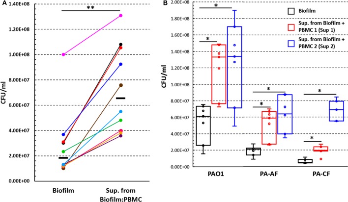
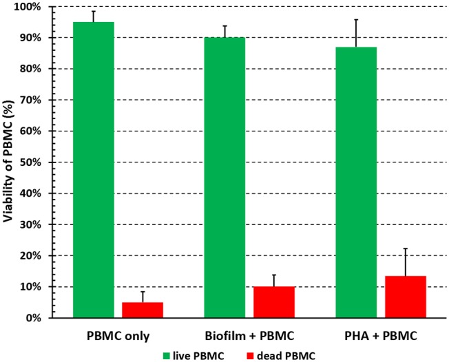
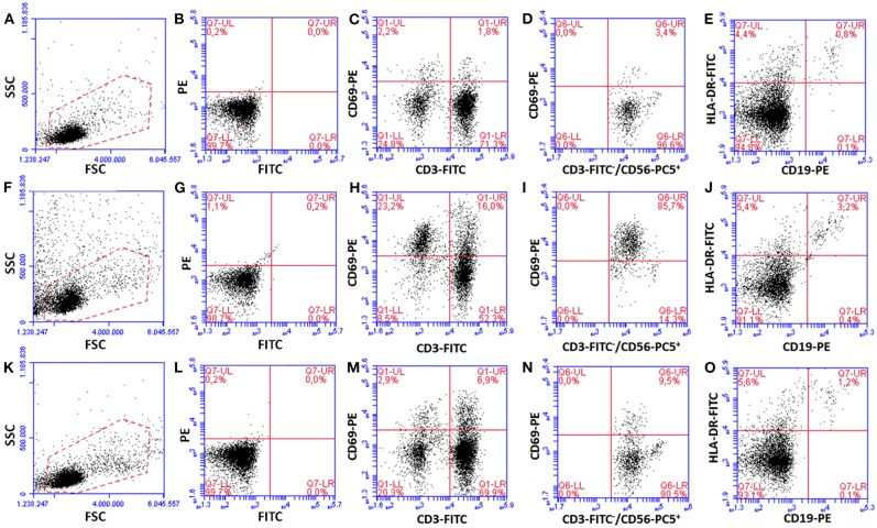
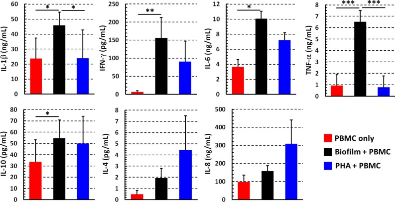
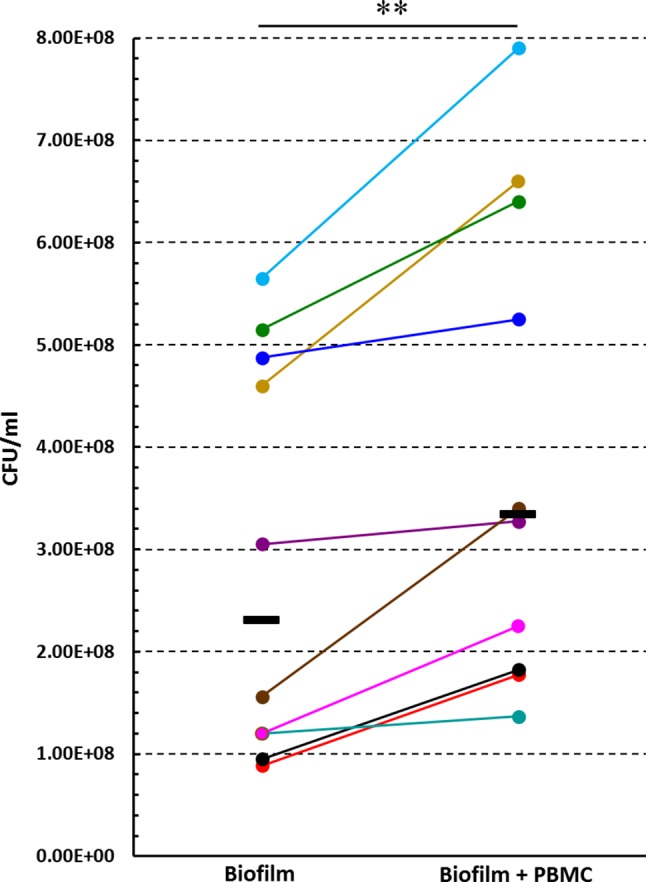
The human immune cell response against bacterial biofilms is a crucial, but still poorly investigated area of research. Herein, we aim to establish an host cell-biofilm interaction model suitable to investigate the peripheral blood mononuclear cell (PBMC) response to biofilms. . biofilms were obtained by incubating bacteria in complete RPMI 1640 medium with 10% human plasma for 24 h. PBMC obtained from healthy donors were added to preformed . biofilms. Following a further 24 h incubation, we assessed (i) PBMC viability and activation; (ii) cytokine profiles in the supernatants; and (iii) CFU counts of biofilm forming bacteria. Cell-death was <10% upon 24 h incubation of PBMC with . biofilms. PBMC incubated for 24 h with preformed . biofilms were significantly more activated compared to PBMC incubated alone. Interestingly, a marked activation of CD56CD3 natural killer (NK) cells was observed that reached 60% of NK cells as an average of different donors. In the culture supernatants of PBMC co-cultured with . biofilms, not only pro-inflammatory (IL-1β, IFN-γ, IL-6, and TNF-α) but also anti-inflammatory (IL-10) cytokines were significantly increased as compared to PBMC incubated alone. Furthermore, incubation of biofilms with PBMC, caused a statistically significant increase in the CFU number of , as compared to biofilms incubated without PBMC. In order to assess whether PBMC products could stimulate the growth of . biofilms, we incubated preformed biofilms with or without supernatants obtained from the co-cultures of PBMC with biofilms. In the presence of the supernatants, the CFU count of biofilm-derived . , was two to seven times higher than those of biofilms incubated without supernatants ( < 0.01). Overall, the results obtained shed light on the reciprocal interaction between human PBMC and . biofilms. . biofilms induced PBMC activation and cytokine secretion but, in turn, the presence of PBMC and/or PBMC-derived components enhanced the number of . biofilm associated bacteria. This may indicate a successful bacterial defensive/persistence strategy against immune response.
人类免疫细胞对细菌生物膜的反应是一个关键但仍未得到充分研究的领域。在此,我们旨在建立一个适合研究外周血单个核细胞(PBMC)对生物膜反应的宿主细胞 - 生物膜相互作用模型。生物膜通过将细菌在含有10%人血浆的完全RPMI 1640培养基中孵育24小时获得。将从健康供体获得的PBMC添加到预先形成的生物膜中。再孵育24小时后,我们评估了:(i)PBMC的活力和活化情况;(ii)上清液中的细胞因子谱;以及(iii)生物膜形成细菌的CFU计数。PBMC与生物膜孵育24小时后细胞死亡率<10%。与单独孵育的PBMC相比,与预先形成的生物膜孵育24小时的PBMC活化程度明显更高。有趣的是,观察到CD56CD3自然杀伤(NK)细胞有明显活化,不同供体的NK细胞平均活化率达到60%。与单独孵育的PBMC相比,与生物膜共培养的PBMC的培养上清液中,不仅促炎细胞因子(IL - 1β、IFN - γ、IL - 6和TNF - α),而且抗炎细胞因子(IL - 10)也显著增加。此外,与未与PBMC孵育的生物膜相比,PBMC与生物膜孵育导致生物膜形成细菌的CFU数量有统计学意义的增加。为了评估PBMC产物是否能刺激生物膜的生长,我们将预先形成的生物膜与有或无PBMC与生物膜共培养上清液一起孵育。在上清液存在的情况下,生物膜来源的CFU计数比未加上清液孵育的生物膜高两到七倍(P<0.01)。总体而言,所获得的结果揭示了人类PBMC与生物膜之间的相互作用。生物膜诱导PBMC活化和细胞因子分泌,但反过来,PBMC和/或PBMC衍生成分的存在增加了生物膜相关细菌的数量。这可能表明细菌针对免疫反应有成功的防御/持续生存策略。