Alvarez Gabriella M, Hackman Daniel A, Miller Adam Bryant, Muscatell Keely A
Department of Psychology & Neuroscience, University of North Carolina at Chapel Hill, Chapel Hill, NC 27599-3270, USA.
USC Suzanne Dworak-Peck School of Social Work, University of Southern California, Los Angeles, CA 90089, USA.
Soc Cogn Affect Neurosci. 2020 Nov 10;15(10):1024-1033. doi: 10.1093/scan/nsaa065.
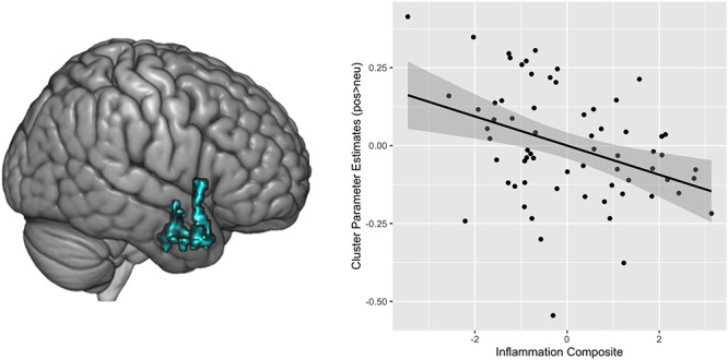
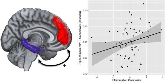
Systemic inflammation is increasingly appreciated as a predictor of health and well-being. Further, inflammation has been shown to influence and be influenced by affective experiences. Although prior work has substantiated associations between inflammatory and affective processes, fewer studies have investigated the neurobiological correlates that underlie links between systemic, low-grade inflammation and affective reactivity. Thus, the current study examined whether markers of systemic inflammation (i.e. interleukin-6, C-reactive protein) are associated with differential patterns of neural activation and connectivity in corticolimbic regions in response to affective images. We investigated this question in a sample of 66 adults (44 women, M age = 54.98 years, range = 35-76) from the Midlife in the United States study. Higher levels of inflammation were associated with lower activity in limbic regions (i.e. amygdala, hippocampus, anterior insula, temporal pole) when viewing positive (vs neutral) images. Higher levels of inflammation were also associated with greater connectivity between the hippocampus and the medial prefrontal cortex in response to positive images. Inflammatory markers were not associated with significant differences in activation or connectivity to negative images. These findings highlight the utility of health neuroscience approaches in demonstrating that physiological processes such as inflammation are related to how our brains respond to affective information.
全身炎症越来越被视为健康和幸福的一个预测指标。此外,炎症已被证明会影响情感体验,并受其影响。尽管先前的研究证实了炎症与情感过程之间的关联,但较少有研究调查全身低度炎症与情感反应之间联系背后的神经生物学相关性。因此,本研究考察了全身炎症标志物(即白细胞介素-6、C反应蛋白)是否与在面对情感图像时皮质边缘区域的神经激活和连接的差异模式相关。我们在美国中年研究的66名成年人(44名女性,年龄中位数=54.98岁,范围=35-76岁)样本中研究了这个问题。当观看积极(与中性相对)图像时,较高水平的炎症与边缘区域(即杏仁核、海马体、前岛叶、颞极)的较低活动相关。较高水平的炎症还与在面对积极图像时海马体与内侧前额叶皮质之间更强的连接性相关。炎症标志物与对消极图像的激活或连接的显著差异无关。这些发现凸显了健康神经科学方法在证明诸如炎症等生理过程与我们大脑对情感信息的反应方式之间的关系方面的效用。