Department of Psychiatry, University of Cambridge, UK.
Department of Clinical Neurosciences, Geneva University Hospitals, Switzerland.
J Alzheimers Dis. 2020;76(1):331-340. doi: 10.3233/JAD-200246.
The changes of cortical structure in Alzheimer's disease (AD) and frontotemporal dementia (FTD) are usually described in terms of atrophy. However, neurodegenerative diseases may also affect the complexity of cortical shape, such as the fractal dimension of the brain surface.
In this study, we aimed at assessing the regional patterns of cortical thickness and fractal dimension changes in a cross-sectional cohort of patients with AD and FTD.
Thirty-two people with symptomatic AD-pathology (clinically probable AD, n = 18, and amyloid-positive mild cognitive impairment, n = 14), 24 with FTD and 28 healthy controls underwent high-resolution 3T structural brain MRI. Using surface-based morphometry, we created vertex-wise cortical thickness and fractal dimension maps for group comparisons and correlations with cognitive measures in AD and FTD.
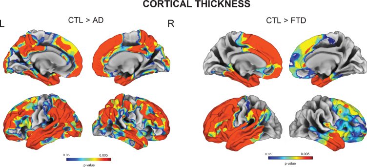
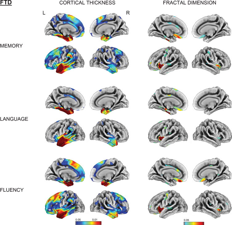
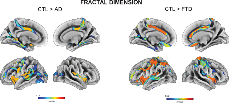
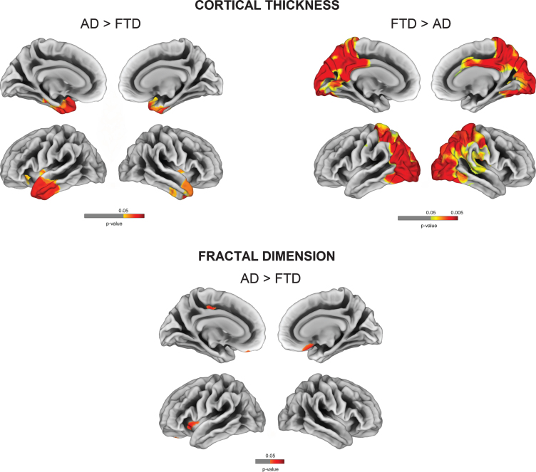
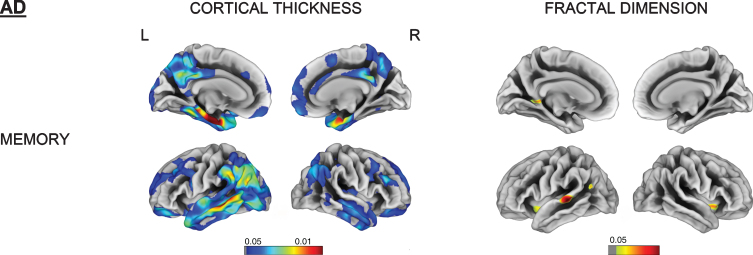
In addition to the well-established pattern of cortical thinning encompassing temporoparietal regions in AD and frontotemporal areas in FTD, we observed reductions of fractal dimension encompassing cingulate areas and insula for both conditions, but specifically involving orbitofrontal cortex and paracentral gyrus for FTD (FDR p < 0.05). Correlational analyses between fractal dimension and cognition showed that these regions were particularly vulnerable with regards to memory and language impairment, especially in FTD.
While the present study demonstrates globally similar patterns of fractal dimension changes in AD and FTD, we observed distinct cortical complexity correlates of cognitive domains impairment. Further studies are required to assess cortical complexity measures at earlier disease stages (e.g., in prodromal/asymptomatic carriers of FTD-related gene mutations) and determine whether fractal dimension represents a sensitive imaging marker for prevention and diagnostic strategies.
阿尔茨海默病(AD)和额颞叶痴呆(FTD)的皮质结构变化通常用萎缩来描述。然而,神经退行性疾病也可能影响皮质形状的复杂性,例如大脑表面的分形维数。
本研究旨在评估 AD 和 FTD 患者横断面队列中皮质厚度和分形维数变化的区域模式。
32 名有症状的 AD 病理患者(临床可能的 AD,n=18,和淀粉样蛋白阳性的轻度认知障碍,n=14)、24 名 FTD 患者和 28 名健康对照者接受了高分辨率 3T 结构脑 MRI 检查。我们使用基于表面的形态测量法,为组间比较创建了顶点级皮质厚度和分形维数图,并将其与 AD 和 FTD 的认知测量值进行了相关性分析。
除了在 AD 中包含颞顶区域和 FTD 中包含额颞区域的公认的皮质变薄模式外,我们还观察到在两种情况下,包括扣带回和岛叶的分形维数减少,但特别是涉及额眶回和旁中央回的 FTD(FDR p<0.05)。分形维数与认知之间的相关分析表明,这些区域在记忆和语言障碍方面特别脆弱,尤其是在 FTD 中。
虽然本研究表明 AD 和 FTD 中分形维数变化具有相似的总体模式,但我们观察到认知领域损伤的皮质复杂度相关性存在差异。需要进一步研究来评估早期疾病阶段的皮质复杂度测量值(例如,在 FTD 相关基因突变的前驱/无症状携带者中),并确定分形维数是否代表预防和诊断策略的敏感影像学标志物。