Biochemistry and Functional Proteomics, Institute of Biology II, Faculty of Biology, University of Freiburg, 79104, Freiburg, Germany.
Department of Molecular Cell Biology, Institute for Cell Biology, University of Bonn, 53121, Bonn, Germany.
Commun Biol. 2020 May 22;3(1):253. doi: 10.1038/s42003-020-0982-5.
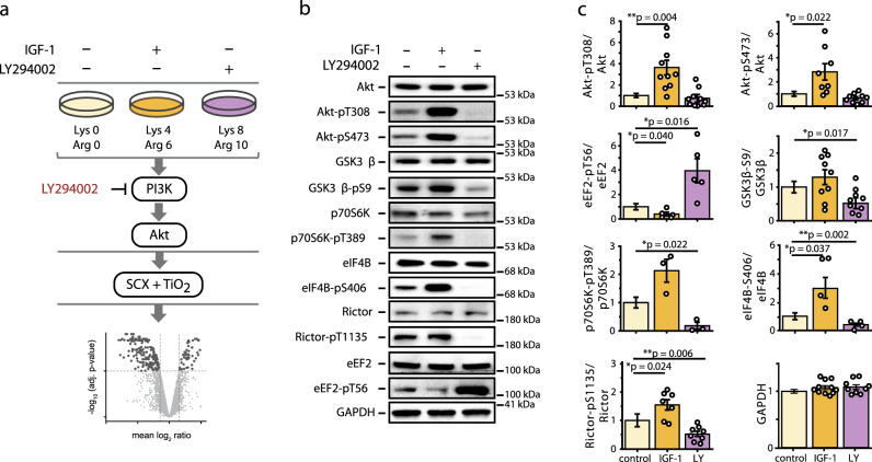
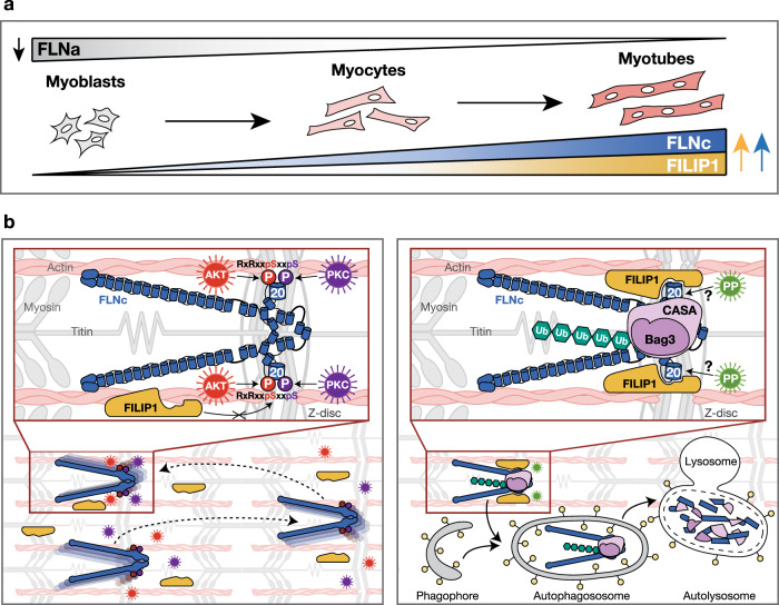
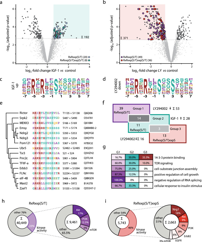
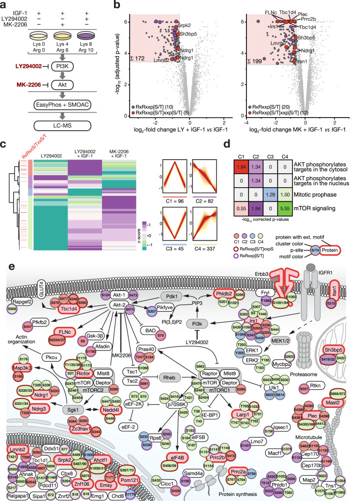
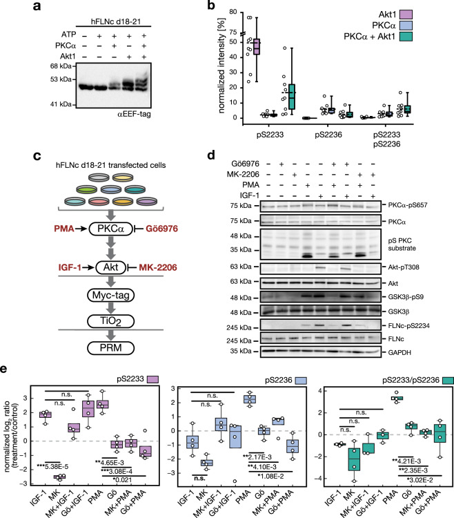
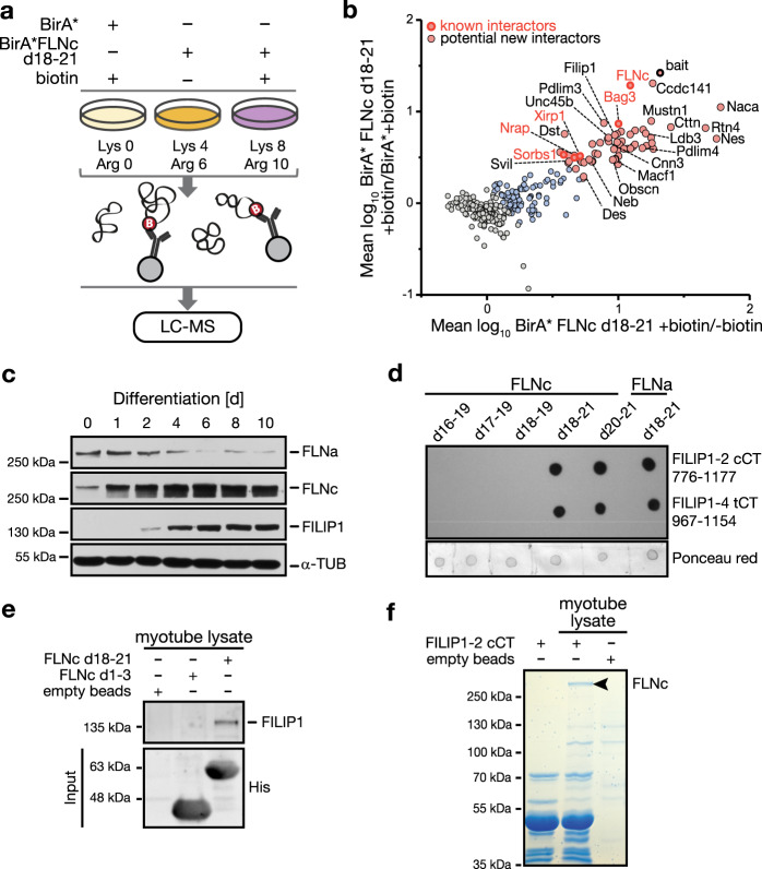
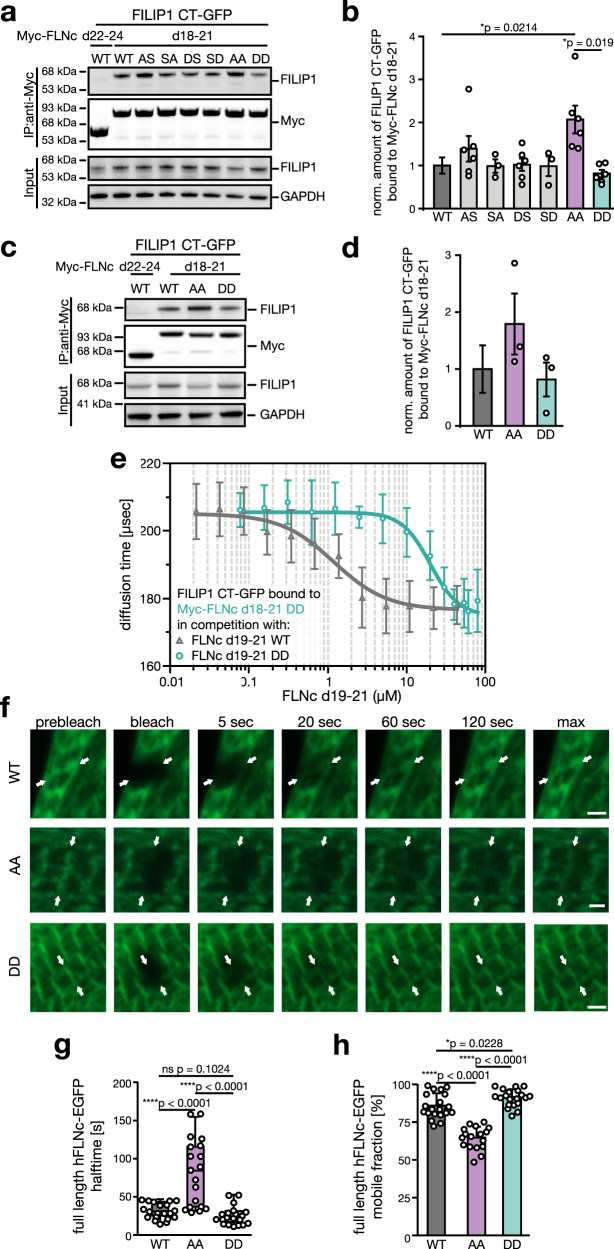
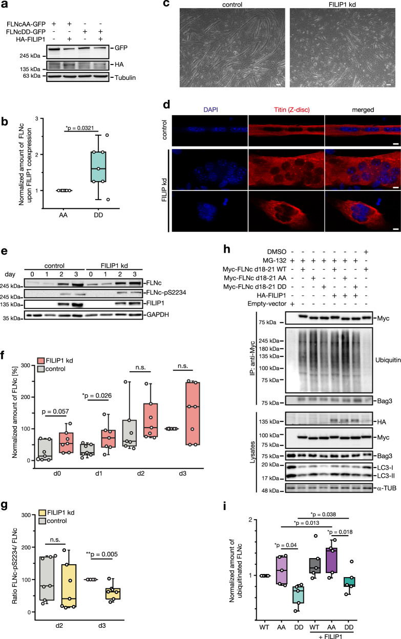
The PI3K/Akt pathway promotes skeletal muscle growth and myogenic differentiation. Although its importance in skeletal muscle biology is well documented, many of its substrates remain to be identified. We here studied PI3K/Akt signaling in contracting skeletal muscle cells by quantitative phosphoproteomics. We identified the extended basophilic phosphosite motif RxRxxp[S/T]xxp[S/T] in various proteins including filamin-C (FLNc). Importantly, this extended motif, located in a unique insert in Ig-like domain 20 of FLNc, is doubly phosphorylated. The protein kinases responsible for this dual-site phosphorylation are Akt and PKCα. Proximity proteomics and interaction analysis identified filamin A-interacting protein 1 (FILIP1) as direct FLNc binding partner. FILIP1 binding induces filamin degradation, thereby negatively regulating its function. Here, dual-site phosphorylation of FLNc not only reduces FILIP1 binding, providing a mechanism to shield FLNc from FILIP1-mediated degradation, but also enables fast dynamics of FLNc necessary for its function as signaling adaptor in cross-striated muscle cells.
PI3K/Akt 通路促进骨骼肌生长和肌生成分化。尽管其在骨骼肌生物学中的重要性已有充分的文献记载,但仍有许多其底物有待鉴定。我们在这里通过定量磷酸蛋白质组学研究了收缩骨骼肌细胞中的 PI3K/Akt 信号。我们在各种蛋白质中鉴定了延伸的碱性磷酸位点模体 RxRxxp[S/T]xxp[S/T],包括细丝蛋白-C (FLNc)。重要的是,这个位于 FLNc 的 Ig 样结构域 20 中的独特插入处的延伸模体被双磷酸化。负责这种双位点磷酸化的蛋白激酶是 Akt 和 PKCα。临近蛋白质组学和相互作用分析鉴定了细丝蛋白 A 相互作用蛋白 1 (FILIP1) 为 FLNc 的直接结合伴侣。FILIP1 结合诱导细丝蛋白降解,从而负调控其功能。在这里,FLNc 的双位点磷酸化不仅减少了 FILIP1 的结合,提供了一种保护 FLNc 免受 FILIP1 介导的降解的机制,而且还使 FLNc 具有快速的动力学,这是其作为横纹肌细胞中信号适配器的功能所必需的。