Department of Biochemistry, Faculty of Science, Mahidol University, Bangkok, Thailand.
Center of Calcium and Bone Research (COCAB), Faculty of Science, Mahidol University, Bangkok, Thailand.
PLoS One. 2020 May 29;15(5):e0234009. doi: 10.1371/journal.pone.0234009. eCollection 2020.
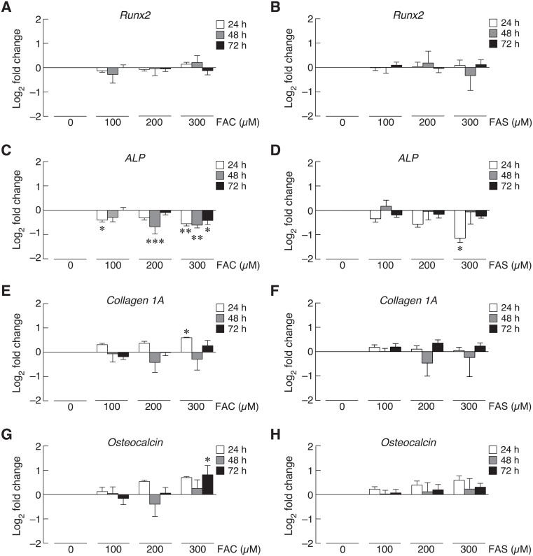
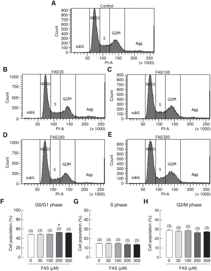
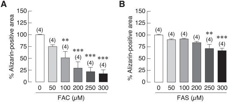
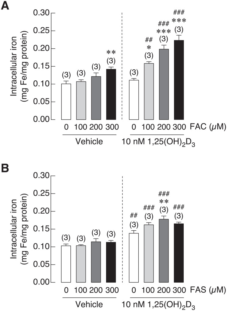
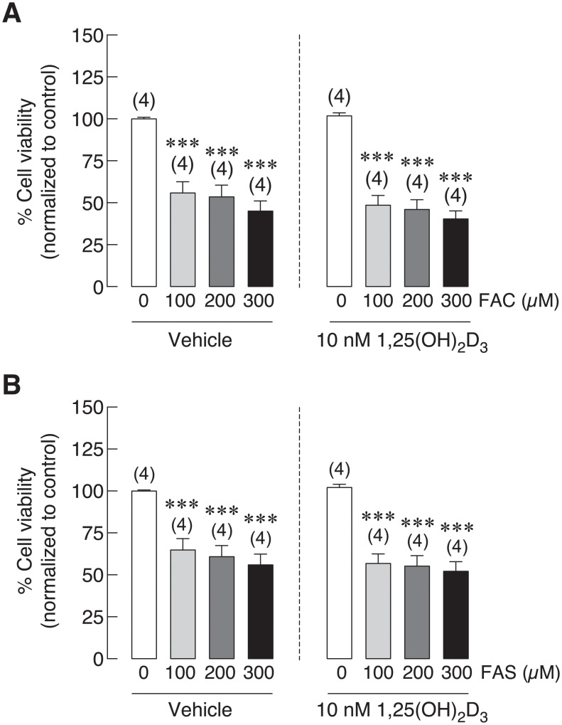
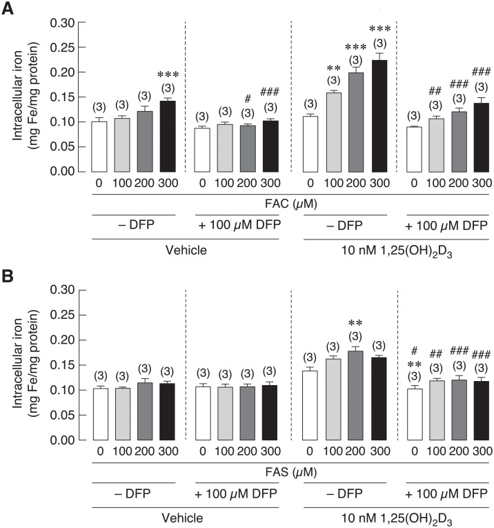
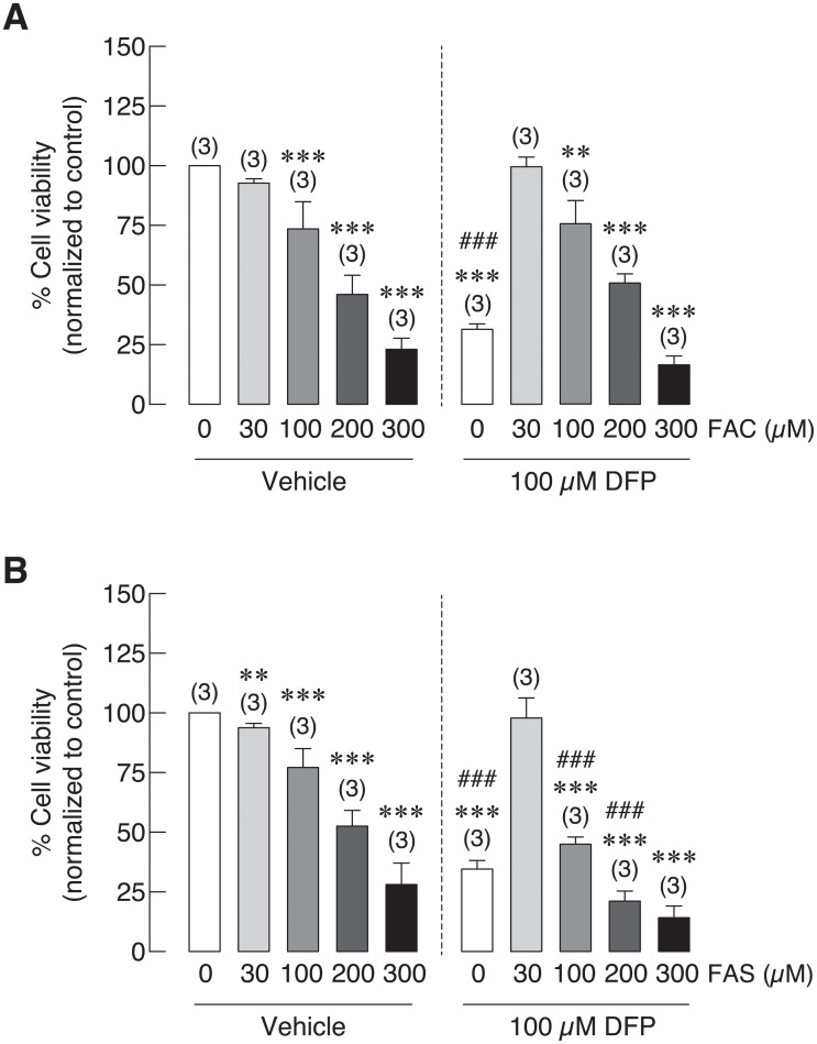
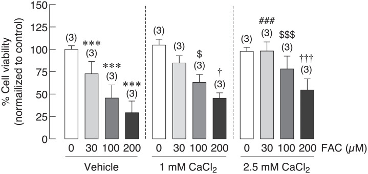
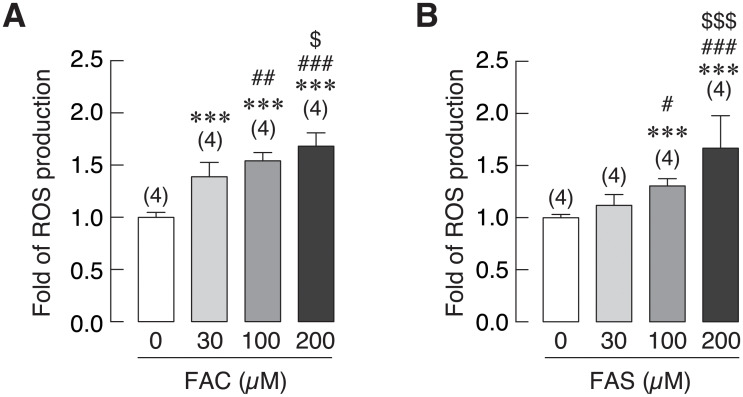
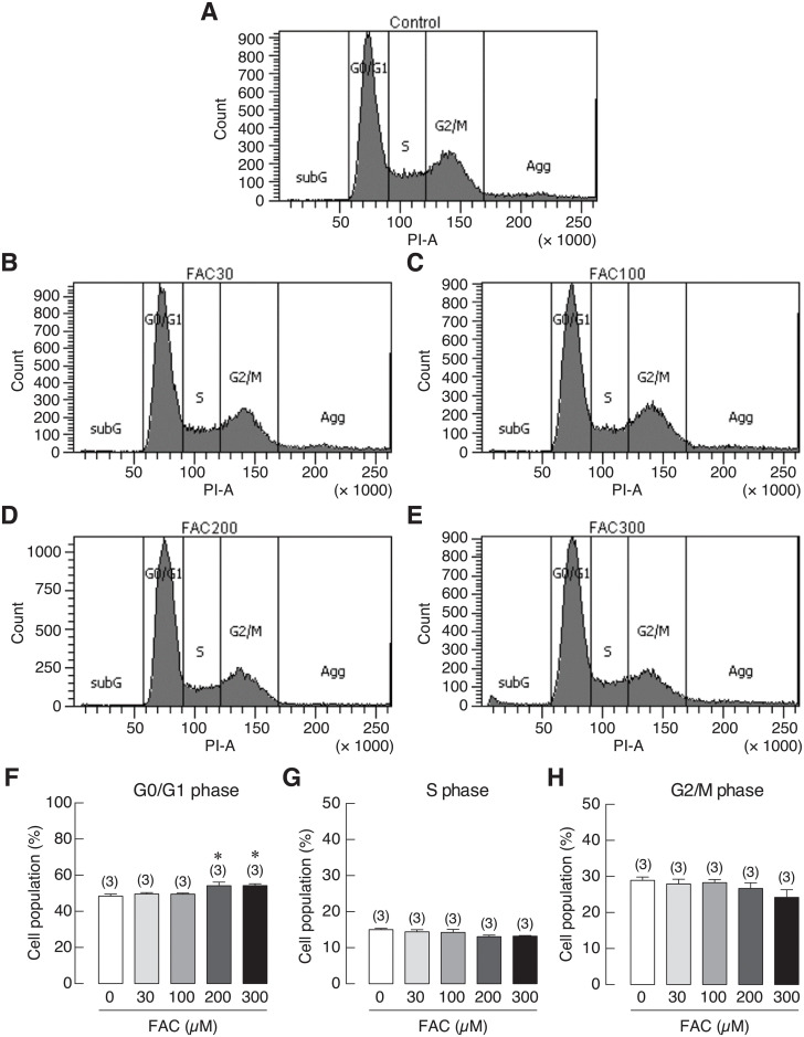
One of the potential contributing factors for iron overload-induced osteoporosis is the iron toxicity on bone forming cells, osteoblasts. In this study, the comparative effects of Fe3+ and Fe2+ on osteoblast differentiation and mineralization were studied in UMR-106 osteoblast cells by using ferric ammonium citrate and ferrous ammonium sulfate as Fe3+ and Fe2+ donors, respectively. Effects of 1,25 dihydroxyvitamin D3 [1,25(OH)2D3] and iron chelator deferiprone on iron uptake ability of osteoblasts were examined, and the potential protective ability of 1,25(OH)2D3, deferiprone and extracellular calcium treatment in osteoblast cell survival under iron overload was also elucidated. The differential effects of Fe3+ and Fe2+ on reactive oxygen species (ROS) production in osteoblasts were also compared. Our results showed that both iron species suppressed alkaline phosphatase gene expression and mineralization with the stronger effects from Fe3+ than Fe2+. 1,25(OH)2D3 significantly increased the intracellular iron but minimally affected osteoblast cell survival under iron overload. Deferiprone markedly decreased intracellular iron in osteoblasts, but it could not recover iron-induced osteoblast cell death. Interestingly, extracellular calcium was able to rescue osteoblasts from iron-induced osteoblast cell death. Additionally, both iron species could induce ROS production and G0/G1 cell cycle arrest in osteoblasts with the stronger effects from Fe3+. In conclusions, Fe3+ and Fe2+ differentially compromised the osteoblast functions and viability, which can be alleviated by an increase in extracellular ionized calcium, but not 1,25(OH)2D3 or iron chelator deferiprone. This study has provided the invaluable information for therapeutic design targeting specific iron specie(s) in iron overload-induced osteoporosis. Moreover, an increase in extracellular calcium could be beneficial for this group of patients.
铁过载诱导骨质疏松的一个潜在致病因素是铁对成骨细胞的毒性。在这项研究中,分别使用柠檬酸铁铵和硫酸亚铁铵作为 Fe3+和 Fe2+供体,研究了 Fe3+和 Fe2+对 UMR-106 成骨细胞分化和矿化的比较影响。研究了 1,25 二羟维生素 D3 [1,25(OH)2D3]和铁螯合剂地拉罗司对成骨细胞摄取铁能力的影响,还阐明了 1,25(OH)2D3、地拉罗司和细胞外钙处理在铁过载下对成骨细胞存活的潜在保护作用。还比较了 Fe3+和 Fe2+对成骨细胞活性氧(ROS)产生的差异影响。我们的结果表明,两种铁均抑制碱性磷酸酶基因表达和矿化,Fe3+的作用强于 Fe2+。1,25(OH)2D3 显著增加细胞内铁,但对铁过载下成骨细胞存活的影响最小。地拉罗司显著降低成骨细胞内铁,但不能恢复铁诱导的成骨细胞死亡。有趣的是,细胞外钙能够挽救铁诱导的成骨细胞死亡。此外,两种铁均能诱导 ROS 产生和 G0/G1 细胞周期停滞,Fe3+的作用更强。总之,Fe3+和 Fe2+ 不同程度地损害成骨细胞功能和活力,增加细胞外离子钙可以缓解,但 1,25(OH)2D3 或铁螯合剂地拉罗司不能缓解。本研究为靶向铁过载诱导骨质疏松中特定铁种类(s)的治疗设计提供了宝贵信息。此外,增加细胞外钙可能对这组患者有益。