• 文献检索
  • 文档翻译
  • 深度研究
  • 学术资讯
  • Suppr Zotero 插件Zotero 插件
  • 邀请有礼
  • 套餐&价格
  • 历史记录
应用&插件
Suppr Zotero 插件Zotero 插件浏览器插件Mac 客户端Windows 客户端微信小程序
定价
高级版会员购买积分包购买API积分包
服务
文献检索文档翻译深度研究API 文档MCP 服务
关于我们
关于 Suppr公司介绍联系我们用户协议隐私条款
关注我们

Suppr 超能文献

核心技术专利:CN118964589B侵权必究
粤ICP备2023148730 号-1Suppr @ 2026

文献检索

告别复杂PubMed语法,用中文像聊天一样搜索,搜遍4000万医学文献。AI智能推荐,让科研检索更轻松。

立即免费搜索

文件翻译

保留排版,准确专业,支持PDF/Word/PPT等文件格式,支持 12+语言互译。

免费翻译文档

深度研究

AI帮你快速写综述,25分钟生成高质量综述,智能提取关键信息,辅助科研写作。

立即免费体验

使用静态和动态[F]-F-DOPA正电子发射断层扫描(PET)参数检测胶质瘤复发或进展患者。

Use of static and dynamic [F]-F-DOPA PET parameters for detecting patients with glioma recurrence or progression.

作者信息

Zaragori Timothée, Ginet Merwan, Marie Pierre-Yves, Roch Véronique, Grignon Rachel, Gauchotte Guillaume, Rech Fabien, Blonski Marie, Lamiral Zohra, Taillandier Luc, Imbert Laëtitia, Verger Antoine

机构信息

Department of Nuclear Medicine & Nancyclotep Imaging platform, Université de Lorraine, CHRU-Nancy, F-54000, Nancy, France.

IADI, INSERM, UMR 1254, Université de Lorraine, F-54000, Nancy, France.

出版信息

EJNMMI Res. 2020 May 29;10(1):56. doi: 10.1186/s13550-020-00645-x.

DOI:10.1186/s13550-020-00645-x
PMID:32472232
原文链接:https://pmc.ncbi.nlm.nih.gov/articles/PMC7260331/
Abstract

BACKGROUND

Static [F]-F-DOPA PET images are currently used for identifying patients with glioma recurrence/progression after treatment, although the additional diagnostic value of dynamic parameters remains unknown in this setting. The aim of this study was to evaluate the performances of static and dynamic [F]-F-DOPA PET parameters for detecting patients with glioma recurrence/progression as well as assess further relationships with patient outcome.

METHODS

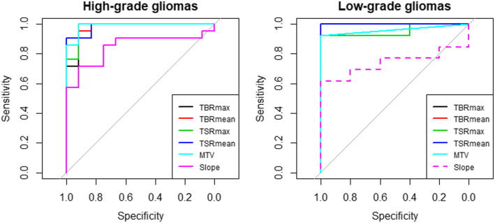
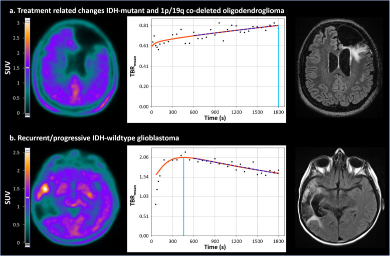
Fifty-one consecutive patients who underwent an [F]-F-DOPA PET for a suspected glioma recurrence/progression at post-resection MRI, were retrospectively included. Static parameters, including mean and maximum tumor-to-normal-brain (TBR) ratios, tumor-to-striatum (TSR) ratios, and metabolic tumor volume (MTV), as well as dynamic parameters with time-to-peak (TTP) values and curve slope, were tested for predicting the following: (1) glioma recurrence/progression at 6 months after the PET exam and (2) survival on longer follow-up.

RESULTS

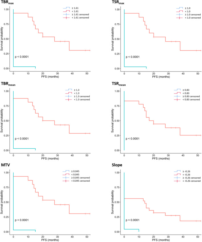
All static parameters were significant predictors of glioma recurrence/progression (accuracy ≥ 94%) with all parameters also associated with mean progression-free survival (PFS) in the overall population (all p < 0.001, 29.7 vs. 0.4 months for TBR, TSR, and MTV). The curve slope was the sole dynamic PET predictor of glioma recurrence/progression (accuracy = 76.5%) and was also associated with mean PFS (p < 0.001, 18.0 vs. 0.4 months). However, no additional information was provided relative to static parameters in multivariate analysis.

CONCLUSION

Although patients with glioma recurrence/progression can be detected by both static and dynamic [F]-F-DOPA PET parameters, most of this diagnostic information can be achieved by conventional static parameters.

摘要

背景

目前,静态[F]-氟多巴正电子发射断层扫描(PET)图像用于识别治疗后胶质瘤复发/进展的患者,尽管在此情况下动态参数的附加诊断价值仍不清楚。本研究的目的是评估静态和动态[F]-氟多巴PET参数检测胶质瘤复发/进展患者的性能,并进一步评估与患者预后的关系。

方法

回顾性纳入51例在切除术后MRI怀疑胶质瘤复发/进展而行[F]-氟多巴PET检查的连续患者。测试静态参数,包括平均和最大肿瘤与正常脑(TBR)比值、肿瘤与纹状体(TSR)比值和代谢肿瘤体积(MTV),以及具有达峰时间(TTP)值和曲线斜率的动态参数,以预测以下情况:(1)PET检查后6个月的胶质瘤复发/进展,以及(2)更长随访期的生存期。

结果

所有静态参数都是胶质瘤复发/进展的显著预测指标(准确率≥94%),所有参数也与总体人群的平均无进展生存期(PFS)相关(所有p<0.001,TBR、TSR和MTV的PFS分别为29.7个月和0.4个月)。曲线斜率是胶质瘤复发/进展的唯一动态PET预测指标(准确率=76.5%),也与平均PFS相关(p<0.001,分别为18.0个月和0.4个月)。然而,在多变量分析中,相对于静态参数没有提供额外信息。

结论

尽管静态和动态[F]-氟多巴PET参数均可检测胶质瘤复发/进展患者,但大多数诊断信息可通过传统静态参数获得。

https://cdn.ncbi.nlm.nih.gov/pmc/blobs/29c3/7260331/960b1f5b83af/13550_2020_645_Fig3_HTML.jpg
https://cdn.ncbi.nlm.nih.gov/pmc/blobs/29c3/7260331/978b94e59c5d/13550_2020_645_Fig1_HTML.jpg
https://cdn.ncbi.nlm.nih.gov/pmc/blobs/29c3/7260331/600bdfb1e413/13550_2020_645_Fig2_HTML.jpg
https://cdn.ncbi.nlm.nih.gov/pmc/blobs/29c3/7260331/960b1f5b83af/13550_2020_645_Fig3_HTML.jpg
https://cdn.ncbi.nlm.nih.gov/pmc/blobs/29c3/7260331/978b94e59c5d/13550_2020_645_Fig1_HTML.jpg
https://cdn.ncbi.nlm.nih.gov/pmc/blobs/29c3/7260331/600bdfb1e413/13550_2020_645_Fig2_HTML.jpg
https://cdn.ncbi.nlm.nih.gov/pmc/blobs/29c3/7260331/960b1f5b83af/13550_2020_645_Fig3_HTML.jpg

相似文献

1
Use of static and dynamic [F]-F-DOPA PET parameters for detecting patients with glioma recurrence or progression.使用静态和动态[F]-F-DOPA正电子发射断层扫描(PET)参数检测胶质瘤复发或进展患者。
EJNMMI Res. 2020 May 29;10(1):56. doi: 10.1186/s13550-020-00645-x.
2
Differentiating high-grade glioma progression from treatment-related changes with dynamic [F]FDOPA PET: a multicentric study.动态 [F]FDOPA PET 鉴别高级别胶质瘤进展与治疗相关性改变:一项多中心研究。
Eur Radiol. 2023 Apr;33(4):2548-2560. doi: 10.1007/s00330-022-09221-4. Epub 2022 Nov 11.
3
Diagnostic and Prognostic Potential of F-FET PET in the Differential Diagnosis of Glioma Recurrence and Treatment-Induced Changes After Chemoradiation Therapy.F-FET PET在胶质瘤复发与放化疗后治疗引起的改变的鉴别诊断中的诊断及预后潜力
Front Oncol. 2021 Oct 4;11:721821. doi: 10.3389/fonc.2021.721821. eCollection 2021.
4
The use of dynamic O-(2-18F-fluoroethyl)-l-tyrosine PET in the diagnosis of patients with progressive and recurrent glioma.动态O-(2-¹⁸F-氟乙基)-L-酪氨酸PET在进展性和复发性胶质瘤患者诊断中的应用
Neuro Oncol. 2015 Sep;17(9):1293-300. doi: 10.1093/neuonc/nov088. Epub 2015 May 24.
5
Grading and outcome prediction of pediatric diffuse astrocytic tumors with diffusion and arterial spin labeling perfusion MRI in comparison with 18F-DOPA PET.弥散张量成像和动脉自旋标记灌注 MRI 对儿童弥漫性星形细胞瘤分级和预后预测与 18F-DOPA PET 的比较
Eur J Nucl Med Mol Imaging. 2017 Nov;44(12):2084-2093. doi: 10.1007/s00259-017-3777-2. Epub 2017 Jul 27.
6
Relevance of Dynamic F-DOPA PET Radiomics for Differentiation of High-Grade Glioma Progression from Treatment-Related Changes.动态F-DOPA PET放射组学在鉴别高级别胶质瘤进展与治疗相关变化中的相关性
Biomedicines. 2021 Dec 16;9(12):1924. doi: 10.3390/biomedicines9121924.
7
Dynamic C-Methionine PET-CT: Prognostic Factors for Disease Progression and Survival in Patients with Suspected Glioma Recurrence.动态C-蛋氨酸PET-CT:疑似胶质瘤复发患者疾病进展和生存的预后因素
Cancers (Basel). 2021 Sep 24;13(19):4777. doi: 10.3390/cancers13194777.
8
F-DOPA uptake does not correlate with IDH mutation status and 1p/19q co-deletion in glioma.F-DOPA摄取与胶质瘤中的异柠檬酸脱氢酶(IDH)突变状态及1p/19q共缺失无关。
Ann Nucl Med. 2019 Apr;33(4):295-302. doi: 10.1007/s12149-018-01328-3. Epub 2019 Jan 3.
9
F-FET PET prior to recurrent high-grade glioma re-irradiation-additional prognostic value of dynamic time-to-peak analysis and early static summation images?复发性高级别胶质瘤再照射前的F-FET PET——动态达峰时间分析和早期静态总和图像的额外预后价值?
J Neurooncol. 2017 Apr;132(2):277-286. doi: 10.1007/s11060-016-2366-8. Epub 2017 Jan 19.
10
Serial 18F-FET PET Imaging of Primarily 18F-FET-Negative Glioma: Does It Make Sense?原发性18F-FET阴性胶质瘤的连续18F-FET PET成像:有意义吗?
J Nucl Med. 2016 Aug;57(8):1177-82. doi: 10.2967/jnumed.115.171033. Epub 2016 Mar 31.

引用本文的文献

1
Case Report: F-FDOPA PET in the clinical management of a dog with an intraventricular tumor suspected to be choroid plexus papilloma.病例报告:氟代多巴正电子发射断层扫描在一只疑似患有脉络丛乳头状瘤的脑室肿瘤犬临床管理中的应用
Front Vet Sci. 2025 Mar 26;12:1477063. doi: 10.3389/fvets.2025.1477063. eCollection 2025.
2
Leveraging machine learning with dynamic 18F-FDG PET/CT: integrating metabolic and flow features for lung cancer differential diagnosis.利用机器学习与动态18F-FDG PET/CT:整合代谢和血流特征用于肺癌鉴别诊断。
Eur J Nucl Med Mol Imaging. 2025 Apr 4. doi: 10.1007/s00259-025-07231-0.
3
A study method using early dynamic acquisition of [F]fluorodopa positron emission tomography for the differential diagnosis between progression and radionecrosis of brain metastases after radiotherapy.

本文引用的文献

1
Reply to Zaragori et al.: "Is IDH mutation status associated with F-FDopa PET uptake".对扎拉戈里等人的回复:“异柠檬酸脱氢酶(IDH)突变状态与氟代多巴正电子发射断层扫描(F-FDopa PET)摄取有关吗”
Ann Nucl Med. 2020 Mar;34(3):230-231. doi: 10.1007/s12149-020-01443-0.
2
Is IDH mutation status associated with F-FDopa PET uptake?异柠檬酸脱氢酶(IDH)突变状态与F-多巴正电子发射断层扫描(PET)摄取有关吗?
Ann Nucl Med. 2020 Mar;34(3):228-229. doi: 10.1007/s12149-020-01442-1. Epub 2020 Jan 30.
3
Integration of dynamic parameters in the analysis of F-FDopa PET imaging improves the prediction of molecular features of gliomas.
一种利用早期动态采集[F]氟多巴正电子发射断层扫描进行放疗后脑转移瘤进展与放射性坏死鉴别诊断的研究方法。
EJNMMI Res. 2024 Oct 9;14(1):93. doi: 10.1186/s13550-024-01158-7.
4
Complex Diagnostic Challenges in Glioblastoma: The Role of F-FDOPA PET Imaging.胶质母细胞瘤的复杂诊断挑战:F-FDOPA PET成像的作用
Pharmaceuticals (Basel). 2024 Sep 15;17(9):1215. doi: 10.3390/ph17091215.
5
Accurate brain pharmacokinetic parametric imaging using the blood input function extracted from the cavernous sinus.利用从海绵窦提取的血液输入函数进行准确的脑药代动力学参数成像。
Am J Nucl Med Mol Imaging. 2024 Aug 25;14(4):272-281. doi: 10.62347/LSYG1380. eCollection 2024.
6
ASL, DSC, DCE perfusion MRI and 18F-DOPA PET/CT in differentiating glioma recurrence from post-treatment changes.磁共振 ASL、DSC、DCE 灌注成像及 18F-DOPA PET/CT 鉴别脑胶质瘤术后复发与治疗后改变
Radiol Med. 2024 Sep;129(9):1382-1393. doi: 10.1007/s11547-024-01862-3. Epub 2024 Aug 8.
7
Recent Update on PET/CT Radiotracers for Imaging Cerebral Glioma.用于脑胶质瘤成像的PET/CT放射性示踪剂的最新进展
Nucl Med Mol Imaging. 2024 Jun;58(4):237-245. doi: 10.1007/s13139-024-00847-4. Epub 2024 Feb 21.
8
Application of PET imaging delta radiomics for predicting progression-free survival in rare high-grade glioma.PET 成像 delta 放射组学在罕见高级别胶质瘤无进展生存期预测中的应用。
Sci Rep. 2024 Feb 8;14(1):3256. doi: 10.1038/s41598-024-53693-x.
9
Comparing [18F]FET PET and [18F]FDOPA PET for glioma recurrence diagnosis: a systematic review and meta-analysis.比较[18F]FET PET和[18F]FDOPA PET用于胶质瘤复发诊断:一项系统评价和荟萃分析。
Front Oncol. 2024 Jan 10;13:1346951. doi: 10.3389/fonc.2023.1346951. eCollection 2023.
10
Evolution and implementation of radiographic response criteria in neuro-oncology.神经肿瘤学中影像学反应标准的演变与实施
Neurooncol Adv. 2023 Sep 13;5(1):vdad118. doi: 10.1093/noajnl/vdad118. eCollection 2023 Jan-Dec.
在F-FDopa PET成像分析中整合动态参数可改善对胶质瘤分子特征的预测。
Eur J Nucl Med Mol Imaging. 2020 Jun;47(6):1381-1390. doi: 10.1007/s00259-019-04509-y. Epub 2019 Sep 16.
4
F-FET PET Imaging in Differentiating Glioma Progression from Treatment-Related Changes: A Single-Center Experience.F-FET PET 成像在鉴别胶质瘤进展与治疗相关变化中的应用:单中心经验。
J Nucl Med. 2020 Apr;61(4):505-511. doi: 10.2967/jnumed.119.234757. Epub 2019 Sep 13.
5
F-DOPA uptake does not correlate with IDH mutation status and 1p/19q co-deletion in glioma.F-DOPA摄取与胶质瘤中的异柠檬酸脱氢酶(IDH)突变状态及1p/19q共缺失无关。
Ann Nucl Med. 2019 Apr;33(4):295-302. doi: 10.1007/s12149-018-01328-3. Epub 2019 Jan 3.
6
Comparisons Between PET With 11C-Methyl-L-Methionine and Arterial Spin Labeling Perfusion Imaging in Recurrent Glioblastomas Treated With Bevacizumab.贝伐珠单抗治疗后复发性胶质母细胞瘤中 11C-甲基-L-蛋氨酸 PET 与动脉自旋标记灌注成像的比较。
Clin Nucl Med. 2019 Mar;44(3):186-193. doi: 10.1097/RLU.0000000000002417.
7
Joint EANM/EANO/RANO practice guidelines/SNMMI procedure standards for imaging of gliomas using PET with radiolabelled amino acids and [F]FDG: version 1.0.EANM/EANO/RANO 联合实践指南/SNMMI 程序标准:使用放射性标记氨基酸和 [F]FDG 的脑胶质瘤 PET 成像:第 1.0 版。
Eur J Nucl Med Mol Imaging. 2019 Mar;46(3):540-557. doi: 10.1007/s00259-018-4207-9. Epub 2018 Dec 5.
8
Influence of Dexamethasone on O-(2-[F]-Fluoroethyl)-L-Tyrosine Uptake in the Human Brain and Quantification of Tumor Uptake.地塞米松对人脑 O-(2-[F]-氟乙基)-L-酪氨酸摄取的影响及肿瘤摄取的定量分析。
Mol Imaging Biol. 2019 Feb;21(1):168-174. doi: 10.1007/s11307-018-1221-z.
9
Diagnosis of glioma recurrence using multiparametric dynamic 18F-fluoroethyl-tyrosine PET-MRI.使用多参数动态 18F-氟乙基酪氨酸 PET-MRI 诊断脑胶质瘤复发。
Eur J Radiol. 2018 Jun;103:32-37. doi: 10.1016/j.ejrad.2018.04.003. Epub 2018 Apr 4.
10
Comparison of O-(2-F-Fluoroethyl)-L-Tyrosine Positron Emission Tomography and Perfusion-Weighted Magnetic Resonance Imaging in the Diagnosis of Patients with Progressive and Recurrent Glioma: A Hybrid Positron Emission Tomography/Magnetic Resonance Study.O-(2-氟乙基)-L-酪氨酸正电子发射断层扫描与灌注加权磁共振成像在进展性和复发性胶质瘤患者诊断中的比较:一项正电子发射断层扫描/磁共振混合研究
World Neurosurg. 2018 May;113:e727-e737. doi: 10.1016/j.wneu.2018.02.139. Epub 2018 Mar 3.