Inui Shohei, Fujikawa Akira, Jitsu Motoyuki, Kunishima Naoaki, Watanabe Sadahiro, Suzuki Yuhi, Umeda Satoshi, Uwabe Yasuhide
Departments of Radiology (S.I., A.F., M.J., N.K., S.W., Y.S., S.U.) and Respiratory Medicine (Y.U.), Japan Self-Defense Forces Central Hospital, Tokyo, Japan.
Radiol Cardiothorac Imaging. 2020 Mar 17;2(2):e200110. doi: 10.1148/ryct.2020200110. eCollection 2020 Apr.
To evaluate the chest CT findings in an environmentally homogeneous cohort from the cruise ship with coronavirus disease 2019 (COVID-19).
This retrospective study comprised 104 cases (mean age, 62 years ± 16 [standard deviation], range, 25-93 years) with COVID-19 confirmed with reverse-transcription polymerase change reaction findings. CT images were reviewed, and the CT severity score was calculated for each lobe and the entire lung. CT findings were compared between asymptomatic and symptomatic cases.
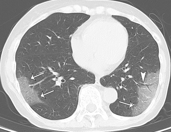
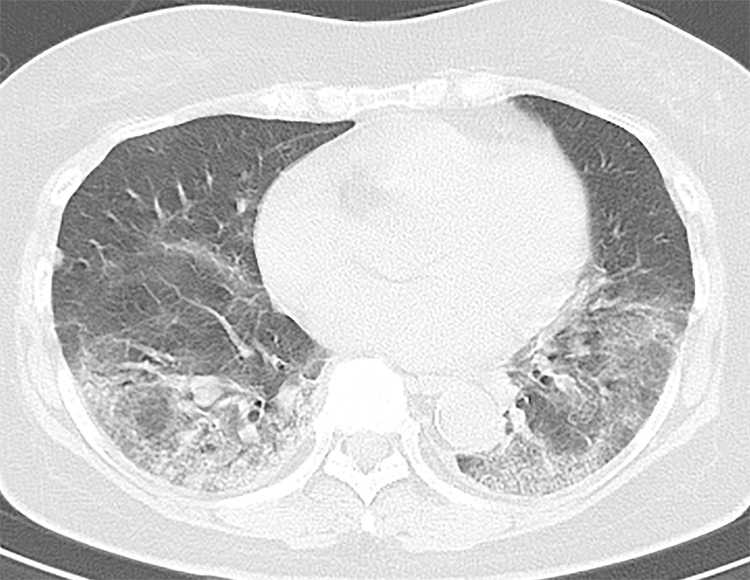
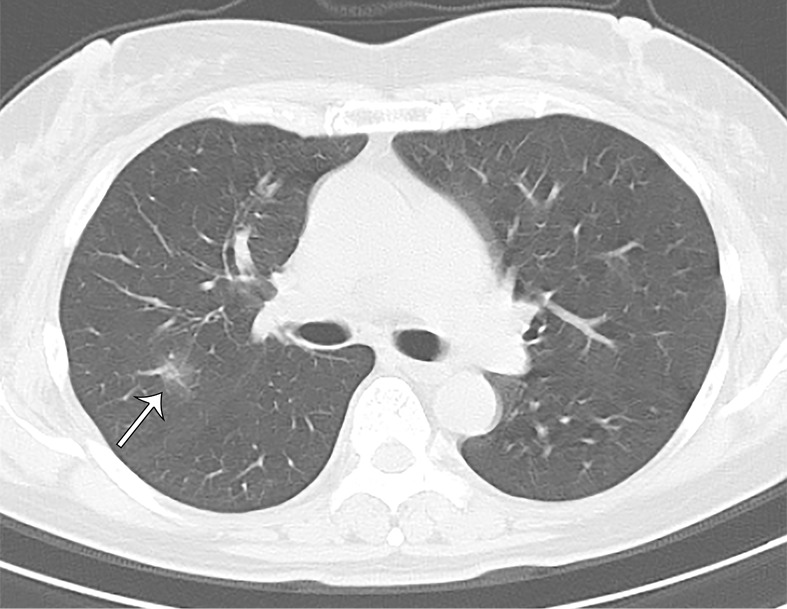
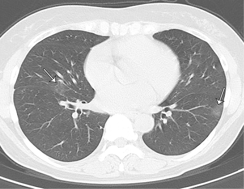
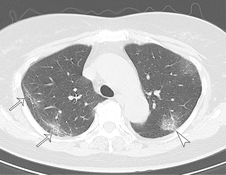
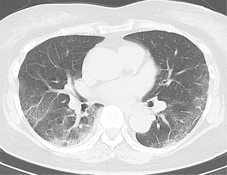
Of 104 cases, 76 (73%) were asymptomatic, 41 (54%) of which had lung opacities on CT. Twenty-eight (27%) cases were symptomatic, 22 (79%) of which had abnormal CT findings. Symptomatic cases showed lung opacities and airway abnormalities on CT more frequently than asymptomatic cases [lung opacity; 22 (79%) vs 41 (54%), airway abnormalities; 14 (50%) vs 15 (20%)]. Asymptomatic cases showed more ground-glass opacity (GGO) over consolidation (83%), while symptomatic cases more frequently showed consolidation over GGO (41%). The CT severity score was higher in symptomatic cases than asymptomatic cases, particularly in the lower lobes [symptomatic vs asymptomatic cases; right lower lobe: 2 ± 1 (0-4) vs 1 ± 1 (0-4); left lower lobe: 2 ± 1 (0-4) vs 1 ± 1 (0-3); total score: 7 ± 5 (1-17) vs 4 ± 2 (1-11)].
This study documented a high incidence of subclinical CT changes in cases with COVID-19. Compared with symptomatic cases, asymptomatic cases showed more GGO over consolidation and milder extension of disease on CT.An earlier incorrect version appeared online. This article was corrected on April 8, 2020.© RSNA, 2020.
评估2019冠状病毒病(COVID-19)游轮上环境同质队列中的胸部CT表现。
本回顾性研究纳入了104例经逆转录聚合酶链反应结果确诊为COVID-19的病例(平均年龄62岁±16[标准差],范围25-93岁)。对CT图像进行了分析,并计算每个肺叶和全肺的CT严重程度评分。比较了无症状和有症状病例的CT表现。
104例中,76例(73%)无症状,其中41例(54%)CT显示肺部有实变影。28例(27%)有症状,其中22例(79%)CT表现异常。有症状病例在CT上比无症状病例更频繁地出现肺部实变影和气道异常[肺部实变影;22例(79%)对41例(54%),气道异常;14例(50%)对15例(20%)]。无症状病例磨玻璃影(GGO)多于实变影(83%),而有症状病例实变影多于磨玻璃影(41%)。有症状病例的CT严重程度评分高于无症状病例,尤其是在下叶[有症状与无症状病例;右下叶:2±1(0-4)对1±1(0-4);左下叶:2±1(0-4)对1±1(0-3);总分:7±5(1-17)对4±2(1-11)]。
本研究记录了COVID-19病例中亚临床CT改变的高发生率。与有症状病例相比,无症状病例在CT上磨玻璃影多于实变影,且疾病累及范围较轻。本文的一个早期错误版本已在线发布。本文于2020年4月8日进行了更正。©RSNA,2020。