• 文献检索
  • 文档翻译
  • 深度研究
  • 学术资讯
  • Suppr Zotero 插件Zotero 插件
  • 邀请有礼
  • 套餐&价格
  • 历史记录
应用&插件
Suppr Zotero 插件Zotero 插件浏览器插件Mac 客户端Windows 客户端微信小程序
定价
高级版会员购买积分包购买API积分包
服务
文献检索文档翻译深度研究API 文档MCP 服务
关于我们
关于 Suppr公司介绍联系我们用户协议隐私条款
关注我们

Suppr 超能文献

核心技术专利:CN118964589B侵权必究
粤ICP备2023148730 号-1Suppr @ 2026

文献检索

告别复杂PubMed语法,用中文像聊天一样搜索,搜遍4000万医学文献。AI智能推荐,让科研检索更轻松。

立即免费搜索

文件翻译

保留排版,准确专业,支持PDF/Word/PPT等文件格式,支持 12+语言互译。

免费翻译文档

深度研究

AI帮你快速写综述,25分钟生成高质量综述,智能提取关键信息,辅助科研写作。

立即免费体验

3T对比增强全心冠状动脉磁共振血管造影与双源计算机断层扫描血管造影检测冠状动脉狭窄的直接比较:单中心经验

A direct comparison of 3 T contrast-enhanced whole-heart coronary cardiovascular magnetic resonance angiography to dual-source computed tomography angiography for detection of coronary artery stenosis: a single-center experience.

作者信息

Sun Bin, Chen Zhiyong, Duan Qing, Xue Yunjing, Chen Lianglong, Zhang Zhongshuai, An Jing

机构信息

Department of Radiology, Union Hospital, Fujian Medical University, 29 Xin-Quan Road, Fuzhou, 350001, People's Republic of China.

Department of Cardiology, Union Hospital, Fujian Medical University, Fuzhou, China.

出版信息

J Cardiovasc Magn Reson. 2020 Jun 1;22(1):40. doi: 10.1186/s12968-020-00630-2.

DOI:10.1186/s12968-020-00630-2
PMID:32475355
原文链接:https://pmc.ncbi.nlm.nih.gov/articles/PMC7262765/
Abstract

BACKGROUND

In recent years, substantial advances have been made in noninvasive cardiac imaging, including cardiac computed tomography (CT) and cardiovascular magnetic resonance (CMR). The purpose of this study was to prospectively compare the diagnostic performance of contrast-enhanced whole heart coronary CMR angiography (CCMRA) to dual-source coronary CT angiography (CCTA) for the diagnosis of significant coronary stenoses (≥50%) in patients with known or suspected coronary artery disease (CAD) referred for conventional x-ray coronary angiography.

METHODS

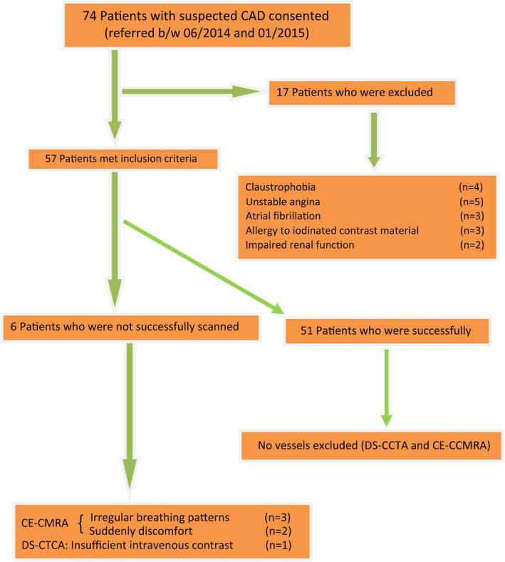
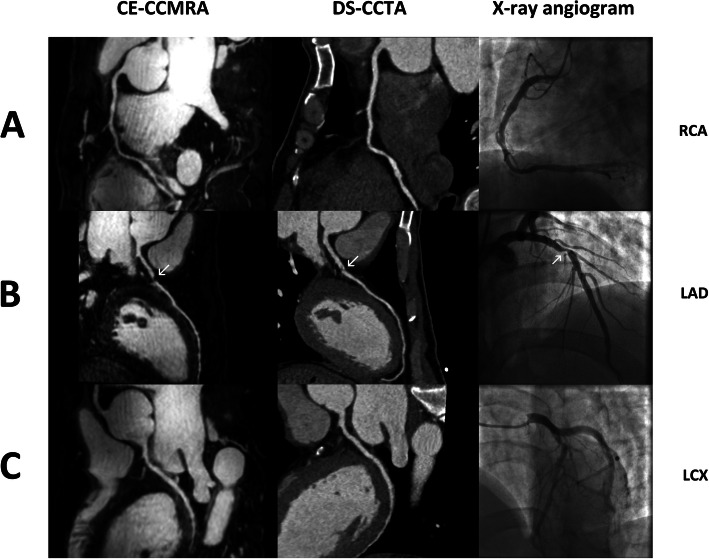
Our objective was to directly compare the diagnostic accuracy of contrast-enhanced whole-heart CCMRA (CE-CCMRA) to dual-source CCTA (DS-CCTA) for the detection of CAD. We prospectively studied 57 symptomatic patients with suspected or known CAD who were scheduled for conventional x-ray coronary angiography. Significant CAD was defined as an x-ray defined diameter reduction of ≥50% in a coronary artery with a reference diameter of ≥1.5 mm.

RESULTS

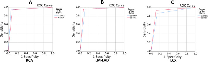
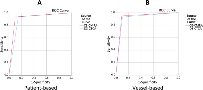
CE-CCMRA and DS-CCTA were completed in 51 (89%) of 57 patients without complications. The acquisition times of CE-CCMRA and DS-CCTA, respectively, were 9.5 ± 3.1 min and 8.3 ± 1.4 s. On patient-based analysis, the sensitivity, specificity, positive and negative predictive value of CE-CCMRA and DS-CCTA were 93.5% versus 93.5%(P > 0.05), 85% versus 90%(P > 0.05), 90.6% versus 93.5%(P > 0.05), and 89.4% versus 90%(P > 0.05), respectively. The area under the curve (AUC) was 0.89 (95% CI: 0.79 to 0.99) for CE-CCMRA and 0.92 (95% CI: 0.83 to 1.00) for DS-CCTA.

CONCLUSIONS

DS-CCTA was found to be superior to CE-CCMRA in the diagnosis of significant coronary stenoses (≥50%) in patients with suspected or known CAD scheduled for conventional x-ray coronary angiography, owing to shorter scanning times and higher spatial resolution. However, CE-CCMRA and DS-CCTA have similar diagnostic accuracies.

摘要

背景

近年来,无创心脏成像技术取得了重大进展,包括心脏计算机断层扫描(CT)和心血管磁共振成像(CMR)。本研究的目的是前瞻性地比较对比增强全心冠状动脉磁共振血管造影(CCMRA)与双源冠状动脉CT血管造影(CCTA)对因常规X线冠状动脉造影而转诊的已知或疑似冠状动脉疾病(CAD)患者显著冠状动脉狭窄(≥50%)的诊断性能。

方法

我们的目标是直接比较对比增强全心CCMRA(CE-CCMRA)与双源CCTA(DS-CCTA)对CAD的诊断准确性。我们前瞻性地研究了57例有症状的疑似或已知CAD患者,他们计划进行常规X线冠状动脉造影。显著CAD定义为在参考直径≥1.5 mm的冠状动脉中,X线显示直径减少≥50%。

结果

57例患者中有51例(89%)完成了CE-CCMRA和DS-CCTA检查,无并发症发生。CE-CCMRA和DS-CCTA的采集时间分别为9.5±3.1分钟和8.3±1.4秒。基于患者的分析中,CE-CCMRA和DS-CCTA的敏感性、特异性、阳性和阴性预测值分别为93.5%对93.5%(P>0.05)、85%对90%(P>0.05)、90.6%对93.5%(P>0.05)和89.4%对90%(P>0.05)。CE-CCMRA的曲线下面积(AUC)为0.89(95%CI:0.79至0.99),DS-CCTA的曲线下面积为0.92(95%CI:0.83至1.00)。

结论

对于计划进行常规X线冠状动脉造影的疑似或已知CAD患者,由于扫描时间较短和空间分辨率较高,发现DS-CCTA在诊断显著冠状动脉狭窄(≥50%)方面优于CE-CCMRA。然而,CE-CCMRA和DS-CCTA具有相似的诊断准确性。

https://cdn.ncbi.nlm.nih.gov/pmc/blobs/124f/7262765/a981f227e6c2/12968_2020_630_Fig5_HTML.jpg
https://cdn.ncbi.nlm.nih.gov/pmc/blobs/124f/7262765/c5dad9900423/12968_2020_630_Fig1_HTML.jpg
https://cdn.ncbi.nlm.nih.gov/pmc/blobs/124f/7262765/3c44b8c24590/12968_2020_630_Fig2_HTML.jpg
https://cdn.ncbi.nlm.nih.gov/pmc/blobs/124f/7262765/ebc2fd7a2195/12968_2020_630_Fig3_HTML.jpg
https://cdn.ncbi.nlm.nih.gov/pmc/blobs/124f/7262765/5b6ac84dcffc/12968_2020_630_Fig4_HTML.jpg
https://cdn.ncbi.nlm.nih.gov/pmc/blobs/124f/7262765/a981f227e6c2/12968_2020_630_Fig5_HTML.jpg

相似文献

1
A direct comparison of 3 T contrast-enhanced whole-heart coronary cardiovascular magnetic resonance angiography to dual-source computed tomography angiography for detection of coronary artery stenosis: a single-center experience.3T对比增强全心冠状动脉磁共振血管造影与双源计算机断层扫描血管造影检测冠状动脉狭窄的直接比较:单中心经验
J Cardiovasc Magn Reson. 2020 Jun 1;22(1):40. doi: 10.1186/s12968-020-00630-2.
2
Dynamic Stress Computed Tomography Perfusion With a Whole-Heart Coverage Scanner in Addition to Coronary Computed Tomography Angiography and Fractional Flow Reserve Computed Tomography Derived.在冠状动脉计算机断层血管造影术和基于计算的血流储备分数计算机断层成像术的基础上,采用全心脏覆盖扫描仪进行动态压力计算机断层灌注。
JACC Cardiovasc Imaging. 2019 Dec;12(12):2460-2471. doi: 10.1016/j.jcmg.2019.02.015. Epub 2019 Apr 17.
3
Value of transluminal attenuation gradient of stress CCTA for diagnosis of haemodynamically significant coronary artery stenosis using wide-area detector CT in patients with coronary artery disease: comparison with stress perfusion CMR.在冠心病患者中使用宽体探测器CT通过应力CCTA的腔内衰减梯度诊断血流动力学显著冠状动脉狭窄的价值:与应力灌注CMR的比较
Cardiovasc J Afr. 2018 Jan/Feb;29(1):16-21. doi: 10.5830/CVJA-2017-026.
4
Stress Computed Tomography Perfusion Versus Fractional Flow Reserve CT Derived in Suspected Coronary Artery Disease: The PERFECTION Study.疑似冠心病患者的应激计算机断层扫描灌注与基于 CT 的血流储备分数:PERFECTION 研究。
JACC Cardiovasc Imaging. 2019 Aug;12(8 Pt 1):1487-1497. doi: 10.1016/j.jcmg.2018.08.023. Epub 2018 Oct 17.
5
Diagnostic performance of static single-scan stress perfusion cardiac computed tomography in detecting hemodynamically significant coronary artery stenosis: a comparison with combined invasive coronary angiography and cardiovascular magnetic resonance-myocardial perfusion imaging.静态单扫描应力灌注心脏计算机断层扫描在检测血流动力学显著冠状动脉狭窄中的诊断性能:与有创冠状动脉造影和心血管磁共振心肌灌注成像联合检查的比较
Acta Radiol. 2018 Oct;59(10):1184-1193. doi: 10.1177/0284185117752553. Epub 2018 Jan 10.
6
Additive value of 3T cardiovascular magnetic resonance coronary angiography for detecting coronary artery disease.3T 心血管磁共振冠状动脉成像检测冠状动脉疾病的附加价值。
J Cardiovasc Magn Reson. 2018 Apr 30;20(1):29. doi: 10.1186/s12968-018-0450-2.
7
Noninvasive diagnosis of ischemia-causing coronary stenosis using CT angiography: diagnostic value of transluminal attenuation gradient and fractional flow reserve computed from coronary CT angiography compared to invasively measured fractional flow reserve.采用 CT 血管造影术无创诊断缺血性冠状动脉狭窄:血管内测量的血流储备分数与 CT 血管造影术计算的管腔衰减梯度和血流储备分数的诊断价值比较。
JACC Cardiovasc Imaging. 2012 Nov;5(11):1088-96. doi: 10.1016/j.jcmg.2012.09.002.
8
High-resolution non-contrast free-breathing coronary cardiovascular magnetic resonance angiography for detection of coronary artery disease: validation against invasive coronary angiography.高分辨率非对比自由呼吸冠状动脉心血管磁共振血管造影术用于检测冠状动脉疾病:与血管内冠状动脉造影术的对照验证。
J Cardiovasc Magn Reson. 2022 Apr 11;24(1):26. doi: 10.1186/s12968-022-00858-0.
9
A comparison of cardiovascular magnetic resonance and single photon emission computed tomography (SPECT) perfusion imaging in left main stem or equivalent coronary artery disease: a CE-MARC substudy.左主干或等效冠状动脉疾病中心血管磁共振与单光子发射计算机断层扫描(SPECT)灌注成像的比较:CE-MARC 子研究。
J Cardiovasc Magn Reson. 2017 Nov 6;19(1):84. doi: 10.1186/s12968-017-0398-7.
10
Prospective comparison of integrated on-site CT-fractional flow reserve and static CT perfusion with coronary CT angiography for detection of flow-limiting coronary stenosis.前瞻性比较整合的 CT 现场分段血流储备分数和 CT 灌注与冠状动脉 CT 血管造影用于检测限制血流的冠状动脉狭窄。
Eur Radiol. 2021 Jul;31(7):5096-5105. doi: 10.1007/s00330-020-07508-y. Epub 2021 Jan 6.

引用本文的文献

1
Determining the scanning range of coronary computed tomography angiography based on deep learning.基于深度学习确定冠状动脉计算机断层扫描血管造影的扫描范围
World J Radiol. 2025 Jul 28;17(7):110394. doi: 10.4329/wjr.v17.i7.110394.
2
Assessing myocardial hyperemia in acute myocarditis with 3T contrast-enhanced whole-heart magnetic resonance imaging: a feasibility study.使用3T对比增强全心磁共振成像评估急性心肌炎中的心肌充血:一项可行性研究。
Quant Imaging Med Surg. 2025 Jul 1;15(7):6414-6425. doi: 10.21037/qims-24-2293. Epub 2025 Jun 24.
3
Fast and automatic coronary artery segmentation using nnU-Net for non-contrast enhanced magnetic resonance coronary angiography.

本文引用的文献

1
Prospectively ECG-triggered high-pitch coronary angiography with third-generation dual-source CT at 70 kVp tube voltage: feasibility, image quality, radiation dose, and effect of iterative reconstruction.前瞻性心电图触发的70 kVp管电压第三代双源CT高螺距冠状动脉造影:可行性、图像质量、辐射剂量及迭代重建的效果
J Cardiovasc Comput Tomogr. 2014 Nov-Dec;8(6):418-25. doi: 10.1016/j.jcct.2014.09.003. Epub 2014 Sep 16.
2
Accuracy of prospectively ECG-triggered very low-dose coronary dual-source CT angiography using iterative reconstruction for the detection of coronary artery stenosis: comparison with invasive catheterization.前瞻性心电图触发的极低剂量冠状动脉双源CT血管造影术联合迭代重建技术检测冠状动脉狭窄的准确性:与有创导管检查的比较
Eur Heart J Cardiovasc Imaging. 2014 Nov;15(11):1238-45. doi: 10.1093/ehjci/jeu113. Epub 2014 Jun 17.
3
使用nnU-Net对非增强磁共振冠状动脉造影进行快速自动冠状动脉分割
Int J Cardiovasc Imaging. 2025 Apr 26. doi: 10.1007/s10554-025-03408-8.
4
Optimization of the acceleration of compressed sensing in whole-heart contrast-free coronary magnetic resonance angiography.全心无对比剂冠状动脉磁共振血管造影中压缩感知加速的优化
J Cardiovasc Magn Reson. 2025 Jan 27;27(1):101845. doi: 10.1016/j.jocmr.2025.101845.
5
Image navigator-based, automated coronary magnetic resonance angiography for the detection of coronary artery stenosis.基于图像导航的自动化冠状动脉磁共振血管造影术用于检测冠状动脉狭窄。
J Cardiovasc Magn Reson. 2024;26(2):101097. doi: 10.1016/j.jocmr.2024.101097. Epub 2024 Sep 16.
6
Self-navigated coronary MR angiography for coronary aneurysm detection in Kawasaki disease at 3T: comparison with conventional diaphragm-navigated coronary MR angiography.3T 下基于自身导航的冠状动脉磁共振血管造影在川崎病冠状动脉瘤检测中的应用:与传统膈肌导航冠状动脉磁共振血管造影的比较。
Eur Radiol. 2024 May;34(5):3400-3410. doi: 10.1007/s00330-023-10350-7. Epub 2023 Oct 20.
7
Diagnostic accuracy of whole heart coronary magnetic resonance angiography: a systematic review and meta-analysis.全心冠状动脉磁共振血管造影的诊断准确性:系统评价和荟萃分析。
J Cardiovasc Magn Reson. 2023 Jun 26;25(1):36. doi: 10.1186/s12968-023-00949-6.
8
Feasibility of 3.0 T balanced fast field echo non-contrast-enhanced whole-heart coronary magnetic resonance angiography.3.0 T平衡式快速场回波非对比增强全心冠状动脉磁共振血管造影的可行性
Cardiovasc Diagn Ther. 2023 Feb 28;13(1):51-60. doi: 10.21037/cdt-22-487. Epub 2022 Dec 29.
9
Analysis of Cardiovascular Disease Angiography Process Based on Rough Set and Internet of Things.基于粗糙集和物联网的心血管疾病血管造影过程分析。
J Healthc Eng. 2022 Jan 18;2022:4123437. doi: 10.1155/2022/4123437. eCollection 2022.
10
Clinical comparison of sub-mm high-resolution non-contrast coronary CMR angiography against coronary CT angiography in patients with low-intermediate risk of coronary artery disease: a single center trial.毫米级高分辨率非对比增强冠状动脉磁共振血管造影与冠状动脉 CT 血管造影在低中度冠状动脉疾病风险患者中的临床比较:一项单中心试验。
J Cardiovasc Magn Reson. 2021 May 17;23(1):57. doi: 10.1186/s12968-021-00758-9.
Diagnostic accuracy of low-dose 256-slice multi-detector coronary CT angiography using iterative reconstruction in patients with suspected coronary artery disease.低剂量 256 层多排探测器冠状动脉 CT 血管成像迭代重建技术在疑似冠心病患者中的诊断准确性。
Eur Radiol. 2014 Jan;24(1):3-11. doi: 10.1007/s00330-013-2969-9. Epub 2013 Jul 26.
4
Diagnostic accuracy of 320-slice computed tomography angiography for detection of coronary artery stenosis: meta-analysis.320层计算机断层扫描血管造影术检测冠状动脉狭窄的诊断准确性:荟萃分析
Int J Cardiol. 2013 Oct 3;168(3):2699-705. doi: 10.1016/j.ijcard.2013.03.023. Epub 2013 Apr 6.
5
3.0T whole-heart coronary magnetic resonance angiography performed with 32-channel cardiac coils: a single-center experience.3.0T 全心脏冠状动脉磁共振血管造影术采用 32 通道心脏线圈:单中心经验。
Circ Cardiovasc Imaging. 2012 Sep 1;5(5):573-9. doi: 10.1161/CIRCIMAGING.112.974972. Epub 2012 Aug 10.
6
Diagnostic accuracy of 1.5-T unenhanced whole-heart coronary MR angiography performed with 32-channel cardiac coils: initial single-center experience.32 通道心脏线圈 1.5T 非增强全心冠状动脉磁共振血管成像的诊断准确性:初步单中心经验。
Radiology. 2011 May;259(2):384-92. doi: 10.1148/radiol.11101323. Epub 2011 Mar 15.
7
Noninvasive detection of coronary artery stenoses with contrast-enhanced whole-heart coronary magnetic resonance angiography at 3.0 T.3.0T对比增强全心冠状动脉磁共振血管造影术对冠状动脉狭窄的无创检测
Cardiology. 2010;117(4):284-90. doi: 10.1159/000323829. Epub 2011 Feb 12.
8
A prospective study for comparison of MR and CT imaging for detection of coronary artery stenosis.一项用于比较 MR 和 CT 成像检测冠状动脉狭窄的前瞻性研究。
JACC Cardiovasc Imaging. 2011 Jan;4(1):50-61. doi: 10.1016/j.jcmg.2010.10.007.
9
Assessment of coronary artery disease using magnetic resonance coronary angiography: a national multicenter trial.采用磁共振冠状动脉成像评估冠状动脉疾病:一项全国多中心试验。
J Am Coll Cardiol. 2010 Sep 14;56(12):983-91. doi: 10.1016/j.jacc.2010.01.071.
10
The diagnostic accuracy of 256-row computed tomographic angiography compared with invasive coronary angiography in patients with suspected coronary artery disease.256 排计算机断层血管造影与有创冠状动脉造影诊断疑似冠心病的准确性比较。
Eur Heart J. 2010 Aug;31(15):1916-23. doi: 10.1093/eurheartj/ehq072. Epub 2010 Mar 16.