Droste R, Chatelain P, Drukker L, Sharma H, Papageorghiou A T, Noble J A
Department of Engineering Science, University of Oxford, Oxford, UK.
Nuffield Department of Women's & Reproductive Health, University of Oxford, Oxford, UK.
Proc IEEE Int Symp Biomed Imaging. 2020 Apr 3;2020:1711-1714. doi: 10.1109/ISBI45749.2020.9098505.
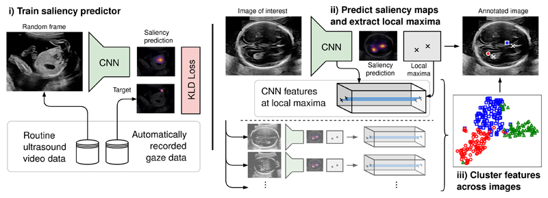
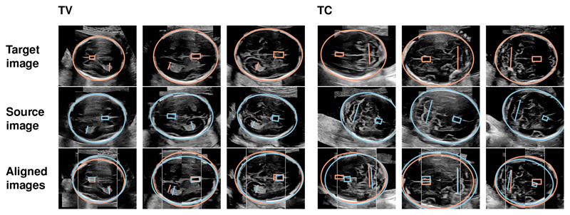
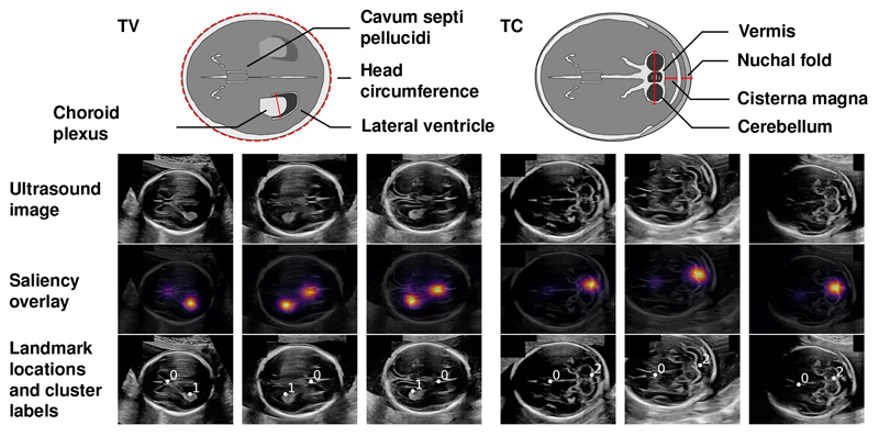
Anatomical landmarks are a crucial prerequisite for many medical imaging tasks. Usually, the set of landmarks for a given task is predefined by experts. The landmark locations for a given image are then annotated manually or via machine learning methods trained on manual annotations. In this paper, in contrast, we present a method to automatically discover and localize anatomical landmarks in medical images. Specifically, we consider landmarks that attract the visual attention of humans, which we term . We illustrate the method for fetal neurosonographic images. First, full-length clinical fetal ultrasound scans are recorded with live sonographer gaze-tracking. Next, a convolutional neural network (CNN) is trained to predict the gaze point distribution (saliency map) of the sonographers on scan video frames. The CNN is then used to predict saliency maps of unseen fetal neurosonographic images, and the landmarks are extracted as the local maxima of these saliency maps. Finally, the landmarks are matched across images by clustering the landmark CNN features. We show that the discovered landmarks can be used within affine image registration, with average landmark alignment errors between 4.1% and 10.9% of the fetal head long axis length.
解剖标志是许多医学成像任务的关键前提条件。通常,给定任务的标志集由专家预先定义。然后,通过人工或基于人工标注训练的机器学习方法对给定图像的标志位置进行标注。相比之下,在本文中,我们提出了一种在医学图像中自动发现和定位解剖标志的方法。具体而言,我们考虑那些吸引人类视觉注意力的标志,我们将其称为 。我们展示了针对胎儿神经超声图像的该方法。首先,通过实时超声检查人员的注视跟踪记录全长临床胎儿超声扫描。接下来,训练一个卷积神经网络(CNN)来预测超声检查人员在扫描视频帧上的注视点分布(显著性图)。然后使用该CNN预测未见过的胎儿神经超声图像的显著性图,并将标志提取为这些显著性图的局部最大值。最后,通过对标志CNN特征进行聚类,在图像之间匹配标志。我们表明,发现的标志可用于仿射图像配准,平均标志对齐误差在胎儿头长轴长度的4.1%至10.9%之间。