Alzubaidi Mahmood, Agus Marco, Alyafei Khalid, Althelaya Khaled A, Shah Uzair, Abd-Alrazaq Alaa, Anbar Mohammed, Makhlouf Michel, Househ Mowafa
College of Science and Engineering, Hamad Bin Khalifa University, Member of Qatar Foundation, Doha, Qatar.
Weil Cornell Medical College-Qatar, Doha, Qatar.
iScience. 2022 Jul 3;25(8):104713. doi: 10.1016/j.isci.2022.104713. eCollection 2022 Aug 19.
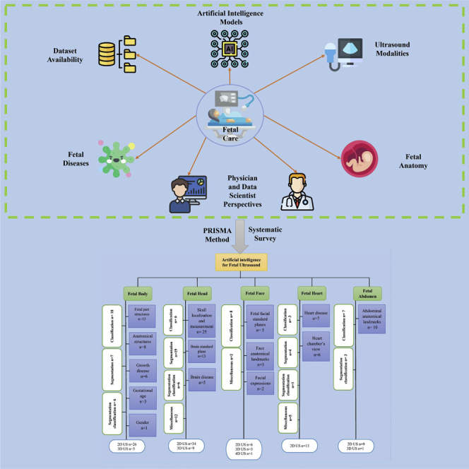
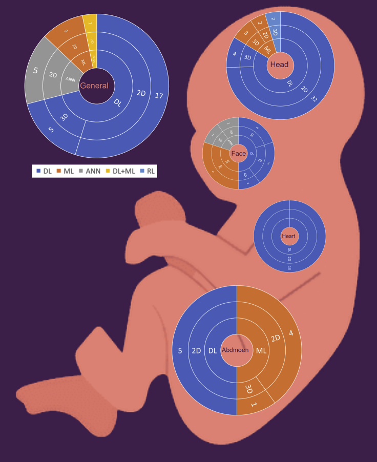
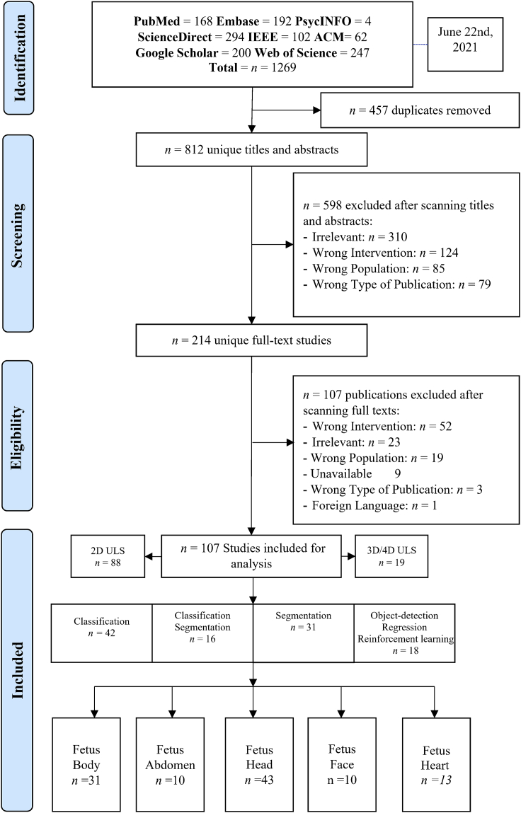
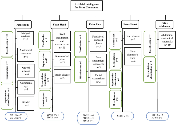
Several reviews have been conducted regarding artificial intelligence (AI) techniques to improve pregnancy outcomes. But they are not focusing on ultrasound images. This survey aims to explore how AI can assist with fetal growth monitoring via ultrasound image. We reported our findings using the guidelines for PRISMA. We conducted a comprehensive search of eight bibliographic databases. Out of 1269 studies 107 are included. We found that 2D ultrasound images were more popular (88) than 3D and 4D ultrasound images (19). Classification is the most used method (42), followed by segmentation (31), classification integrated with segmentation (16) and other miscellaneous methods such as object-detection, regression, and reinforcement learning (18). The most common areas that gained traction within the pregnancy domain were the fetus head (43), fetus body (31), fetus heart (13), fetus abdomen (10), and the fetus face (10). This survey will promote the development of improved AI models for fetal clinical applications.
已经有几项关于利用人工智能(AI)技术改善妊娠结局的综述。但它们并未聚焦于超声图像。本调查旨在探讨人工智能如何通过超声图像辅助胎儿生长监测。我们按照系统评价和Meta分析的首选报告项目(PRISMA)指南报告了我们的研究结果。我们对八个文献数据库进行了全面检索。在1269项研究中,纳入了107项。我们发现二维超声图像(88项)比三维和四维超声图像(19项)更受欢迎。分类是最常用的方法(42项),其次是分割(31项)、分类与分割相结合(16项)以及其他杂项方法,如目标检测、回归和强化学习(18项)。在妊娠领域最受关注的常见部位是胎儿头部(43项)、胎儿身体(31项)、胎儿心脏(13项)、胎儿腹部(10项)和胎儿面部(10项)。这项调查将推动用于胎儿临床应用的改进型人工智能模型的发展。